SlideShare a Scribd company logo
1 of 80
Radiological imaging of endometriosis.
Dr/ ABD ALLAH NAZEER. MD.
Endometriosis is defined as the presence of endometrial
epithelium and stroma in an ectopic site outside the uterine
cavity . Endometriosis occurs in 10% of the female population
and almost, exclusively, in women of reproductive age . The
most common symptoms are dysmenorrhea, dyspareunia,
pelvic pain, and infertility although endometriosis may be
asymptomatic .
Adenomyosis is the presence of endometrial glands and stroma
within the myometrium and it is the cause of uterine
enlargement, menorrhagia, and dysmenorrhea and. The most
common sites for endometrial implantation within the pelvis
are the ovaries, broad and round ligaments, fallopian tubes,
and cervix. Endometriosis can affect the woman in the
reproductive age; however, it cannot be accurately diagnosed
by clinical criteria alone. In addition, the transabdominal and
transvaginal US are not as accurate as MRI.
Superficial endometriosis.
In superficial endometriosis – also known as Sampson's syndrome -
superficial plaques are scattered across the peritoneum, ovaries and
uterine ligaments. These patients tend to have minor symptoms and
usually also less structural changes in the pelvis. At laparoscopy, these
implants may be seen as superficial powder-burn or gunshot lesions.
In deep pelvic endometriosis - also called Cullen's syndrome - there is
subperitoneal infiltration of endometrial deposits. The symptoms are
more severe and related to the localization and depth of invasion. MRI
is of use for the diagnosis of deep infiltrating endometriotic lesions and
for the assessment of disease extension. Preoperative mapping of
disease extension is important to decide whether surgical intervention
is indicated, and if so, for planning complete surgical excision.
Deep pelvic endometriosis.
Endometriomas - also known as chocolate cysts
Develop when superficial endometriotic lesions on the
surface of the ovary invaginate.
Blood produced by such an implant during each
menstrual cycle cannot escape and will accumulate
within the ovary, forming a cyst known as an
endometrioma.
Endometriomas present as complex cystic masses,
often thick-walled with a homogeneous content.
On transvaginal ultrasound, endometriomas may be
seen as thick-walled cysts with low level echoes.
On the left a transvaginal ultrasound image and the
corresponding laparoscopic image during cystectomy.
Sites.
Pelvic:
-Uterine= adenomyosis(50%).
Extra-uterine:
-Ovary 30%.
- Pelvic peritoneum 10%.
-Fallopian tube.
-Vagina.
-Bladder &rectum.
-Pelvic colon.
- Ligaments.
Extra-pelvic
Umbilicus.
Scars(Laparotomy).
Lung &pleura.
Others.
Symptomatology.
- Pelvic pain(65%).
. Dysmenorrhea, especially suggestive of
endometriosis the pain if it occurs after
years of pain free menstruation.
. Deep dyspareunia.
. Chronic pelvic pain.
. Ovulation pain with menstrual irregularity.
. Other types of pain- Sciatica.
-Infertility(35%).
GIT symptoms:
. Diarrhea.
. Constipation.
. Changes in bowel habits.
. Rectal bleeding.
. Pain with defecation.
. Tenesmus.
. Abdominal distension.
. Small caliber stool.
.Colicky abdominal pain.
. Bowel obstruction.
. Bowel perforation.
Urinary symptoms:
-Urgency.
- Frequency.
- Supra-pubic pain.
- Flank pain.
- Urge incontinence.
- Dyspareunia.
- Hematuria.
- Dysuria.
- Backache.
- Hydronephrosis.
Normal anatomy of the female pelvis. (a, b) Axial (a) and
sagittal (b) T2-weighted MR images show the uterosacral
ligaments (arrows in a), rectovaginal pouch (* in b), and
retrocervical area (outlined in b). (c, d) Laparoscopic images
show the uterosacral ligaments (arrows in c), rectovaginal
pouch (* in c), and retrocervical area (outlined in d). (e) Drawing
of the female pelvis (oblique coronal view, superoinferior
orientation) shows the ureters (arrows), uterosacral ligaments
(arrowheads), cardinal ligaments and parametrium (black *),
and round ligaments (white *). Note that the ureters course
lateral to the uterosacral ligaments and immediately caudal to
the cardinal and broad ligaments.
Normal anatomy of the female pelvis.
Ultrasound examination:
Sonographic examinations took place on two perpendicular planes
using PHILIPS HD11 instrument. Pelvic transabdominal US was
performed by using a 3.5-MHz transducer and transvaginal US
examination with 8-MHz transducer. During each Sonographic
examination, the uterine borders, whether regular or irregular, uterine
size, myometrial echotexture and the presence of focal areas with ill-
defined borders or abnormal echotexture were described. When these
areas were present, the following criteria of adenomyosis were
evaluated: presence of heterogeneity, increased or decreased areas of
echogenicity, or presence of myometrial cysts. Another type of
ultrasound that we used in our study is trans-rectal ultrasound which is
necessary in the evaluation of deep infiltrating endometriosis involving
recto-vaginal and utero-sacral ligaments but unfortunately we have no
cases that have the deep infiltration of these regions.
Radiological imaging.
MRI protocols:
MRI performed using T2 weighted images required 4 mm slices in
the sagittal, coronal, and axial planes related to the orientation of
the uterine cavity that used fast spin echo sequences in all the three
planes. T1 weighted spin echo and gradient echo images were
obtained both with and without fat saturation for all patients. High-
resolution body coils were used for data acquisition, and exams
were completed in 30–45 min. T1 weighted fat saturated and T2
weighted images for four major criteria:
1.Borders, size and uterine symmetry.
2.Maximal junctional zone (JZ max) thickness and/or the presence
of an ill-defined, relatively homogenous, low signal intensity,
myometrial area.
3.Maximal (JZ max) to myometrial thickness ratio using the maximal
thickness of JZ and the corresponding thickness of the entire
myometrium obtained at the same level.
4.High intensity spots within the myometrium, the presence of T1
bright/T2 dark signals, suggesting the existence of endometriosis in
any of the anatomical locations.
Sagittal T2-weighted image with fat suppression of 27-year-old woman in second
part of her cycle. Uterus is evaluated between isthmus and end of uterine cavity
(white lines). Junctional zone (short arrows) should be measured from several
sites on anterior and posterior walls. Junctional zone measure can be compared
with entire thickness of myometrium (long arrows) evaluated at same site.
Role of the Junctional Zone Ultrasound studies have been paramount in
understanding the role of the junctional zone, which is intricately linked with its
structural organization and biochemical properties. Pelvic ultrasound, performed
transabdominally in the 1980s and later endovaginally in the 1990s, has permitted
observation of contractions of the junctional zone occurring in the nongravid uterus.
Video recordings have identified these contractions with a speed of 1.2–1.7 mm/s
with a frequency of 3–5 contractions/min. Contractions are classified according to
their direction: from the uterine cervix toward the body, from the body to the cervix,
or both simultaneously . These junctional contractions have been secondarily
described on MRI with particular dynamic sequences. They appear as thin bands
perpendicular to the junctional zone and in low signal intensity on T2-weighted
images. This signal has been partly explained by the venous vasoconstriction that
reduces the quantity of circulating blood in the myometrium. In 2007, Kido et al. used
MRI to illustrate the influence of oral contraceptives on uterine contractions. It was
clearly shown that these contractions have an amplitude, a frequency, and a
direction that correlate with the phase of the menstrual cycle. Thus, in the first part
of the cycle, contractions occur from the cervix toward the body and increase in
intensity until ovulation. These contractions participate directly in the transport of
spermatozoids toward the ovum as reported by Kunz and colleagues by utilizing inert
particles. confirmed this hypothesis by studying the strong link between contractions
issued from the junctional zone and fertility.
Adenomyosis is the presence of endometrial glandular cells and cells of the
chorion more than 2.5 mm from the endometrium-myometrium interface. A
hypertrophic reaction of the smooth-muscle cells surrounding the ectopic
glands is another important element of diagnosis . Involvement of the
myometrium can include the entire interface, termed “diffuse adenomyosis,”
or it can present in a limited area, termed “focal adenomyosis.” These two
forms have similar frequency on hysterectomy samples but are rarely
associated. Adenomyoma, a localized confluence of adenomyotic glands
forming a mass, constitutes an unusual form of adenomyosis.
The actual prevalence of adenomyosis remains unknown. Risk factors for
adenomyosis—most notably, the existence of prior endouterine surgery—
were isolated. Multiparity is another risk factor, which appears
contradictory to the not so well-established link between adenomyosis and
infertility. Adenomyosis preferentially affects women between 40 and 50
years old; however, most forms are asymptomatic, explaining perhaps the
lower rate of detection in young women. A positive diagnosis of
adenomyosis cannot be made reliably on the basis of clinical examination
alone. Supporting evidence, including MRI findings, is necessary to entertain
and confirm the diagnosis.
Radiographic features:
Imaging features are variable and in many instances very subtle. Three
(some say four) forms can be distinguished:
diffuse adenomyosis: most common
focal adenomyosis and adenomyoma: some consider these as separate
(see below).
cystic adenomyosis and adenomyotic cyst: rare
Adenomyosis is usually relatively generalized, affecting large portions of
the uterus (typically the posterior wall), but sparing the cervix. Despite
often marked enlargement of the uterus, the overall contour is usually
preserved.
In some cases it may also be localized, forming a mass. In such cases the
term adenomyoma may be used, although there appears to be some
disagreement about whether the terms focal adenomyosis and
adenomyoma refer to exactly the same entity (please refer to the article
on adenomyoma for further discussion).
A rare variant is cystic adenomyosis which is believed to be the result of
repeated focal hemorrhages resulting in cystic spaces filled with altered
blood products.
Pelvic ultrasound:
Ultrasound is usually the first and often the only imaging modality employed
to investigate menorrhagia and dysmenorrhea. Unfortunately, the sonographic
features of adenomyosis are variable, and may be absent. The reported
sensitivity and specificity of trans-abdominal ultrasound are 32-63 % and 95-97
% respectively .
The spectrum of findings includes:
normal appearing uterus, focal or diffuse bulkiness, typically of the posterior
wall heterogeneous echogenicity (heterogenous myometrial echotexture)
hyperechoic: islands of endometrial glands
hypoechoic: associated muscle hypertrophy
a "venetian blind" appearance may be seen due to subendometrial
echogenic linear striations & acoustic shadowing where endometrial tissues
cause a hyperplastic reaction.
small myometrial: sub endometrial cysts: thickening of the transition zone
sometimes can be visualized as a hypoechoic halo surrounding the endometrial
layer of 12 mm or more thickness subendometrial echogenic linear striations
subendometrial echogenic nodules
When an adenomyoma is present, then appearances may closely mimic those
of a uterine fibroid, which may also co-exist.
Hysterosalpingogram (HSG):
May show diverticula extending into the myometrium .
CT is unable to diagnose adenomyosis: but may suggest its presence when
uterine enlargement is present. Distinguishing between adenomyosis and uterine
fibroids on CT is difficult if not impossible, although presence of calcifications
strongly favours the latter .
Pelvic MRI: MRI is the modality of choice to diagnose and characterize
adenomyosis, and T2 weighted images (sagittal and axial) are most useful. It has a
very diagnostic accuracy with a sensitivity of 78-88% and a specificity of 67-93% .
The most easily recognised feature is thickening of the junctional zone of the uterus
to more than 12 mm, either diffusely or focally (normal junctional zone measures
no more than 5 mm)
T2: typically a region of adenomyosis appears as an ill-defined ovoid/diffuse
region of thickening often with small high T2 signal regions representing
small regions of cystic change the region may also have a striated
appearance.
T1: Foci of high T1 signal are often seen, indicating menstrual hemorrhage into the
ectopic endometrial tissues.
T1 C+ (Gd): contrast enhanced MR evaluation is usually not indicated in adenomyosis,
however if performed, it shows enhancement of the ectopic endometrial glands.
HSG with adenomyosis of the uterus.
CT with adenomyosis of the uterus.
42-year-old woman who presented with chronic pelvic pain. Coronal (A) and
axial (B) T2-weighted images reveal cystic area surrounded by low-signal-
intensity ring (arrow). Overall appearance on MRI is that of cystic adenomyoma.
36-year-old woman who underwent MRI evaluation for polymyomatous uterus
discovered on ultrasound. On sagittal T2-weighted image, posterior myoma
(black arrow) is visible in frank hyposignal. Focal adenomyosis (white arrow) is
visible on anterior uterine wall, presenting as ill-defined area in hyposignal-
containing cysts. Hyperintense linear or reticulated streaks extend from
endometrium into markedly thickened anterior myometrium (arrowheads).
Diffuse adenomyosis of the uterus.
Adenomyosis of the uterus.
Diffuse adenomyosis of the uterus.
Focal uterine adenomyosis.
Adenomyosis with endometrioma.
MRI (sagittal T2WI) shows adenomyosis with abnormal increase in thickness
of junctional zone (12 mm) and abnormal high T2 SI of the myometrium
represents abnormal stromal glands inside the myometrium. B: MRI (coronal
T2WI) shows the same finding as well as bilateral ovarian simple cysts.
Sagittal transvaginal sonography demonstrates
myometrial anechoic lacunae specific of
adenomyosis involving the ventral and the dorsal
myometrium (A). Transversal transabdominal
examination shows a very large asymmetric uterus
with thickening of the dorsal myometrium.. (B).
Transvaginal sonography demonstrates diffuse
adenomyosis involving the ventral myometrium and
a dorsal subserous leiomyoma. Sagittal T2-
weighted magnetic resonance image through the
uterus shows numerous high-intensity spots in the
inner myometrium, thickening of junctional zone
(JZ) (13 mm) and ratio max >40% (D). Axial T2-
weighted magnetic resonance image demonstrates
diffuse thickening of JZ both ventrally and dorsally,
consistent with severe adenomyosis. Numerous foci
of high signal representing the heterotopic
endometrium are present (E). Sagittal T2-weighted
magnetic resonance image through the uterus (F):
there is an ill-defined mass centred around the
endometrium. Several foci of increased signal
consistent with heterotopic endometrium are
present in the inner myometrium without mass
effect on the endometrial cavity. In contrast,
leiomyoma is very hypointense and has well-
defined borders (F).
Linear striations, sonographic findings suggestive of
adenomyosis, in a 47-year-old woman with pelvic pain and
dysmenorrhea. A, Sagittal transvaginal sonogram shows an
enlarged uterus. A heterogeneous echo texture involving the
anterior myometrium is shown with linear striated shadowing
(arrowheads). B, Sagittal fat-suppressed T2-weighted MRI
shows marked adenomyosis involving the anterior uterine wall
manifested as a thickened junctional zone with scattered
myometrial cysts (arrows). C, Sagittal T1-weighted gradient
echo MRI (TR, 270 milliseconds; TE, 5 milliseconds) obtained
after administration of gadolinium dimeglumine shows
enhancement of the endometrium and myometrium.
Myometrial cysts (arrows) do not show enhancement
Polypoid Adenomyoma
An adenomyoma is occasionally located in a submucosal or subserosal
position. A submucosal adenomyoma manifesting as a polypoid mass
protruding into the endometrial cavity has been termed “polypoid
adenomyoma. This lesion usually arises from the uterine corpus,
occasionally from the lower uterine segment and cervix. It can be seen as a
pedunculated or sessile mass. Its appearance on MRI as an uncommon
variant of adenomyosis has been described. Use of the term polypoid
adenomyoma has been controversial. Although some authors have
discussed this lesion as a distinct entity, the gynecologic pathologic
literature reflects the opinion that the term polypoid adenomyoma refers
to a polypoidal form of a circumscribed adenomyotic mass.
Atypical Polypoid Adenomyoma
This is an uncommon variant of polypoid adenomyoma characterized by
atypical endometrial glands and often squamous metaplasia and a cellular
smooth muscle stroma. Before its delineation, this lesion was often
considered malignant. The distinction between the typical and atypical
adenomyoma depends on histologic examination because the radiologic
findings are similar.
Polypoid adenomyomas. A, Sagittal transvaginal
sonogram in a 30-year-old woman with a history of
menometrorrhagia shows an indistinct endometrial echo
complex with variable thickness; a cyst is shown adjacent to
it (arrow). Sonohysterography was performed for further
evaluation of these findings. B, Sagittal image from the
sonohysterogram reveals an intracavitary mass containing a
cystic focus (arrow). The echo texture of this mass, unlike an
endometrial polyp, is similar to that of the myometrium. No
endometrial abnormality was seen. C, Transvaginal
sonogram in a 39-year-old woman with menorrhagia shows
a large prolapsing adenomyoma distending the cervical
canal (arrow). The echo texture of the mass is similar to that
of the myometrium; it is, therefore, sonographically
indistinguishable from a myoma. The arrowhead points to
the posterior vaginal wall.
Cystic adenomyoma in a 37-year-old woman with a history of
infertility. A, Multiplanar display from 3D TVS of the uterus
shows a cystic mass within the right side of the uterine corpus
containing low-level echoes. The bottom left image is a
coronal plane through the endometrial cavity showing 2
normal cornua. Top left, axial; top right, sagittal; bottom left,
coronal. B, Axial T1-weighted gradient echo opposed phase
MRI (TR, 225 milliseconds; TE, 2 milliseconds) shows a
hyperintense lesion within the myometrium. Lack of a
chemical shift artifact around the lesion confirms the absence
of fat within. C, Axial T2-weighted MRI (TR, 4000 milliseconds;
TE, 100 milliseconds) image shows an intermediate to
hyperintense signal in the cystic mass (arrow), showing
shading, consistent with chronic hemorrhage.
Multiple fibroids and diffuse adenomyosis in a 50-year-old woman. A, Sagittal
transabdominal sonogram shows a bulky uterus due to multiple fibroids. The
endometrial cavity is distorted by the presence of fibroids (arrows). B, Sagittal T2-
weighted MRI shows diffuse adenomyosis with marked thickening of the junctional zone
(arrow) and the presence of multiple fibroids (arrowheads). In the presence of large
multiple fibroids, diagnosing adenomyosis on sonography can be a difficult task.
Ultrasound of the pelvis
showed left ovarian cystic
lesion with internal echoes
and two hyperechogenic
areas inside corresponding
to hemorrhagic content of
the cyst. C: Axial fat sat
MRI showed hyperintense
lesion at left ovary. D: The
lesion showed drop of SI at
T2WI MRI which is
pathognomonic of
endometrioma. E, F:
Sagittal and coronal T2WI
MRI showed low SI of the
lesion which is
pathologically proved
endometrioma by
laparoscopic examination
and biopsy.
Solid fibrotic thickening of the uterosacral ligament in a 40-year-old woman with
endometriosis and pelvic pain. (a) Axial T2-weighted MR image shows low-signal-
intensity, mass like thickening of the proximal right uterosacral ligament (arrows). The
normal-appearing sacral portion of the ligament (arrowheads) is also depicted. (b)
Sagittal T2-weighted MR image shows band like fibrotic adhesions above the ligament
(arrows) and shading within the superior right ovarian endometriomas (arrowheads).
Endometriosis of the vesicouterine
pouch in a 27-year-old woman with chronic
pelvic pain. Sagittal T2-weighted (a),
sagittal fat-suppressed T1-weighted (b),
and oblique coronal T2-weighted (c) MR
images depict irregular thickening of the
upper external aspect of the bladder wall
(arrowheads in a, arrows in c) that partially
obliterates the vesicouterine recesses. Tiny
bloody foci (arrow in b) are also seen,
adding specificity to the finding.
Endometriotic involvement of
the vesicovaginal septum in a
37-year-old woman who had
undergone hysterectomy and
presented with chronic dysuria.
US showed a focal mass bulging
into the posterior vesical wall.
(a) Sagittal T2-weighted MR
image shows a hypointense
nodule (arrows) in the
vesicovaginal septum. (b) On an
axial fat-suppressed T1-
weighted MR image, the lesion
(arrowhead) has high signal
intensity, which did not
decrease on subsequent
images. This finding indicates
proteinaceous or bloody
content and causes the lesion to
resemble an endometrioma.
Intrinsic bladder endometriosis in a
22-year-old woman who had undergone a
cesarean section and presented with cyclic
voiding symptoms but no hematuria. (a)
Axial T2-weighted MR image shows
hypointense irregular focal wall
thickening (arrow) on the left
posterolateral aspect of the bladder. Note
the relative sparing of the perivesical fat
(*) and the absence of enlarged lymph
nodes. (b) Axial fat-suppressed T1-
weighted MR image reveals that the area
of wall thickening contains small
intermingled hyperintense foci (arrow), a
finding that indicates bloody content and
that was crucial for differentiating
between endometriosis and cancer. The
patient underwent cystoscopy to exclude
bladder cancer, and the tentative
diagnosis of intrinsic bladder
endometriosis was confirmed.
Bladder endometriosis with urethral involvement in a 31-year-
old woman. (a, b) Sagittal T2-weighted (a) and fat-suppressed
T1-weighted (b) MR images obtained during the patient’s
menstrual period show massive irregular thickening of the
anteroinferior bladder wall (arrow in a) that extends to the
vesical trigone and the upper urethra and is associated with tiny
interspersed bloody foci (arrowhead in b), findings that are
consistent with a diagnosis of bladder endometriosis. Note the
smooth outer contour of the bladder wall, as well as the normal-
appearing prevesical fat (arrowheads in a). Such massive focal
wall thickening with no signs of extravesical extension or
perivesical fat stranding makes a diagnosis of bladder cancer
less likely. (c) Axial T2-weighted MR image shows irregular
hypointense thickening on the anterior
contour of the urethra (*), a finding that indicates endometriotic
involvement by direct extension from the bladder lesion.
Bladder endometriosis with ureteral
involvement in a 33-year-old woman who
presented with cyclic dysuria and pelvic pain,
but no signs of hematuria or weight loss. (a)
Axial T2-weighted MR image reveals
hypointense irregular focal thickening located
in the left posterolateral aspect of the bladder
wall (arrows) and extending into the
perivesical fat (arrowheads). (b) Sagittal T2-
weighted MR image shows the perivesical and
bladder wall thickening (arrow), as well as
upstream ureteral dilatation (arrowheads). (c)
Oblique coronal T2-weighted MR image
shows an infiltrating lesion (*) encasing the
lower third of the ureter (arrows). (d) Three-
dimensional contrast-enhanced coronal
reformatted fat-suppressed T1-weighted MR
image shows a dilated left ureter (solid
arrows) and a deep infiltrating endometriotic
lesion involving the wall of the bladder (B)
and the lower third of the ureter
(arrowheads). There is also a hyperintense
component in the bladder submucosa (open
arrow), a finding that indicates bloody
content. C = cervix, U = uterus.
Solid endometriosis of the bladder and right round ligament in a 40-year-old woman. (a, b) Sagittal (a) and axial (b)
T2-weighted MR images show a poorly marginated, low-signal-intensity mass (arrowheads) in the right posterior
bladder wall that extends posteriorly, obliterating the vesicouterine pouch. Intralesional 1–4-mm high-signal-intensity
foci (arrows in a) represent ectopic endometrial glands. The right round ligament is thickened (arrows in b) as it
courses inferiorly toward the canal of Nuck. (c, d) Axial T1-weighted gradient-echo fat-suppressed MR images
obtained before (c) and after (d) the administration of a gadolinium-based contrast material show T1 hyperintensity
in some of the endometrial glands within the posterior bladder lesion (arrow in c) and solid enhancement of both the
bladder mass (arrowheads in d) and the thickened round ligament (arrow in d).
Ureteral endometriosis in a 31-year-old woman
with chronic pelvic pain and left hydronephrosis.
(a) Axial T2-weighted MR image shows
hypointense irregular and spiculated thickening
and stranding of the perivesical fat on the left
side (arrows). The ureter (arrowhead) traverses
the involved area, a finding that is suggestive of
infiltrating endometriosis. (b) Para sagittal T2-
weighted MR image reveals moderate dilatation
of the left ureter (arrowheads) to the level of the
insertion of the ipsilateral uterosacral ligament
(arrow), which is irregular and thickened,
indicating associated deep endometriosis. (c)
Fat-suppressed T1-weighted MR image shows a
cystic lesion (arrow) whose high signal intensity
indicates proteinaceous or bloody content. The
lesion resembles an endometrioma located
medial to hypointense thickening of the
perivesical fat. (d) Three-dimensional contrast-
enhanced fat-suppressed excretory phase T1-
weighted MR urogram shows the repercussions
of the findings in a–c to the urinary system. The
left ureter is severely narrowed in its lower third
(arrowhead), with associated upstream
dilatation up to the renal pelvis (*).The lesion
(arrow) is seen adjacent to the narrowed ureter
and represents an endometrioma.
Endometriotic involvement of
the retrocervical area in a 33-
year-old woman with
dyspareunia. At earlier MR
imaging examinations, an
endometrial implant was seen in
this area. (a) Sagittal T2-weighted
MR image shows an irregular,
solid, nodular hypointense mass
(arrow) slightly inferior to the
uterine torus, with intermingled
hyperintense foci (arrowhead). (b)
On an axial fat-suppressed T1-
weighted MR image, the
intermingled foci (arrow) remain
hyperintense, a finding
that indicates bloody content.
Solid invasive endometriosis of the rectosigmoid colon in a 40-year-old woman. (a) Lateral image from
a double-contrast barium examination shows circumferential narrowing of the rectosigmoid colon,
with mass effect, speculation, and pleating of the anterior margin (arrow). (b) Sagittal T2-weighted
fast spin-echo MR image shows extraluminal findings suggestive of solid invasive endometriosis. The
mushroom cap sign represents the low-signal-intensity core of fibrotic endometriosis and hypertrophic
muscularis propria (*) capped by high-signal-intensity mucosa (straight arrows). Invasion of the
serosal surface of the uterus and obliteration of the cul-de-sac (arrowheads) are seen, with high-
signal-intensity 1–3-mm foci (curved arrow) representing ectopic endometrial glands.
Endometriotic involvement of the uterine ligaments in a 31-year-old woman with
chronic pelvic pain and a known history of endometriosis. Axial gadolinium-
enhanced fat-suppressed late-phase T1-weighted MR image shows homogeneous
late enhancement of thickened round uterine ligaments (arrows) due to
involvement of their uterine insertions by an endometriotic lesion.
Endometriotic involvement of the posterior pelvic compartment in a 29-year-old woman
with hypermenorrhea, dyspareunia, and dysmenorrhea. Earlier MR imaging showed
retrocervical endometriosis with fibrotic obliteration of the rectovaginal pouch. Sagittal (a)
and axial (b) T2-weighted MR images show an irregular hypointense mass (arrowhead)
that extends from the posterior cervix inferiorly to the vaginal fornix. Note the presence of
a subserous leiomyoma in the anterior uterine wall (black arrow in a), focal adenomyosis in
the corporal anterior uterine wall (white arrow), and a right follicular ovarian cyst (* in b).
Endometriotic involvement of the
uterosacral ligaments in a 31-year-old
woman who presented with pelvic pain,
dyspareunia, and painful defecation.
Earlier MR imaging showed involvement
of these structures. (a, b) Coronal (a) and
axial (b) T2-weighted images show
hypointense irregular spiculated nodular
thickening of the right uterosacral
ligament (white arrow in a), with minimal
intermingled hyperintense foci. These
findings create an arciform abnormality
(arrowheads in b) that extends to the
anterior rectal wall, where there
is focal parietal thickening (black arrow).
(c) Axial fat-suppressed T1-weighted MR
image shows hyperintense foci (solid
arrows), a finding that indicates bloody
content. Open arrow indicates focal
parietal thickening. (d) Laparoscopic
image obtained in a different patient
shows involvement of the uterosacral
ligaments (arrows).
Isolated endometriotic involvement of the vagina in a 38-year-old woman who
had undergone hysterectomy and presented with dyspareunia and chronic pelvic
pain. Earlier MR imaging showed vaginal involvement. (a) Axial T2-weighted MR
image shows a hypointense nodule (arrows) in the posterior upper third of the
vagina. (b) Axial T1-weighted MR image shows minimal intermingled
hyperintense foci (arrowheads), a finding that indicates bloody content.
Endometriotic involvement of the ischioanal fossa in a 32-year-old woman who presented
with right hip pain. The patient had no history of perineal surgery. (a) Axial T2-weighted MR
image shows solid, predominantly glandular endometriosis (arrows) in the ischioanal fossa
located close to the right ischiatic tuberosity and involving the internal obturator muscle and
anal sphincter. (b, c) Axial T1-weighted (b) and fat-saturated T1-weighted (c) MR images show
intermingled hyperintense foci (arrow), a finding that indicates bloody content. Arrowhead in
b indicates cortical sclerosis. (d) Coronal T1-weighted MR image shows significant bone
reaction (cortical sclerosis) in the medial aspect of the right ischiatic tuberosity (arrowhead).
Sigmoid endometriosis in a 30-year-old woman
who experienced painful defecation and
hematochezia during her menstrual period. (a, b)
Sagittal (a) and coronal (b) T2- weighted MR
images obtained after the administration of a
saline rectal enema and intravaginal aqueous gel
show hypointense nodular thickening of the
sigmoid wall (arrows) adhering to the posterior
uterine serosa (arrowhead in a). (c) Sagittal
gadolinium-enhanced fat suppressed late-phase
T1-weighted MR image shows enhancement of the
lesion (arrow). The findings in a–c are consistent
with a diagnosis of sigmoid endometriosis.
Cyclic sciatica in a 27-year-old woman.
Earlier MR imaging showed sciatic nerve
involvement. (a) Axial MR image shows
hypointense irregular spiculated soft-tissue
thickening (arrows) in the left sciatic notch.
(b) Axial fat-suppressed T1-weighted MR
image shows minimal intermingled
hyperintense foci (arrows), a finding that
indicates bloody content. (c) Axial
gadolinium-enhanced fat suppressed
late-phase T1-weighted MR image shows
enhancement of the lesion (arrows).
Deep endometriosis at various sites in a 35-year-old
woman who presented with chronic pelvic pain,
infertility, and dysuria. Earlier MR imaging showed
endometriotic involvement of the urinary bladder,
rectovaginal septum, and retrocervical area. (a, b)
Sagittal (a) and axial (b) T2-weighted MR images
demonstrate hypointense focal thickening of the
right posterolateral wall of the bladder (black
arrow) with punctate intermingled bleeding,
along with an extraperitoneal, irregular hypointense
nodule (white arrows) that mainly involves the
retrocervical area and the upper portion of the
rectovaginal septum. (c) Axial fat-saturated T1-
weighted MR image shows the nodule. Arrowhead
= punctate bleeding.
37 year old woman with a history of dysmenorrhea. Images: (a) coronal T2-
weighted and (b) axial T1 with fat suppression and gadolinium. Enlarged right
ovary with multiple cysts (*) and an endometrioma with intermediate signal on T2
and hyperintense on T1 with fat suppression and gadolinium (à black).
Relatively hyperechoic
SOL at right ovary. B: MRI
(axial T1WI) shows
hyperintense content of
the right ovary. C: Axial
T2WI shows low SI of the
cyst of the right ovary
(shading sign). D: Axial
T1WI with fat sat shows
hyperintense cystic right
ovarian lesion
(hemorrhage). E, F: MRI
(sagittal and coronal
T2WI) revealed low SI of
right ovarian lesion.
37 year old woman. Infertility study. Images: (a) Sagittal T2-weighted and (b) coronal
T2 and (c) axial T2. Bilateral ovarian endometrioma and left hydrosalpinx (à white).
Endometrioma at ultrasound and laparoscopy.
T2- and fat sat T1-images of an endometrioma with hypointensity on T2 (shading),
fluid-fluid levels on T2 (left) and hyperintense blood on T1WI with fat sat (right).
Endometrioma.
Endometrioma of the left ovary in a
33-year-old woman. (a, b) Axial T1-
weighted in-phase (a) and opposed-
phase fat-suppressed (b) gradient-
echo MR images show a T1-
hyperintense left ovarian lesion, a
feature suggestive of endometrioma.
(c) Axial T2-weighted MR image
shows lower signal intensity in the
endometrioma than in the adjacent
normal ovarian follicles (arrows). (d,
e) Diffusion-weighted MR images
obtained with b values of 50 (d) and
800 (e) sec/mm2 show low to
intermediate signal intensity in both
the endometrioma and the normal
follicles. (f) ADC map from diffusion-
weighted MR imaging shows
restricted diffusion in the
endometrioma relative to that in the
adjacent ovarian follicles.
Left ovarian endometriomas, bilateral hematosalpinges,
and fibrotic solid invasive endometriosis in the posterior
uterus of a 47-year-old woman with severe pelvic pain. (a)
Axial T1-weighted in-phase gradient-echo MR image shows
two foci of high signal intensity in the left ovary (arrow) and
multiple foci of high signal intensity in the left anterior and
midline posterior pelvis (arrowheads). (b) Axial opposed-
phase T1-weighted fat-suppressed gradient-echo MR image
shows persistent high signal intensity in all three regions, a
finding that helps exclude the presence of a fat-containing
mature cystic teratoma. The improved dynamic range
achieved with fat suppression facilitates visualization of a
subcentimeter endometrioma of the anterior left ovary
(arrow). (c) Axial T2-weighted fast spin-echo MR image
shows low signal intensity within the left ovarian
endometriomas (solid arrow) and lesser degrees of T2 signal
hypointensity within the dependent portions of the loculi in
the left anterior and midline posterior pelvis (arrowheads).
The posterior uterus is markedly thickened with low-signal-
intensity soft tissue (F) containing scattered 2–4-mm foci of
high signal intensity (open arrows). Although the tissue has
the appearance of adenomyosis, it is separated from the
posterior junctional zone (JZ). These findings are suggestive
of fibrotic endometriosis. (d, e) Sagittal (d) and coronal fat-
suppressed (e) T2-weighted MR images show dilatation of
the right and left fallopian tubes, respectively (arrows). In d,
extension of fibrotic endometriosis (F) into the posterior
uterus is seen. The patient later underwent hysterectomy
and bilateral salpingo-oophorectomy, at which the MR
imaging findings were confirmed.
Right ovarian endometrioma in a 35-year-old woman that
might have been misinterpreted as a mature cystic teratoma
with reliance on short inversion time inversion-recovery
(STIR) imaging alone to detect Intralesional fat. (a) Axial T1-
weighted spin-echo MR image shows a high-signal-intensity
mass (arrow) within the right adnexa. (b) Coronal STIR MR
image shows that the mass (arrow) has low signal intensity
similar to that of suppressed fat. (c) Axial fat-suppressed
gradient-echo MR image shows high signal intensity of the
mass (arrow), a finding that helps confirm that it is not
composed of fat. The low signal intensity of the mass on the
STIR image could be secondary to either T1- or T2-
shortening effects but cannot be considered indicative of fat
content. To avoid this pitfall of STIR MR imaging of the
female pelvis, MR systems capable of performing chemical-
shift fat suppression should be used.
Bilateral endometriomas, solid endometriosis of the posterior
uterus, and left-sided peritoneal inclusion cyst in a 43-year-old
woman. (a, b) Axial T1-weighted in-phase (a) and fat-suppressed
opposed-phase (b) gradient-echo MR images show bilateral
hyperintense adnexal lesions (arrows) that retain high signal
intensity with fat suppression. A peritoneal inclusion cyst (*) is also
shown posterior to the uterus (U). Susceptibility artifacts anterior to
the rectus muscles (arrowheads in a) indicate that the patient has
undergone a previous surgical procedure. (c) Axial T2-weighted MR
image shows shading within the bilateral endometriomas (straight
arrows). The normal endometrial complex and junctional zone are
shown in the anterior uterus (arrowheads). The posterior uterus is
markedly enlarged by poorly marginated soft tissue (E) with low T2
signal intensity and scattered internal 1–2-mm foci of higher signal
intensity (curved arrow) representing infiltration of solid
endometriosis into the uterine wall. Loculated fluid posterior to the
left ovary represents the peritoneal inclusion cyst
53 year old woman. Background history of hysterectomy with cyclic hematuria. Images: (a)
Sagittal T2-weighted, (b) coronal T2, (c) axial T2 and (d) Cystoscopy. On the bladder roof at
left (à white) a mass of 1.5 x 1.1 cm can be seen, this is an intermediate T2 signal with
undefined borders infiltrating the bladder wall, being located in the bladder lumen (à black).
In the cystoscopy can be observed, in left top wall, a sessile uplifted lesion of approximately
1 cm. The remainder of the bladder mucosa appears normal and without lesions.
31 year old woman with a history of dysmenorrhea. Images: (a) coronal T2-weighted (b) axial
T1 and (c) coronal T2. Left ovarian endometrioma (à white) of 9.3 x 7.6 cm attached to
the uterine torus by a deep endometriosis foci (à white curved). Infiltration of the distal ureter
by fibrotic endometrial implant (à) that is associated with hydroureteronephrosis (*).
46 year old woman. Nodule of the abdominal wall under study. Images: (a) sagittal T2-weighted
and (b) axial T1 and (c) axial T1 with fat-suppression and gadolinium. Solid node 20 x 12mm on
abdominal wall at the hypogastrium level lateralized to the left, heterogeneous signal (with
hypointense focus) on T2 and isointense on T1 (white circle) with discrete and homogeneous
enhancement with contrast. External marker (*). Pathology compatible with endometriosis foci.
33 year old woman. Palpable nodule in relation to episiotomy. Images: (a) axial
T2-weighted and (b) axial T1. Lobulated nodule in the right perianal region on
episiotomy site (à white). Slightly hyperintense on T1 and isointense on T2.
Transvaginal US revealed abnormal increase in thickness of myometrium in
a retroverted uterus. B: MRI (sagittal T2WI) shows retroverted uterus with
abnormal increase thickness in junctional zone (10 mm) and diffuse
abnormal high T2 SI of myometrium abnormal stromal glands. C: MRI
(coronal T2WI) shows the same findings as image B. Examination.
Decidualized endometriosis in a 36-
year-old woman in the 12th week of
pregnancy. (a, b) Sagittal T2-weighted
fast spin-echo MR images show an
intrauterine gestational sac, a focal
contraction (C) of the posterior
myometrium, and an anterior placenta
(P). The bilobed low-signal-intensity
mass above the uterus represents an
endometrioma (E). Mural nodules with
higher signal intensity within the
endometrioma (arrows in b) represent
decidualized endometriosis. (c, d) Axial
T1-weighted fat-suppressed (c) and T2-
weighted fast spin-echo (d) MR images
show the typical signal intensity of an
endometrioma, with T1 hyperintensity
and T2 shading, and the higher T2
signal intensity of decidualized
endometriosis (arrow in d).
Hematometrocolpos, hematosalpinx,
and endometriomas in a 15-year-old girl
with an obstructing transverse vaginal
septum. (a) Coronal T2-weighted fat-
suppressed MR image shows dilatation
of the endometrium (E), endocervix (C),
and proximal vagina (V) due to a
transverse vaginal septum (not shown).
(b, c) Axial T1-weighted fat-suppressed
(b) and T2-weighted fast spin-echo (c)
MR images obtained at the level of the
uterus show the distended vaginal canal
(V) and segments of a dilated right
fallopian tube (arrow). These structures
have similar high T1 signal intensity and
intermediate T2 signal intensity. (d)
Axial T1-weighted fat-suppressed MR
image obtained at the level of the right
ovary shows multiple subcentimeter
high-signal-intensity endometriomas
(arrows), findings confirmed at
resection of the vaginal septum.
Endometrioid cystadenocarcinoma of the left ovary
in a 51-year-old woman with long-standing
endometriosis. (a) Axial T2-weighted MR image
shows a cystic pelvic mass that also contains solid
soft tissue (arrow). The signal of the cystic
component was slightly hypointense to that of the
bladder (not shown). (b, c) Axial T1-weighted fat-
suppressed gradient-echo MR images obtained
before (b) and after (c) the administration of a
gadolinium-based contrast material show that the
cystic component has T1 hyperintensity, a finding
suggestive of proteinaceous or hemorrhagic
content. The signal intensity of the solid
component of the mass, relative to that of the
cystic portion, is lower in b and higher in c. (d) MR
image obtained by subtracting the unenhanced
image dataset from the contrast-enhanced image
dataset facilitates the detection of enhancing
tissue (arrow) by increasing the dynamic range
(61) and eliminating the high signal intensity of
nonenhancing hemorrhagic and proteinaceous
tissue. (e, f) Unenhanced T1-weighted fat-
suppressed (e) and gadolinium-enhanced
subtraction (f) MR images obtained at a lower
level show additional enhancing nodular
components (arrows in f) in the tumor. A 1-cm right
ovarian endometrioma (arrow in e) is also seen.
Endometriosis involving appendix. A, Mucocele formation in 38-year-old woman with chronic right
lower quadrant pain. CT image obtained with oral and IV contrast material shows lobulated, low
attenuation cystic mass (arrow) in right lower quadrant. Surgery and pathology confirmed mucocele
secondary to endometriotic implant with obstruction of appendix. B, Acute appendicitis in 24-year-old
woman with right lower quadrant pain and fever. CT image obtained with oral and IV contrast material
shows dilated, fluid-filled appendix (black arrow) compatible with acute appendicitis. Soft-tissue density
at base of appendix (white arrow) was confirmed as endometriotic implant at pathology.
Infected endometrioma in 38-year-old woman with pelvic pain and fever of unknown source. A, CT
image obtained initially shows large pelvic mass (arrows) that is uniformly low attenuation. Ovarian
origin of mass was suspected. B, Transvaginal sonogram confirms that mass originates from right
ovary. There were homogeneous low-level echoes and diagnosis of endometrioma was made. No
other source of patient’s fever was identified and she was taken to surgery for further evaluation of
mass. Intraoperatively, 16 × 12 cm mass was densely adherent to adjacent structures. Cyst puncture
yielded 800 mL of foul-smelling, purulent contents. Pathology confirmed endometrioma with
superimposed acute and chronic inflammation and salpingitis. Cursors delineate mass.
Thank You.

More Related Content

What's hot

Presentation1.pptx, radiological imaging of beign breast diseases
Presentation1.pptx, radiological imaging of beign breast diseasesPresentation1.pptx, radiological imaging of beign breast diseases
Presentation1.pptx, radiological imaging of beign breast diseasesAbdellah Nazeer
 
Uterine artery embolisation
Uterine artery embolisationUterine artery embolisation
Uterine artery embolisationmeducationdotnet
 
First trimester ultrasound Dr. Muhammad Bin Zulfiqar
First trimester ultrasound Dr. Muhammad Bin ZulfiqarFirst trimester ultrasound Dr. Muhammad Bin Zulfiqar
First trimester ultrasound Dr. Muhammad Bin ZulfiqarDr. Muhammad Bin Zulfiqar
 
Presentation1.pptx, radiological imaging of uterine lesions.
Presentation1.pptx, radiological imaging of uterine lesions.Presentation1.pptx, radiological imaging of uterine lesions.
Presentation1.pptx, radiological imaging of uterine lesions.Abdellah Nazeer
 
Obstetrics doppler ultrasound
Obstetrics doppler ultrasoundObstetrics doppler ultrasound
Obstetrics doppler ultrasoundBharti Gahtori
 
Ultrasound Imaging of Placenta
Ultrasound Imaging of PlacentaUltrasound Imaging of Placenta
Ultrasound Imaging of PlacentaVishwanath R S
 
MR BASED IMAGING OF FEMALE PELVIC FLOOR
MR BASED IMAGING OF FEMALE PELVIC FLOORMR BASED IMAGING OF FEMALE PELVIC FLOOR
MR BASED IMAGING OF FEMALE PELVIC FLOORSumiya Arshad
 
Imaging in obstetrics & gynaecology part 2
Imaging in obstetrics & gynaecology part 2Imaging in obstetrics & gynaecology part 2
Imaging in obstetrics & gynaecology part 2drmcbansal
 
Presentation1.pptx, radiological imaging of ectopic pregancy.
Presentation1.pptx, radiological imaging of ectopic pregancy.Presentation1.pptx, radiological imaging of ectopic pregancy.
Presentation1.pptx, radiological imaging of ectopic pregancy.Abdellah Nazeer
 
Fetal Neurosonogram
Fetal Neurosonogram Fetal Neurosonogram
Fetal Neurosonogram nasrat1949
 
Presentation1.pptx, radiological imaging of scrotal diseases.
Presentation1.pptx, radiological imaging of scrotal diseases.Presentation1.pptx, radiological imaging of scrotal diseases.
Presentation1.pptx, radiological imaging of scrotal diseases.Abdellah Nazeer
 
First trimester ultrasound
First trimester ultrasoundFirst trimester ultrasound
First trimester ultrasoundRoshan Valentine
 
Ultrasound of breast
Ultrasound of  breastUltrasound of  breast
Ultrasound of breastLALIT KARKI
 
Placenta ultrasound
Placenta ultrasoundPlacenta ultrasound
Placenta ultrasoundDoaa Gadalla
 
USG AND DOPPLER IN DIAGNOSIS AND MANAGEMENT OF IUGR
USG AND DOPPLER IN DIAGNOSIS AND MANAGEMENT OF IUGRUSG AND DOPPLER IN DIAGNOSIS AND MANAGEMENT OF IUGR
USG AND DOPPLER IN DIAGNOSIS AND MANAGEMENT OF IUGRshiv lasune
 
Interventional ultrasound in obstetrics dr rabi
Interventional ultrasound in obstetrics dr rabiInterventional ultrasound in obstetrics dr rabi
Interventional ultrasound in obstetrics dr rabiRabi Satpathy
 
Imaging in infertility
Imaging in infertilityImaging in infertility
Imaging in infertilityRamanGhimire3
 

What's hot (20)

Presentation1.pptx, radiological imaging of beign breast diseases
Presentation1.pptx, radiological imaging of beign breast diseasesPresentation1.pptx, radiological imaging of beign breast diseases
Presentation1.pptx, radiological imaging of beign breast diseases
 
Uterine artery embolisation
Uterine artery embolisationUterine artery embolisation
Uterine artery embolisation
 
First trimester ultrasound Dr. Muhammad Bin Zulfiqar
First trimester ultrasound Dr. Muhammad Bin ZulfiqarFirst trimester ultrasound Dr. Muhammad Bin Zulfiqar
First trimester ultrasound Dr. Muhammad Bin Zulfiqar
 
Presentation1.pptx, radiological imaging of uterine lesions.
Presentation1.pptx, radiological imaging of uterine lesions.Presentation1.pptx, radiological imaging of uterine lesions.
Presentation1.pptx, radiological imaging of uterine lesions.
 
Obstetrics doppler ultrasound
Obstetrics doppler ultrasoundObstetrics doppler ultrasound
Obstetrics doppler ultrasound
 
Role of ultrasound in ovarian lesions
Role of ultrasound in ovarian lesionsRole of ultrasound in ovarian lesions
Role of ultrasound in ovarian lesions
 
Ultrasound Imaging of Placenta
Ultrasound Imaging of PlacentaUltrasound Imaging of Placenta
Ultrasound Imaging of Placenta
 
Fetal brain usg 1
Fetal brain usg   1Fetal brain usg   1
Fetal brain usg 1
 
MR BASED IMAGING OF FEMALE PELVIC FLOOR
MR BASED IMAGING OF FEMALE PELVIC FLOORMR BASED IMAGING OF FEMALE PELVIC FLOOR
MR BASED IMAGING OF FEMALE PELVIC FLOOR
 
Imaging in obstetrics & gynaecology part 2
Imaging in obstetrics & gynaecology part 2Imaging in obstetrics & gynaecology part 2
Imaging in obstetrics & gynaecology part 2
 
Presentation1.pptx, radiological imaging of ectopic pregancy.
Presentation1.pptx, radiological imaging of ectopic pregancy.Presentation1.pptx, radiological imaging of ectopic pregancy.
Presentation1.pptx, radiological imaging of ectopic pregancy.
 
Fetal Neurosonogram
Fetal Neurosonogram Fetal Neurosonogram
Fetal Neurosonogram
 
Presentation1.pptx, radiological imaging of scrotal diseases.
Presentation1.pptx, radiological imaging of scrotal diseases.Presentation1.pptx, radiological imaging of scrotal diseases.
Presentation1.pptx, radiological imaging of scrotal diseases.
 
First trimester ultrasound
First trimester ultrasoundFirst trimester ultrasound
First trimester ultrasound
 
Ultrasound of breast
Ultrasound of  breastUltrasound of  breast
Ultrasound of breast
 
Placenta ultrasound
Placenta ultrasoundPlacenta ultrasound
Placenta ultrasound
 
USG AND DOPPLER IN DIAGNOSIS AND MANAGEMENT OF IUGR
USG AND DOPPLER IN DIAGNOSIS AND MANAGEMENT OF IUGRUSG AND DOPPLER IN DIAGNOSIS AND MANAGEMENT OF IUGR
USG AND DOPPLER IN DIAGNOSIS AND MANAGEMENT OF IUGR
 
Interventional ultrasound in obstetrics dr rabi
Interventional ultrasound in obstetrics dr rabiInterventional ultrasound in obstetrics dr rabi
Interventional ultrasound in obstetrics dr rabi
 
Imaging in infertility
Imaging in infertilityImaging in infertility
Imaging in infertility
 
Antenatal doppler
Antenatal dopplerAntenatal doppler
Antenatal doppler
 

Viewers also liked

Endometriosis by Ultrasound
Endometriosis by UltrasoundEndometriosis by Ultrasound
Endometriosis by UltrasoundAqib Umair
 
Adenomyosis and its Sonographic Appearance
Adenomyosis and its Sonographic AppearanceAdenomyosis and its Sonographic Appearance
Adenomyosis and its Sonographic AppearanceAqib Umair
 
Uterine Fibroids on Ultrasound
Uterine Fibroids on UltrasoundUterine Fibroids on Ultrasound
Uterine Fibroids on UltrasoundAqib Umair
 
Extragenital endometriosis
Extragenital endometriosisExtragenital endometriosis
Extragenital endometriosisBulent Urman
 
Presentation1, radiological imaging of anal carcinoma.
Presentation1, radiological imaging of anal carcinoma.Presentation1, radiological imaging of anal carcinoma.
Presentation1, radiological imaging of anal carcinoma.Abdellah Nazeer
 
Endometriosis and laparoscopy when and how
Endometriosis and laparoscopy when and howEndometriosis and laparoscopy when and how
Endometriosis and laparoscopy when and howKawita Bapat
 
Endometrial polyp, hyperplasia, carcinoma
Endometrial polyp, hyperplasia, carcinomaEndometrial polyp, hyperplasia, carcinoma
Endometrial polyp, hyperplasia, carcinomaMohammad Manzoor
 
34 DAVID SUTTON PICTURES THE GYNAECOLOGICAL IMAGING
34 DAVID SUTTON PICTURES  THE GYNAECOLOGICAL IMAGING34 DAVID SUTTON PICTURES  THE GYNAECOLOGICAL IMAGING
34 DAVID SUTTON PICTURES THE GYNAECOLOGICAL IMAGINGDr. Muhammad Bin Zulfiqar
 
Endometriosis and INFERTILITY an update Dr. Sharda Jain / Dr. Jyoti Agarwal, ...
Endometriosis and INFERTILITY an update Dr. Sharda Jain / Dr. Jyoti Agarwal, ...Endometriosis and INFERTILITY an update Dr. Sharda Jain / Dr. Jyoti Agarwal, ...
Endometriosis and INFERTILITY an update Dr. Sharda Jain / Dr. Jyoti Agarwal, ...Lifecare Centre
 
Presentation1, radiological imaging of ear microcia.
Presentation1, radiological imaging of ear microcia.Presentation1, radiological imaging of ear microcia.
Presentation1, radiological imaging of ear microcia.Abdellah Nazeer
 
Us ofthemyometrium
Us ofthemyometriumUs ofthemyometrium
Us ofthemyometriumnermine amin
 
A to zinc using dietary supplements wisely 3-5-2011
A  to zinc  using dietary supplements wisely 3-5-2011A  to zinc  using dietary supplements wisely 3-5-2011
A to zinc using dietary supplements wisely 3-5-2011North Memorial Health Care
 

Viewers also liked (20)

Endometriosis by Ultrasound
Endometriosis by UltrasoundEndometriosis by Ultrasound
Endometriosis by Ultrasound
 
Adenomyosis and its Sonographic Appearance
Adenomyosis and its Sonographic AppearanceAdenomyosis and its Sonographic Appearance
Adenomyosis and its Sonographic Appearance
 
Uterine Fibroids on Ultrasound
Uterine Fibroids on UltrasoundUterine Fibroids on Ultrasound
Uterine Fibroids on Ultrasound
 
Diagnosis and management of endometriosis pathophysiology to practice
Diagnosis and management of endometriosis pathophysiology to practiceDiagnosis and management of endometriosis pathophysiology to practice
Diagnosis and management of endometriosis pathophysiology to practice
 
Extragenital endometriosis
Extragenital endometriosisExtragenital endometriosis
Extragenital endometriosis
 
Endometrial Carcinoma
Endometrial Carcinoma Endometrial Carcinoma
Endometrial Carcinoma
 
Presentation1, radiological imaging of anal carcinoma.
Presentation1, radiological imaging of anal carcinoma.Presentation1, radiological imaging of anal carcinoma.
Presentation1, radiological imaging of anal carcinoma.
 
Sonohysterography
SonohysterographySonohysterography
Sonohysterography
 
Endometriosis and laparoscopy when and how
Endometriosis and laparoscopy when and howEndometriosis and laparoscopy when and how
Endometriosis and laparoscopy when and how
 
Endometriosis
EndometriosisEndometriosis
Endometriosis
 
Endometrial polyp, hyperplasia, carcinoma
Endometrial polyp, hyperplasia, carcinomaEndometrial polyp, hyperplasia, carcinoma
Endometrial polyp, hyperplasia, carcinoma
 
Endometrial carcinoma - SSJ,CALICUT MEDICAL COLLEGE
Endometrial carcinoma - SSJ,CALICUT MEDICAL COLLEGEEndometrial carcinoma - SSJ,CALICUT MEDICAL COLLEGE
Endometrial carcinoma - SSJ,CALICUT MEDICAL COLLEGE
 
34 DAVID SUTTON PICTURES THE GYNAECOLOGICAL IMAGING
34 DAVID SUTTON PICTURES  THE GYNAECOLOGICAL IMAGING34 DAVID SUTTON PICTURES  THE GYNAECOLOGICAL IMAGING
34 DAVID SUTTON PICTURES THE GYNAECOLOGICAL IMAGING
 
Endometriosis and INFERTILITY an update Dr. Sharda Jain / Dr. Jyoti Agarwal, ...
Endometriosis and INFERTILITY an update Dr. Sharda Jain / Dr. Jyoti Agarwal, ...Endometriosis and INFERTILITY an update Dr. Sharda Jain / Dr. Jyoti Agarwal, ...
Endometriosis and INFERTILITY an update Dr. Sharda Jain / Dr. Jyoti Agarwal, ...
 
Endometriosis
EndometriosisEndometriosis
Endometriosis
 
Endometriosis
EndometriosisEndometriosis
Endometriosis
 
Presentation1, radiological imaging of ear microcia.
Presentation1, radiological imaging of ear microcia.Presentation1, radiological imaging of ear microcia.
Presentation1, radiological imaging of ear microcia.
 
Us ofthemyometrium
Us ofthemyometriumUs ofthemyometrium
Us ofthemyometrium
 
A to zinc using dietary supplements wisely 3-5-2011
A  to zinc  using dietary supplements wisely 3-5-2011A  to zinc  using dietary supplements wisely 3-5-2011
A to zinc using dietary supplements wisely 3-5-2011
 
Antepartum ctg
Antepartum ctgAntepartum ctg
Antepartum ctg
 

Similar to Presentation1.pptx, radiological imaging of endometriosis.

Endometriosis.ppt
Endometriosis.pptEndometriosis.ppt
Endometriosis.pptabdelnaser5
 
urethral mass DD.pptx
urethral mass DD.pptxurethral mass DD.pptx
urethral mass DD.pptxIslamMansy4
 
1657561952968317.pptx
1657561952968317.pptx1657561952968317.pptx
1657561952968317.pptxArpanUpreti2
 
Cancer of the Female Genital Tract
Cancer of the Female Genital TractCancer of the Female Genital Tract
Cancer of the Female Genital TractEneutron
 
Primary Endometrial Stromal Sarcoma arising from Cervix
Primary Endometrial Stromal Sarcoma arising from CervixPrimary Endometrial Stromal Sarcoma arising from Cervix
Primary Endometrial Stromal Sarcoma arising from Cervixiosrjce
 
Endometrial pathologies
Endometrial pathologiesEndometrial pathologies
Endometrial pathologiesairwave12
 
mri adenomyosis.pptx
mri adenomyosis.pptxmri adenomyosis.pptx
mri adenomyosis.pptxssuser00df861
 
15c.Endometriosis
15c.Endometriosis15c.Endometriosis
15c.EndometriosisDeep Deep
 
Gynaecology Thicknend Endometrium leceture 3 part 2.pptx
Gynaecology Thicknend Endometrium leceture 3 part 2.pptxGynaecology Thicknend Endometrium leceture 3 part 2.pptx
Gynaecology Thicknend Endometrium leceture 3 part 2.pptxRadiantree
 
ADENOMYOSIS AND INFERTILITY: UPDATE
ADENOMYOSIS AND INFERTILITY: UPDATEADENOMYOSIS AND INFERTILITY: UPDATE
ADENOMYOSIS AND INFERTILITY: UPDATEOsama Warda
 
A rare case of rectus sheath endometriosis at caesarean section scar
A rare case  of rectus sheath endometriosis at caesarean section scarA rare case  of rectus sheath endometriosis at caesarean section scar
A rare case of rectus sheath endometriosis at caesarean section scarMishra Sunita
 
Female urethral diverticula
Female urethral diverticulaFemale urethral diverticula
Female urethral diverticulaNawaz Ali
 

Similar to Presentation1.pptx, radiological imaging of endometriosis. (20)

Managing adenomyosis
Managing adenomyosisManaging adenomyosis
Managing adenomyosis
 
Endometriosis.ppt
Endometriosis.pptEndometriosis.ppt
Endometriosis.ppt
 
urethral mass DD.pptx
urethral mass DD.pptxurethral mass DD.pptx
urethral mass DD.pptx
 
1657561952968317.pptx
1657561952968317.pptx1657561952968317.pptx
1657561952968317.pptx
 
Adenomyosis
AdenomyosisAdenomyosis
Adenomyosis
 
adenomyosis
adenomyosisadenomyosis
adenomyosis
 
Cancer of the Female Genital Tract
Cancer of the Female Genital TractCancer of the Female Genital Tract
Cancer of the Female Genital Tract
 
Diagnosing adenomyosis .pptx
Diagnosing adenomyosis .pptxDiagnosing adenomyosis .pptx
Diagnosing adenomyosis .pptx
 
Primary Endometrial Stromal Sarcoma arising from Cervix
Primary Endometrial Stromal Sarcoma arising from CervixPrimary Endometrial Stromal Sarcoma arising from Cervix
Primary Endometrial Stromal Sarcoma arising from Cervix
 
Endometrial pathologies
Endometrial pathologiesEndometrial pathologies
Endometrial pathologies
 
mri adenomyosis.pptx
mri adenomyosis.pptxmri adenomyosis.pptx
mri adenomyosis.pptx
 
15c.Endometriosis
15c.Endometriosis15c.Endometriosis
15c.Endometriosis
 
Endometriosis
EndometriosisEndometriosis
Endometriosis
 
MRI Case (Adenomyosis).pptx
MRI Case (Adenomyosis).pptxMRI Case (Adenomyosis).pptx
MRI Case (Adenomyosis).pptx
 
Gynaecology Thicknend Endometrium leceture 3 part 2.pptx
Gynaecology Thicknend Endometrium leceture 3 part 2.pptxGynaecology Thicknend Endometrium leceture 3 part 2.pptx
Gynaecology Thicknend Endometrium leceture 3 part 2.pptx
 
ADENOMYOSIS AND INFERTILITY: UPDATE
ADENOMYOSIS AND INFERTILITY: UPDATEADENOMYOSIS AND INFERTILITY: UPDATE
ADENOMYOSIS AND INFERTILITY: UPDATE
 
A rare case of rectus sheath endometriosis at caesarean section scar
A rare case  of rectus sheath endometriosis at caesarean section scarA rare case  of rectus sheath endometriosis at caesarean section scar
A rare case of rectus sheath endometriosis at caesarean section scar
 
fibroids
fibroidsfibroids
fibroids
 
Endometriosis
EndometriosisEndometriosis
Endometriosis
 
Female urethral diverticula
Female urethral diverticulaFemale urethral diverticula
Female urethral diverticula
 

More from Abdellah Nazeer

Muculoskeletal Pediatic Imaging..pptx
Muculoskeletal Pediatic Imaging..pptxMuculoskeletal Pediatic Imaging..pptx
Muculoskeletal Pediatic Imaging..pptxAbdellah Nazeer
 
Presentation1, Ultrasound of the bowel loops and the lymph nodes..pptx
Presentation1, Ultrasound of the bowel loops and the lymph nodes..pptxPresentation1, Ultrasound of the bowel loops and the lymph nodes..pptx
Presentation1, Ultrasound of the bowel loops and the lymph nodes..pptxAbdellah Nazeer
 
Presentation1 Short cases MD..pptx
Presentation1 Short cases MD..pptxPresentation1 Short cases MD..pptx
Presentation1 Short cases MD..pptxAbdellah Nazeer
 
Presentation1, MD MCQ Cases..pptx
Presentation1, MD MCQ Cases..pptxPresentation1, MD MCQ Cases..pptx
Presentation1, MD MCQ Cases..pptxAbdellah Nazeer
 
Presentation1, Short Cases Quiz..pptx
Presentation1, Short Cases Quiz..pptxPresentation1, Short Cases Quiz..pptx
Presentation1, Short Cases Quiz..pptxAbdellah Nazeer
 
Presentation1, radiological imaging of lateral hindfoot impingement.
Presentation1, radiological imaging of lateral hindfoot impingement.Presentation1, radiological imaging of lateral hindfoot impingement.
Presentation1, radiological imaging of lateral hindfoot impingement.Abdellah Nazeer
 
Presentation2, radiological anatomy of the liver and spleen.
Presentation2, radiological anatomy of the liver and spleen.Presentation2, radiological anatomy of the liver and spleen.
Presentation2, radiological anatomy of the liver and spleen.Abdellah Nazeer
 
Presentation1, artifacts and pitfalls of the wrist and elbow joints.
Presentation1, artifacts and pitfalls of the wrist and elbow joints.Presentation1, artifacts and pitfalls of the wrist and elbow joints.
Presentation1, artifacts and pitfalls of the wrist and elbow joints.Abdellah Nazeer
 
Presentation1, artifact and pitfalls of the knee, hip and ankle joints.
Presentation1, artifact and pitfalls of the knee, hip and ankle joints.Presentation1, artifact and pitfalls of the knee, hip and ankle joints.
Presentation1, artifact and pitfalls of the knee, hip and ankle joints.Abdellah Nazeer
 
Presentation1, radiological imaging of artifact and pitfalls in shoulder join...
Presentation1, radiological imaging of artifact and pitfalls in shoulder join...Presentation1, radiological imaging of artifact and pitfalls in shoulder join...
Presentation1, radiological imaging of artifact and pitfalls in shoulder join...Abdellah Nazeer
 
Presentation1, radiological imaging of internal abdominal hernia.
Presentation1, radiological imaging of internal abdominal hernia.Presentation1, radiological imaging of internal abdominal hernia.
Presentation1, radiological imaging of internal abdominal hernia.Abdellah Nazeer
 
Presentation11, radiological imaging of ovarian torsion.
Presentation11, radiological imaging of ovarian torsion.Presentation11, radiological imaging of ovarian torsion.
Presentation11, radiological imaging of ovarian torsion.Abdellah Nazeer
 
Presentation1, musculoskeletal anatomy.
Presentation1, musculoskeletal anatomy.Presentation1, musculoskeletal anatomy.
Presentation1, musculoskeletal anatomy.Abdellah Nazeer
 
Presentation1, new mri techniques in the diagnosis and monitoring of multiple...
Presentation1, new mri techniques in the diagnosis and monitoring of multiple...Presentation1, new mri techniques in the diagnosis and monitoring of multiple...
Presentation1, new mri techniques in the diagnosis and monitoring of multiple...Abdellah Nazeer
 
Presentation1, radiological application of diffusion weighted mri in neck mas...
Presentation1, radiological application of diffusion weighted mri in neck mas...Presentation1, radiological application of diffusion weighted mri in neck mas...
Presentation1, radiological application of diffusion weighted mri in neck mas...Abdellah Nazeer
 
Presentation1, radiological application of diffusion weighted images in breas...
Presentation1, radiological application of diffusion weighted images in breas...Presentation1, radiological application of diffusion weighted images in breas...
Presentation1, radiological application of diffusion weighted images in breas...Abdellah Nazeer
 
Presentation1, radiological application of diffusion weighted images in abdom...
Presentation1, radiological application of diffusion weighted images in abdom...Presentation1, radiological application of diffusion weighted images in abdom...
Presentation1, radiological application of diffusion weighted images in abdom...Abdellah Nazeer
 
Presentation1, radiological application of diffusion weighted imges in neuror...
Presentation1, radiological application of diffusion weighted imges in neuror...Presentation1, radiological application of diffusion weighted imges in neuror...
Presentation1, radiological application of diffusion weighted imges in neuror...Abdellah Nazeer
 
Presentation1, mr physics.
Presentation1, mr physics.Presentation1, mr physics.
Presentation1, mr physics.Abdellah Nazeer
 
Presentation1. ct physics.
Presentation1. ct physics.Presentation1. ct physics.
Presentation1. ct physics.Abdellah Nazeer
 

More from Abdellah Nazeer (20)

Muculoskeletal Pediatic Imaging..pptx
Muculoskeletal Pediatic Imaging..pptxMuculoskeletal Pediatic Imaging..pptx
Muculoskeletal Pediatic Imaging..pptx
 
Presentation1, Ultrasound of the bowel loops and the lymph nodes..pptx
Presentation1, Ultrasound of the bowel loops and the lymph nodes..pptxPresentation1, Ultrasound of the bowel loops and the lymph nodes..pptx
Presentation1, Ultrasound of the bowel loops and the lymph nodes..pptx
 
Presentation1 Short cases MD..pptx
Presentation1 Short cases MD..pptxPresentation1 Short cases MD..pptx
Presentation1 Short cases MD..pptx
 
Presentation1, MD MCQ Cases..pptx
Presentation1, MD MCQ Cases..pptxPresentation1, MD MCQ Cases..pptx
Presentation1, MD MCQ Cases..pptx
 
Presentation1, Short Cases Quiz..pptx
Presentation1, Short Cases Quiz..pptxPresentation1, Short Cases Quiz..pptx
Presentation1, Short Cases Quiz..pptx
 
Presentation1, radiological imaging of lateral hindfoot impingement.
Presentation1, radiological imaging of lateral hindfoot impingement.Presentation1, radiological imaging of lateral hindfoot impingement.
Presentation1, radiological imaging of lateral hindfoot impingement.
 
Presentation2, radiological anatomy of the liver and spleen.
Presentation2, radiological anatomy of the liver and spleen.Presentation2, radiological anatomy of the liver and spleen.
Presentation2, radiological anatomy of the liver and spleen.
 
Presentation1, artifacts and pitfalls of the wrist and elbow joints.
Presentation1, artifacts and pitfalls of the wrist and elbow joints.Presentation1, artifacts and pitfalls of the wrist and elbow joints.
Presentation1, artifacts and pitfalls of the wrist and elbow joints.
 
Presentation1, artifact and pitfalls of the knee, hip and ankle joints.
Presentation1, artifact and pitfalls of the knee, hip and ankle joints.Presentation1, artifact and pitfalls of the knee, hip and ankle joints.
Presentation1, artifact and pitfalls of the knee, hip and ankle joints.
 
Presentation1, radiological imaging of artifact and pitfalls in shoulder join...
Presentation1, radiological imaging of artifact and pitfalls in shoulder join...Presentation1, radiological imaging of artifact and pitfalls in shoulder join...
Presentation1, radiological imaging of artifact and pitfalls in shoulder join...
 
Presentation1, radiological imaging of internal abdominal hernia.
Presentation1, radiological imaging of internal abdominal hernia.Presentation1, radiological imaging of internal abdominal hernia.
Presentation1, radiological imaging of internal abdominal hernia.
 
Presentation11, radiological imaging of ovarian torsion.
Presentation11, radiological imaging of ovarian torsion.Presentation11, radiological imaging of ovarian torsion.
Presentation11, radiological imaging of ovarian torsion.
 
Presentation1, musculoskeletal anatomy.
Presentation1, musculoskeletal anatomy.Presentation1, musculoskeletal anatomy.
Presentation1, musculoskeletal anatomy.
 
Presentation1, new mri techniques in the diagnosis and monitoring of multiple...
Presentation1, new mri techniques in the diagnosis and monitoring of multiple...Presentation1, new mri techniques in the diagnosis and monitoring of multiple...
Presentation1, new mri techniques in the diagnosis and monitoring of multiple...
 
Presentation1, radiological application of diffusion weighted mri in neck mas...
Presentation1, radiological application of diffusion weighted mri in neck mas...Presentation1, radiological application of diffusion weighted mri in neck mas...
Presentation1, radiological application of diffusion weighted mri in neck mas...
 
Presentation1, radiological application of diffusion weighted images in breas...
Presentation1, radiological application of diffusion weighted images in breas...Presentation1, radiological application of diffusion weighted images in breas...
Presentation1, radiological application of diffusion weighted images in breas...
 
Presentation1, radiological application of diffusion weighted images in abdom...
Presentation1, radiological application of diffusion weighted images in abdom...Presentation1, radiological application of diffusion weighted images in abdom...
Presentation1, radiological application of diffusion weighted images in abdom...
 
Presentation1, radiological application of diffusion weighted imges in neuror...
Presentation1, radiological application of diffusion weighted imges in neuror...Presentation1, radiological application of diffusion weighted imges in neuror...
Presentation1, radiological application of diffusion weighted imges in neuror...
 
Presentation1, mr physics.
Presentation1, mr physics.Presentation1, mr physics.
Presentation1, mr physics.
 
Presentation1. ct physics.
Presentation1. ct physics.Presentation1. ct physics.
Presentation1. ct physics.
 

Presentation1.pptx, radiological imaging of endometriosis.

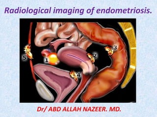
  • 1. Radiological imaging of endometriosis. Dr/ ABD ALLAH NAZEER. MD.
  • 2. Endometriosis is defined as the presence of endometrial epithelium and stroma in an ectopic site outside the uterine cavity . Endometriosis occurs in 10% of the female population and almost, exclusively, in women of reproductive age . The most common symptoms are dysmenorrhea, dyspareunia, pelvic pain, and infertility although endometriosis may be asymptomatic . Adenomyosis is the presence of endometrial glands and stroma within the myometrium and it is the cause of uterine enlargement, menorrhagia, and dysmenorrhea and. The most common sites for endometrial implantation within the pelvis are the ovaries, broad and round ligaments, fallopian tubes, and cervix. Endometriosis can affect the woman in the reproductive age; however, it cannot be accurately diagnosed by clinical criteria alone. In addition, the transabdominal and transvaginal US are not as accurate as MRI.
  • 3. Superficial endometriosis. In superficial endometriosis – also known as Sampson's syndrome - superficial plaques are scattered across the peritoneum, ovaries and uterine ligaments. These patients tend to have minor symptoms and usually also less structural changes in the pelvis. At laparoscopy, these implants may be seen as superficial powder-burn or gunshot lesions. In deep pelvic endometriosis - also called Cullen's syndrome - there is subperitoneal infiltration of endometrial deposits. The symptoms are more severe and related to the localization and depth of invasion. MRI is of use for the diagnosis of deep infiltrating endometriotic lesions and for the assessment of disease extension. Preoperative mapping of disease extension is important to decide whether surgical intervention is indicated, and if so, for planning complete surgical excision. Deep pelvic endometriosis.
  • 4. Endometriomas - also known as chocolate cysts Develop when superficial endometriotic lesions on the surface of the ovary invaginate. Blood produced by such an implant during each menstrual cycle cannot escape and will accumulate within the ovary, forming a cyst known as an endometrioma. Endometriomas present as complex cystic masses, often thick-walled with a homogeneous content. On transvaginal ultrasound, endometriomas may be seen as thick-walled cysts with low level echoes. On the left a transvaginal ultrasound image and the corresponding laparoscopic image during cystectomy.
  • 5.
  • 6. Sites. Pelvic: -Uterine= adenomyosis(50%). Extra-uterine: -Ovary 30%. - Pelvic peritoneum 10%. -Fallopian tube. -Vagina. -Bladder &rectum. -Pelvic colon. - Ligaments. Extra-pelvic Umbilicus. Scars(Laparotomy). Lung &pleura. Others.
  • 7.
  • 8. Symptomatology. - Pelvic pain(65%). . Dysmenorrhea, especially suggestive of endometriosis the pain if it occurs after years of pain free menstruation. . Deep dyspareunia. . Chronic pelvic pain. . Ovulation pain with menstrual irregularity. . Other types of pain- Sciatica. -Infertility(35%).
  • 9. GIT symptoms: . Diarrhea. . Constipation. . Changes in bowel habits. . Rectal bleeding. . Pain with defecation. . Tenesmus. . Abdominal distension. . Small caliber stool. .Colicky abdominal pain. . Bowel obstruction. . Bowel perforation. Urinary symptoms: -Urgency. - Frequency. - Supra-pubic pain. - Flank pain. - Urge incontinence. - Dyspareunia. - Hematuria. - Dysuria. - Backache. - Hydronephrosis.
  • 10. Normal anatomy of the female pelvis. (a, b) Axial (a) and sagittal (b) T2-weighted MR images show the uterosacral ligaments (arrows in a), rectovaginal pouch (* in b), and retrocervical area (outlined in b). (c, d) Laparoscopic images show the uterosacral ligaments (arrows in c), rectovaginal pouch (* in c), and retrocervical area (outlined in d). (e) Drawing of the female pelvis (oblique coronal view, superoinferior orientation) shows the ureters (arrows), uterosacral ligaments (arrowheads), cardinal ligaments and parametrium (black *), and round ligaments (white *). Note that the ureters course lateral to the uterosacral ligaments and immediately caudal to the cardinal and broad ligaments. Normal anatomy of the female pelvis.
  • 11. Ultrasound examination: Sonographic examinations took place on two perpendicular planes using PHILIPS HD11 instrument. Pelvic transabdominal US was performed by using a 3.5-MHz transducer and transvaginal US examination with 8-MHz transducer. During each Sonographic examination, the uterine borders, whether regular or irregular, uterine size, myometrial echotexture and the presence of focal areas with ill- defined borders or abnormal echotexture were described. When these areas were present, the following criteria of adenomyosis were evaluated: presence of heterogeneity, increased or decreased areas of echogenicity, or presence of myometrial cysts. Another type of ultrasound that we used in our study is trans-rectal ultrasound which is necessary in the evaluation of deep infiltrating endometriosis involving recto-vaginal and utero-sacral ligaments but unfortunately we have no cases that have the deep infiltration of these regions. Radiological imaging.
  • 12. MRI protocols: MRI performed using T2 weighted images required 4 mm slices in the sagittal, coronal, and axial planes related to the orientation of the uterine cavity that used fast spin echo sequences in all the three planes. T1 weighted spin echo and gradient echo images were obtained both with and without fat saturation for all patients. High- resolution body coils were used for data acquisition, and exams were completed in 30–45 min. T1 weighted fat saturated and T2 weighted images for four major criteria: 1.Borders, size and uterine symmetry. 2.Maximal junctional zone (JZ max) thickness and/or the presence of an ill-defined, relatively homogenous, low signal intensity, myometrial area. 3.Maximal (JZ max) to myometrial thickness ratio using the maximal thickness of JZ and the corresponding thickness of the entire myometrium obtained at the same level. 4.High intensity spots within the myometrium, the presence of T1 bright/T2 dark signals, suggesting the existence of endometriosis in any of the anatomical locations.
  • 13.
  • 14. Sagittal T2-weighted image with fat suppression of 27-year-old woman in second part of her cycle. Uterus is evaluated between isthmus and end of uterine cavity (white lines). Junctional zone (short arrows) should be measured from several sites on anterior and posterior walls. Junctional zone measure can be compared with entire thickness of myometrium (long arrows) evaluated at same site.
  • 15. Role of the Junctional Zone Ultrasound studies have been paramount in understanding the role of the junctional zone, which is intricately linked with its structural organization and biochemical properties. Pelvic ultrasound, performed transabdominally in the 1980s and later endovaginally in the 1990s, has permitted observation of contractions of the junctional zone occurring in the nongravid uterus. Video recordings have identified these contractions with a speed of 1.2–1.7 mm/s with a frequency of 3–5 contractions/min. Contractions are classified according to their direction: from the uterine cervix toward the body, from the body to the cervix, or both simultaneously . These junctional contractions have been secondarily described on MRI with particular dynamic sequences. They appear as thin bands perpendicular to the junctional zone and in low signal intensity on T2-weighted images. This signal has been partly explained by the venous vasoconstriction that reduces the quantity of circulating blood in the myometrium. In 2007, Kido et al. used MRI to illustrate the influence of oral contraceptives on uterine contractions. It was clearly shown that these contractions have an amplitude, a frequency, and a direction that correlate with the phase of the menstrual cycle. Thus, in the first part of the cycle, contractions occur from the cervix toward the body and increase in intensity until ovulation. These contractions participate directly in the transport of spermatozoids toward the ovum as reported by Kunz and colleagues by utilizing inert particles. confirmed this hypothesis by studying the strong link between contractions issued from the junctional zone and fertility.
  • 16. Adenomyosis is the presence of endometrial glandular cells and cells of the chorion more than 2.5 mm from the endometrium-myometrium interface. A hypertrophic reaction of the smooth-muscle cells surrounding the ectopic glands is another important element of diagnosis . Involvement of the myometrium can include the entire interface, termed “diffuse adenomyosis,” or it can present in a limited area, termed “focal adenomyosis.” These two forms have similar frequency on hysterectomy samples but are rarely associated. Adenomyoma, a localized confluence of adenomyotic glands forming a mass, constitutes an unusual form of adenomyosis. The actual prevalence of adenomyosis remains unknown. Risk factors for adenomyosis—most notably, the existence of prior endouterine surgery— were isolated. Multiparity is another risk factor, which appears contradictory to the not so well-established link between adenomyosis and infertility. Adenomyosis preferentially affects women between 40 and 50 years old; however, most forms are asymptomatic, explaining perhaps the lower rate of detection in young women. A positive diagnosis of adenomyosis cannot be made reliably on the basis of clinical examination alone. Supporting evidence, including MRI findings, is necessary to entertain and confirm the diagnosis.
  • 17. Radiographic features: Imaging features are variable and in many instances very subtle. Three (some say four) forms can be distinguished: diffuse adenomyosis: most common focal adenomyosis and adenomyoma: some consider these as separate (see below). cystic adenomyosis and adenomyotic cyst: rare Adenomyosis is usually relatively generalized, affecting large portions of the uterus (typically the posterior wall), but sparing the cervix. Despite often marked enlargement of the uterus, the overall contour is usually preserved. In some cases it may also be localized, forming a mass. In such cases the term adenomyoma may be used, although there appears to be some disagreement about whether the terms focal adenomyosis and adenomyoma refer to exactly the same entity (please refer to the article on adenomyoma for further discussion). A rare variant is cystic adenomyosis which is believed to be the result of repeated focal hemorrhages resulting in cystic spaces filled with altered blood products.
  • 18. Pelvic ultrasound: Ultrasound is usually the first and often the only imaging modality employed to investigate menorrhagia and dysmenorrhea. Unfortunately, the sonographic features of adenomyosis are variable, and may be absent. The reported sensitivity and specificity of trans-abdominal ultrasound are 32-63 % and 95-97 % respectively . The spectrum of findings includes: normal appearing uterus, focal or diffuse bulkiness, typically of the posterior wall heterogeneous echogenicity (heterogenous myometrial echotexture) hyperechoic: islands of endometrial glands hypoechoic: associated muscle hypertrophy a "venetian blind" appearance may be seen due to subendometrial echogenic linear striations & acoustic shadowing where endometrial tissues cause a hyperplastic reaction. small myometrial: sub endometrial cysts: thickening of the transition zone sometimes can be visualized as a hypoechoic halo surrounding the endometrial layer of 12 mm or more thickness subendometrial echogenic linear striations subendometrial echogenic nodules When an adenomyoma is present, then appearances may closely mimic those of a uterine fibroid, which may also co-exist.
  • 19. Hysterosalpingogram (HSG): May show diverticula extending into the myometrium . CT is unable to diagnose adenomyosis: but may suggest its presence when uterine enlargement is present. Distinguishing between adenomyosis and uterine fibroids on CT is difficult if not impossible, although presence of calcifications strongly favours the latter . Pelvic MRI: MRI is the modality of choice to diagnose and characterize adenomyosis, and T2 weighted images (sagittal and axial) are most useful. It has a very diagnostic accuracy with a sensitivity of 78-88% and a specificity of 67-93% . The most easily recognised feature is thickening of the junctional zone of the uterus to more than 12 mm, either diffusely or focally (normal junctional zone measures no more than 5 mm) T2: typically a region of adenomyosis appears as an ill-defined ovoid/diffuse region of thickening often with small high T2 signal regions representing small regions of cystic change the region may also have a striated appearance. T1: Foci of high T1 signal are often seen, indicating menstrual hemorrhage into the ectopic endometrial tissues. T1 C+ (Gd): contrast enhanced MR evaluation is usually not indicated in adenomyosis, however if performed, it shows enhancement of the ectopic endometrial glands.
  • 20. HSG with adenomyosis of the uterus.
  • 21. CT with adenomyosis of the uterus.
  • 22. 42-year-old woman who presented with chronic pelvic pain. Coronal (A) and axial (B) T2-weighted images reveal cystic area surrounded by low-signal- intensity ring (arrow). Overall appearance on MRI is that of cystic adenomyoma.
  • 23. 36-year-old woman who underwent MRI evaluation for polymyomatous uterus discovered on ultrasound. On sagittal T2-weighted image, posterior myoma (black arrow) is visible in frank hyposignal. Focal adenomyosis (white arrow) is visible on anterior uterine wall, presenting as ill-defined area in hyposignal- containing cysts. Hyperintense linear or reticulated streaks extend from endometrium into markedly thickened anterior myometrium (arrowheads).
  • 24. Diffuse adenomyosis of the uterus.
  • 26. Diffuse adenomyosis of the uterus.
  • 29. MRI (sagittal T2WI) shows adenomyosis with abnormal increase in thickness of junctional zone (12 mm) and abnormal high T2 SI of the myometrium represents abnormal stromal glands inside the myometrium. B: MRI (coronal T2WI) shows the same finding as well as bilateral ovarian simple cysts.
  • 30. Sagittal transvaginal sonography demonstrates myometrial anechoic lacunae specific of adenomyosis involving the ventral and the dorsal myometrium (A). Transversal transabdominal examination shows a very large asymmetric uterus with thickening of the dorsal myometrium.. (B). Transvaginal sonography demonstrates diffuse adenomyosis involving the ventral myometrium and a dorsal subserous leiomyoma. Sagittal T2- weighted magnetic resonance image through the uterus shows numerous high-intensity spots in the inner myometrium, thickening of junctional zone (JZ) (13 mm) and ratio max >40% (D). Axial T2- weighted magnetic resonance image demonstrates diffuse thickening of JZ both ventrally and dorsally, consistent with severe adenomyosis. Numerous foci of high signal representing the heterotopic endometrium are present (E). Sagittal T2-weighted magnetic resonance image through the uterus (F): there is an ill-defined mass centred around the endometrium. Several foci of increased signal consistent with heterotopic endometrium are present in the inner myometrium without mass effect on the endometrial cavity. In contrast, leiomyoma is very hypointense and has well- defined borders (F).
  • 31. Linear striations, sonographic findings suggestive of adenomyosis, in a 47-year-old woman with pelvic pain and dysmenorrhea. A, Sagittal transvaginal sonogram shows an enlarged uterus. A heterogeneous echo texture involving the anterior myometrium is shown with linear striated shadowing (arrowheads). B, Sagittal fat-suppressed T2-weighted MRI shows marked adenomyosis involving the anterior uterine wall manifested as a thickened junctional zone with scattered myometrial cysts (arrows). C, Sagittal T1-weighted gradient echo MRI (TR, 270 milliseconds; TE, 5 milliseconds) obtained after administration of gadolinium dimeglumine shows enhancement of the endometrium and myometrium. Myometrial cysts (arrows) do not show enhancement
  • 32. Polypoid Adenomyoma An adenomyoma is occasionally located in a submucosal or subserosal position. A submucosal adenomyoma manifesting as a polypoid mass protruding into the endometrial cavity has been termed “polypoid adenomyoma. This lesion usually arises from the uterine corpus, occasionally from the lower uterine segment and cervix. It can be seen as a pedunculated or sessile mass. Its appearance on MRI as an uncommon variant of adenomyosis has been described. Use of the term polypoid adenomyoma has been controversial. Although some authors have discussed this lesion as a distinct entity, the gynecologic pathologic literature reflects the opinion that the term polypoid adenomyoma refers to a polypoidal form of a circumscribed adenomyotic mass. Atypical Polypoid Adenomyoma This is an uncommon variant of polypoid adenomyoma characterized by atypical endometrial glands and often squamous metaplasia and a cellular smooth muscle stroma. Before its delineation, this lesion was often considered malignant. The distinction between the typical and atypical adenomyoma depends on histologic examination because the radiologic findings are similar.
  • 33. Polypoid adenomyomas. A, Sagittal transvaginal sonogram in a 30-year-old woman with a history of menometrorrhagia shows an indistinct endometrial echo complex with variable thickness; a cyst is shown adjacent to it (arrow). Sonohysterography was performed for further evaluation of these findings. B, Sagittal image from the sonohysterogram reveals an intracavitary mass containing a cystic focus (arrow). The echo texture of this mass, unlike an endometrial polyp, is similar to that of the myometrium. No endometrial abnormality was seen. C, Transvaginal sonogram in a 39-year-old woman with menorrhagia shows a large prolapsing adenomyoma distending the cervical canal (arrow). The echo texture of the mass is similar to that of the myometrium; it is, therefore, sonographically indistinguishable from a myoma. The arrowhead points to the posterior vaginal wall.
  • 34. Cystic adenomyoma in a 37-year-old woman with a history of infertility. A, Multiplanar display from 3D TVS of the uterus shows a cystic mass within the right side of the uterine corpus containing low-level echoes. The bottom left image is a coronal plane through the endometrial cavity showing 2 normal cornua. Top left, axial; top right, sagittal; bottom left, coronal. B, Axial T1-weighted gradient echo opposed phase MRI (TR, 225 milliseconds; TE, 2 milliseconds) shows a hyperintense lesion within the myometrium. Lack of a chemical shift artifact around the lesion confirms the absence of fat within. C, Axial T2-weighted MRI (TR, 4000 milliseconds; TE, 100 milliseconds) image shows an intermediate to hyperintense signal in the cystic mass (arrow), showing shading, consistent with chronic hemorrhage.
  • 35. Multiple fibroids and diffuse adenomyosis in a 50-year-old woman. A, Sagittal transabdominal sonogram shows a bulky uterus due to multiple fibroids. The endometrial cavity is distorted by the presence of fibroids (arrows). B, Sagittal T2- weighted MRI shows diffuse adenomyosis with marked thickening of the junctional zone (arrow) and the presence of multiple fibroids (arrowheads). In the presence of large multiple fibroids, diagnosing adenomyosis on sonography can be a difficult task.
  • 36. Ultrasound of the pelvis showed left ovarian cystic lesion with internal echoes and two hyperechogenic areas inside corresponding to hemorrhagic content of the cyst. C: Axial fat sat MRI showed hyperintense lesion at left ovary. D: The lesion showed drop of SI at T2WI MRI which is pathognomonic of endometrioma. E, F: Sagittal and coronal T2WI MRI showed low SI of the lesion which is pathologically proved endometrioma by laparoscopic examination and biopsy.
  • 37.
  • 38.
  • 39. Solid fibrotic thickening of the uterosacral ligament in a 40-year-old woman with endometriosis and pelvic pain. (a) Axial T2-weighted MR image shows low-signal- intensity, mass like thickening of the proximal right uterosacral ligament (arrows). The normal-appearing sacral portion of the ligament (arrowheads) is also depicted. (b) Sagittal T2-weighted MR image shows band like fibrotic adhesions above the ligament (arrows) and shading within the superior right ovarian endometriomas (arrowheads).
  • 40. Endometriosis of the vesicouterine pouch in a 27-year-old woman with chronic pelvic pain. Sagittal T2-weighted (a), sagittal fat-suppressed T1-weighted (b), and oblique coronal T2-weighted (c) MR images depict irregular thickening of the upper external aspect of the bladder wall (arrowheads in a, arrows in c) that partially obliterates the vesicouterine recesses. Tiny bloody foci (arrow in b) are also seen, adding specificity to the finding.
  • 41. Endometriotic involvement of the vesicovaginal septum in a 37-year-old woman who had undergone hysterectomy and presented with chronic dysuria. US showed a focal mass bulging into the posterior vesical wall. (a) Sagittal T2-weighted MR image shows a hypointense nodule (arrows) in the vesicovaginal septum. (b) On an axial fat-suppressed T1- weighted MR image, the lesion (arrowhead) has high signal intensity, which did not decrease on subsequent images. This finding indicates proteinaceous or bloody content and causes the lesion to resemble an endometrioma.
  • 42.
  • 43. Intrinsic bladder endometriosis in a 22-year-old woman who had undergone a cesarean section and presented with cyclic voiding symptoms but no hematuria. (a) Axial T2-weighted MR image shows hypointense irregular focal wall thickening (arrow) on the left posterolateral aspect of the bladder. Note the relative sparing of the perivesical fat (*) and the absence of enlarged lymph nodes. (b) Axial fat-suppressed T1- weighted MR image reveals that the area of wall thickening contains small intermingled hyperintense foci (arrow), a finding that indicates bloody content and that was crucial for differentiating between endometriosis and cancer. The patient underwent cystoscopy to exclude bladder cancer, and the tentative diagnosis of intrinsic bladder endometriosis was confirmed.
  • 44. Bladder endometriosis with urethral involvement in a 31-year- old woman. (a, b) Sagittal T2-weighted (a) and fat-suppressed T1-weighted (b) MR images obtained during the patient’s menstrual period show massive irregular thickening of the anteroinferior bladder wall (arrow in a) that extends to the vesical trigone and the upper urethra and is associated with tiny interspersed bloody foci (arrowhead in b), findings that are consistent with a diagnosis of bladder endometriosis. Note the smooth outer contour of the bladder wall, as well as the normal- appearing prevesical fat (arrowheads in a). Such massive focal wall thickening with no signs of extravesical extension or perivesical fat stranding makes a diagnosis of bladder cancer less likely. (c) Axial T2-weighted MR image shows irregular hypointense thickening on the anterior contour of the urethra (*), a finding that indicates endometriotic involvement by direct extension from the bladder lesion.
  • 45. Bladder endometriosis with ureteral involvement in a 33-year-old woman who presented with cyclic dysuria and pelvic pain, but no signs of hematuria or weight loss. (a) Axial T2-weighted MR image reveals hypointense irregular focal thickening located in the left posterolateral aspect of the bladder wall (arrows) and extending into the perivesical fat (arrowheads). (b) Sagittal T2- weighted MR image shows the perivesical and bladder wall thickening (arrow), as well as upstream ureteral dilatation (arrowheads). (c) Oblique coronal T2-weighted MR image shows an infiltrating lesion (*) encasing the lower third of the ureter (arrows). (d) Three- dimensional contrast-enhanced coronal reformatted fat-suppressed T1-weighted MR image shows a dilated left ureter (solid arrows) and a deep infiltrating endometriotic lesion involving the wall of the bladder (B) and the lower third of the ureter (arrowheads). There is also a hyperintense component in the bladder submucosa (open arrow), a finding that indicates bloody content. C = cervix, U = uterus.
  • 46. Solid endometriosis of the bladder and right round ligament in a 40-year-old woman. (a, b) Sagittal (a) and axial (b) T2-weighted MR images show a poorly marginated, low-signal-intensity mass (arrowheads) in the right posterior bladder wall that extends posteriorly, obliterating the vesicouterine pouch. Intralesional 1–4-mm high-signal-intensity foci (arrows in a) represent ectopic endometrial glands. The right round ligament is thickened (arrows in b) as it courses inferiorly toward the canal of Nuck. (c, d) Axial T1-weighted gradient-echo fat-suppressed MR images obtained before (c) and after (d) the administration of a gadolinium-based contrast material show T1 hyperintensity in some of the endometrial glands within the posterior bladder lesion (arrow in c) and solid enhancement of both the bladder mass (arrowheads in d) and the thickened round ligament (arrow in d).
  • 47. Ureteral endometriosis in a 31-year-old woman with chronic pelvic pain and left hydronephrosis. (a) Axial T2-weighted MR image shows hypointense irregular and spiculated thickening and stranding of the perivesical fat on the left side (arrows). The ureter (arrowhead) traverses the involved area, a finding that is suggestive of infiltrating endometriosis. (b) Para sagittal T2- weighted MR image reveals moderate dilatation of the left ureter (arrowheads) to the level of the insertion of the ipsilateral uterosacral ligament (arrow), which is irregular and thickened, indicating associated deep endometriosis. (c) Fat-suppressed T1-weighted MR image shows a cystic lesion (arrow) whose high signal intensity indicates proteinaceous or bloody content. The lesion resembles an endometrioma located medial to hypointense thickening of the perivesical fat. (d) Three-dimensional contrast- enhanced fat-suppressed excretory phase T1- weighted MR urogram shows the repercussions of the findings in a–c to the urinary system. The left ureter is severely narrowed in its lower third (arrowhead), with associated upstream dilatation up to the renal pelvis (*).The lesion (arrow) is seen adjacent to the narrowed ureter and represents an endometrioma.
  • 48.
  • 49. Endometriotic involvement of the retrocervical area in a 33- year-old woman with dyspareunia. At earlier MR imaging examinations, an endometrial implant was seen in this area. (a) Sagittal T2-weighted MR image shows an irregular, solid, nodular hypointense mass (arrow) slightly inferior to the uterine torus, with intermingled hyperintense foci (arrowhead). (b) On an axial fat-suppressed T1- weighted MR image, the intermingled foci (arrow) remain hyperintense, a finding that indicates bloody content.
  • 50. Solid invasive endometriosis of the rectosigmoid colon in a 40-year-old woman. (a) Lateral image from a double-contrast barium examination shows circumferential narrowing of the rectosigmoid colon, with mass effect, speculation, and pleating of the anterior margin (arrow). (b) Sagittal T2-weighted fast spin-echo MR image shows extraluminal findings suggestive of solid invasive endometriosis. The mushroom cap sign represents the low-signal-intensity core of fibrotic endometriosis and hypertrophic muscularis propria (*) capped by high-signal-intensity mucosa (straight arrows). Invasion of the serosal surface of the uterus and obliteration of the cul-de-sac (arrowheads) are seen, with high- signal-intensity 1–3-mm foci (curved arrow) representing ectopic endometrial glands.
  • 51. Endometriotic involvement of the uterine ligaments in a 31-year-old woman with chronic pelvic pain and a known history of endometriosis. Axial gadolinium- enhanced fat-suppressed late-phase T1-weighted MR image shows homogeneous late enhancement of thickened round uterine ligaments (arrows) due to involvement of their uterine insertions by an endometriotic lesion.
  • 52. Endometriotic involvement of the posterior pelvic compartment in a 29-year-old woman with hypermenorrhea, dyspareunia, and dysmenorrhea. Earlier MR imaging showed retrocervical endometriosis with fibrotic obliteration of the rectovaginal pouch. Sagittal (a) and axial (b) T2-weighted MR images show an irregular hypointense mass (arrowhead) that extends from the posterior cervix inferiorly to the vaginal fornix. Note the presence of a subserous leiomyoma in the anterior uterine wall (black arrow in a), focal adenomyosis in the corporal anterior uterine wall (white arrow), and a right follicular ovarian cyst (* in b).
  • 53. Endometriotic involvement of the uterosacral ligaments in a 31-year-old woman who presented with pelvic pain, dyspareunia, and painful defecation. Earlier MR imaging showed involvement of these structures. (a, b) Coronal (a) and axial (b) T2-weighted images show hypointense irregular spiculated nodular thickening of the right uterosacral ligament (white arrow in a), with minimal intermingled hyperintense foci. These findings create an arciform abnormality (arrowheads in b) that extends to the anterior rectal wall, where there is focal parietal thickening (black arrow). (c) Axial fat-suppressed T1-weighted MR image shows hyperintense foci (solid arrows), a finding that indicates bloody content. Open arrow indicates focal parietal thickening. (d) Laparoscopic image obtained in a different patient shows involvement of the uterosacral ligaments (arrows).
  • 54. Isolated endometriotic involvement of the vagina in a 38-year-old woman who had undergone hysterectomy and presented with dyspareunia and chronic pelvic pain. Earlier MR imaging showed vaginal involvement. (a) Axial T2-weighted MR image shows a hypointense nodule (arrows) in the posterior upper third of the vagina. (b) Axial T1-weighted MR image shows minimal intermingled hyperintense foci (arrowheads), a finding that indicates bloody content.
  • 55.
  • 56. Endometriotic involvement of the ischioanal fossa in a 32-year-old woman who presented with right hip pain. The patient had no history of perineal surgery. (a) Axial T2-weighted MR image shows solid, predominantly glandular endometriosis (arrows) in the ischioanal fossa located close to the right ischiatic tuberosity and involving the internal obturator muscle and anal sphincter. (b, c) Axial T1-weighted (b) and fat-saturated T1-weighted (c) MR images show intermingled hyperintense foci (arrow), a finding that indicates bloody content. Arrowhead in b indicates cortical sclerosis. (d) Coronal T1-weighted MR image shows significant bone reaction (cortical sclerosis) in the medial aspect of the right ischiatic tuberosity (arrowhead).
  • 57. Sigmoid endometriosis in a 30-year-old woman who experienced painful defecation and hematochezia during her menstrual period. (a, b) Sagittal (a) and coronal (b) T2- weighted MR images obtained after the administration of a saline rectal enema and intravaginal aqueous gel show hypointense nodular thickening of the sigmoid wall (arrows) adhering to the posterior uterine serosa (arrowhead in a). (c) Sagittal gadolinium-enhanced fat suppressed late-phase T1-weighted MR image shows enhancement of the lesion (arrow). The findings in a–c are consistent with a diagnosis of sigmoid endometriosis.
  • 58. Cyclic sciatica in a 27-year-old woman. Earlier MR imaging showed sciatic nerve involvement. (a) Axial MR image shows hypointense irregular spiculated soft-tissue thickening (arrows) in the left sciatic notch. (b) Axial fat-suppressed T1-weighted MR image shows minimal intermingled hyperintense foci (arrows), a finding that indicates bloody content. (c) Axial gadolinium-enhanced fat suppressed late-phase T1-weighted MR image shows enhancement of the lesion (arrows).
  • 59. Deep endometriosis at various sites in a 35-year-old woman who presented with chronic pelvic pain, infertility, and dysuria. Earlier MR imaging showed endometriotic involvement of the urinary bladder, rectovaginal septum, and retrocervical area. (a, b) Sagittal (a) and axial (b) T2-weighted MR images demonstrate hypointense focal thickening of the right posterolateral wall of the bladder (black arrow) with punctate intermingled bleeding, along with an extraperitoneal, irregular hypointense nodule (white arrows) that mainly involves the retrocervical area and the upper portion of the rectovaginal septum. (c) Axial fat-saturated T1- weighted MR image shows the nodule. Arrowhead = punctate bleeding.
  • 60. 37 year old woman with a history of dysmenorrhea. Images: (a) coronal T2- weighted and (b) axial T1 with fat suppression and gadolinium. Enlarged right ovary with multiple cysts (*) and an endometrioma with intermediate signal on T2 and hyperintense on T1 with fat suppression and gadolinium (à black).
  • 61. Relatively hyperechoic SOL at right ovary. B: MRI (axial T1WI) shows hyperintense content of the right ovary. C: Axial T2WI shows low SI of the cyst of the right ovary (shading sign). D: Axial T1WI with fat sat shows hyperintense cystic right ovarian lesion (hemorrhage). E, F: MRI (sagittal and coronal T2WI) revealed low SI of right ovarian lesion.
  • 62. 37 year old woman. Infertility study. Images: (a) Sagittal T2-weighted and (b) coronal T2 and (c) axial T2. Bilateral ovarian endometrioma and left hydrosalpinx (à white).
  • 63. Endometrioma at ultrasound and laparoscopy.
  • 64. T2- and fat sat T1-images of an endometrioma with hypointensity on T2 (shading), fluid-fluid levels on T2 (left) and hyperintense blood on T1WI with fat sat (right).
  • 66. Endometrioma of the left ovary in a 33-year-old woman. (a, b) Axial T1- weighted in-phase (a) and opposed- phase fat-suppressed (b) gradient- echo MR images show a T1- hyperintense left ovarian lesion, a feature suggestive of endometrioma. (c) Axial T2-weighted MR image shows lower signal intensity in the endometrioma than in the adjacent normal ovarian follicles (arrows). (d, e) Diffusion-weighted MR images obtained with b values of 50 (d) and 800 (e) sec/mm2 show low to intermediate signal intensity in both the endometrioma and the normal follicles. (f) ADC map from diffusion- weighted MR imaging shows restricted diffusion in the endometrioma relative to that in the adjacent ovarian follicles.
  • 67. Left ovarian endometriomas, bilateral hematosalpinges, and fibrotic solid invasive endometriosis in the posterior uterus of a 47-year-old woman with severe pelvic pain. (a) Axial T1-weighted in-phase gradient-echo MR image shows two foci of high signal intensity in the left ovary (arrow) and multiple foci of high signal intensity in the left anterior and midline posterior pelvis (arrowheads). (b) Axial opposed- phase T1-weighted fat-suppressed gradient-echo MR image shows persistent high signal intensity in all three regions, a finding that helps exclude the presence of a fat-containing mature cystic teratoma. The improved dynamic range achieved with fat suppression facilitates visualization of a subcentimeter endometrioma of the anterior left ovary (arrow). (c) Axial T2-weighted fast spin-echo MR image shows low signal intensity within the left ovarian endometriomas (solid arrow) and lesser degrees of T2 signal hypointensity within the dependent portions of the loculi in the left anterior and midline posterior pelvis (arrowheads). The posterior uterus is markedly thickened with low-signal- intensity soft tissue (F) containing scattered 2–4-mm foci of high signal intensity (open arrows). Although the tissue has the appearance of adenomyosis, it is separated from the posterior junctional zone (JZ). These findings are suggestive of fibrotic endometriosis. (d, e) Sagittal (d) and coronal fat- suppressed (e) T2-weighted MR images show dilatation of the right and left fallopian tubes, respectively (arrows). In d, extension of fibrotic endometriosis (F) into the posterior uterus is seen. The patient later underwent hysterectomy and bilateral salpingo-oophorectomy, at which the MR imaging findings were confirmed.
  • 68. Right ovarian endometrioma in a 35-year-old woman that might have been misinterpreted as a mature cystic teratoma with reliance on short inversion time inversion-recovery (STIR) imaging alone to detect Intralesional fat. (a) Axial T1- weighted spin-echo MR image shows a high-signal-intensity mass (arrow) within the right adnexa. (b) Coronal STIR MR image shows that the mass (arrow) has low signal intensity similar to that of suppressed fat. (c) Axial fat-suppressed gradient-echo MR image shows high signal intensity of the mass (arrow), a finding that helps confirm that it is not composed of fat. The low signal intensity of the mass on the STIR image could be secondary to either T1- or T2- shortening effects but cannot be considered indicative of fat content. To avoid this pitfall of STIR MR imaging of the female pelvis, MR systems capable of performing chemical- shift fat suppression should be used.
  • 69. Bilateral endometriomas, solid endometriosis of the posterior uterus, and left-sided peritoneal inclusion cyst in a 43-year-old woman. (a, b) Axial T1-weighted in-phase (a) and fat-suppressed opposed-phase (b) gradient-echo MR images show bilateral hyperintense adnexal lesions (arrows) that retain high signal intensity with fat suppression. A peritoneal inclusion cyst (*) is also shown posterior to the uterus (U). Susceptibility artifacts anterior to the rectus muscles (arrowheads in a) indicate that the patient has undergone a previous surgical procedure. (c) Axial T2-weighted MR image shows shading within the bilateral endometriomas (straight arrows). The normal endometrial complex and junctional zone are shown in the anterior uterus (arrowheads). The posterior uterus is markedly enlarged by poorly marginated soft tissue (E) with low T2 signal intensity and scattered internal 1–2-mm foci of higher signal intensity (curved arrow) representing infiltration of solid endometriosis into the uterine wall. Loculated fluid posterior to the left ovary represents the peritoneal inclusion cyst
  • 70. 53 year old woman. Background history of hysterectomy with cyclic hematuria. Images: (a) Sagittal T2-weighted, (b) coronal T2, (c) axial T2 and (d) Cystoscopy. On the bladder roof at left (à white) a mass of 1.5 x 1.1 cm can be seen, this is an intermediate T2 signal with undefined borders infiltrating the bladder wall, being located in the bladder lumen (à black). In the cystoscopy can be observed, in left top wall, a sessile uplifted lesion of approximately 1 cm. The remainder of the bladder mucosa appears normal and without lesions.
  • 71. 31 year old woman with a history of dysmenorrhea. Images: (a) coronal T2-weighted (b) axial T1 and (c) coronal T2. Left ovarian endometrioma (à white) of 9.3 x 7.6 cm attached to the uterine torus by a deep endometriosis foci (à white curved). Infiltration of the distal ureter by fibrotic endometrial implant (à) that is associated with hydroureteronephrosis (*).
  • 72. 46 year old woman. Nodule of the abdominal wall under study. Images: (a) sagittal T2-weighted and (b) axial T1 and (c) axial T1 with fat-suppression and gadolinium. Solid node 20 x 12mm on abdominal wall at the hypogastrium level lateralized to the left, heterogeneous signal (with hypointense focus) on T2 and isointense on T1 (white circle) with discrete and homogeneous enhancement with contrast. External marker (*). Pathology compatible with endometriosis foci.
  • 73. 33 year old woman. Palpable nodule in relation to episiotomy. Images: (a) axial T2-weighted and (b) axial T1. Lobulated nodule in the right perianal region on episiotomy site (à white). Slightly hyperintense on T1 and isointense on T2.
  • 74. Transvaginal US revealed abnormal increase in thickness of myometrium in a retroverted uterus. B: MRI (sagittal T2WI) shows retroverted uterus with abnormal increase thickness in junctional zone (10 mm) and diffuse abnormal high T2 SI of myometrium abnormal stromal glands. C: MRI (coronal T2WI) shows the same findings as image B. Examination.
  • 75. Decidualized endometriosis in a 36- year-old woman in the 12th week of pregnancy. (a, b) Sagittal T2-weighted fast spin-echo MR images show an intrauterine gestational sac, a focal contraction (C) of the posterior myometrium, and an anterior placenta (P). The bilobed low-signal-intensity mass above the uterus represents an endometrioma (E). Mural nodules with higher signal intensity within the endometrioma (arrows in b) represent decidualized endometriosis. (c, d) Axial T1-weighted fat-suppressed (c) and T2- weighted fast spin-echo (d) MR images show the typical signal intensity of an endometrioma, with T1 hyperintensity and T2 shading, and the higher T2 signal intensity of decidualized endometriosis (arrow in d).
  • 76. Hematometrocolpos, hematosalpinx, and endometriomas in a 15-year-old girl with an obstructing transverse vaginal septum. (a) Coronal T2-weighted fat- suppressed MR image shows dilatation of the endometrium (E), endocervix (C), and proximal vagina (V) due to a transverse vaginal septum (not shown). (b, c) Axial T1-weighted fat-suppressed (b) and T2-weighted fast spin-echo (c) MR images obtained at the level of the uterus show the distended vaginal canal (V) and segments of a dilated right fallopian tube (arrow). These structures have similar high T1 signal intensity and intermediate T2 signal intensity. (d) Axial T1-weighted fat-suppressed MR image obtained at the level of the right ovary shows multiple subcentimeter high-signal-intensity endometriomas (arrows), findings confirmed at resection of the vaginal septum.
  • 77. Endometrioid cystadenocarcinoma of the left ovary in a 51-year-old woman with long-standing endometriosis. (a) Axial T2-weighted MR image shows a cystic pelvic mass that also contains solid soft tissue (arrow). The signal of the cystic component was slightly hypointense to that of the bladder (not shown). (b, c) Axial T1-weighted fat- suppressed gradient-echo MR images obtained before (b) and after (c) the administration of a gadolinium-based contrast material show that the cystic component has T1 hyperintensity, a finding suggestive of proteinaceous or hemorrhagic content. The signal intensity of the solid component of the mass, relative to that of the cystic portion, is lower in b and higher in c. (d) MR image obtained by subtracting the unenhanced image dataset from the contrast-enhanced image dataset facilitates the detection of enhancing tissue (arrow) by increasing the dynamic range (61) and eliminating the high signal intensity of nonenhancing hemorrhagic and proteinaceous tissue. (e, f) Unenhanced T1-weighted fat- suppressed (e) and gadolinium-enhanced subtraction (f) MR images obtained at a lower level show additional enhancing nodular components (arrows in f) in the tumor. A 1-cm right ovarian endometrioma (arrow in e) is also seen.
  • 78. Endometriosis involving appendix. A, Mucocele formation in 38-year-old woman with chronic right lower quadrant pain. CT image obtained with oral and IV contrast material shows lobulated, low attenuation cystic mass (arrow) in right lower quadrant. Surgery and pathology confirmed mucocele secondary to endometriotic implant with obstruction of appendix. B, Acute appendicitis in 24-year-old woman with right lower quadrant pain and fever. CT image obtained with oral and IV contrast material shows dilated, fluid-filled appendix (black arrow) compatible with acute appendicitis. Soft-tissue density at base of appendix (white arrow) was confirmed as endometriotic implant at pathology.
  • 79. Infected endometrioma in 38-year-old woman with pelvic pain and fever of unknown source. A, CT image obtained initially shows large pelvic mass (arrows) that is uniformly low attenuation. Ovarian origin of mass was suspected. B, Transvaginal sonogram confirms that mass originates from right ovary. There were homogeneous low-level echoes and diagnosis of endometrioma was made. No other source of patient’s fever was identified and she was taken to surgery for further evaluation of mass. Intraoperatively, 16 × 12 cm mass was densely adherent to adjacent structures. Cyst puncture yielded 800 mL of foul-smelling, purulent contents. Pathology confirmed endometrioma with superimposed acute and chronic inflammation and salpingitis. Cursors delineate mass.