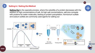
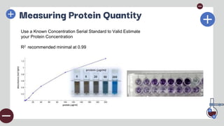
Proteomics Basic World:
Characterization and
Identification
Leonardo Tejo Gunawan,S.Si.,M.Biotek
Hanyang Biomedical Research Institute
Lab Division of StemCell Biology and Regenerative Medicine
2024.05.18
Introduction
Proteins are fundamental to virtually
every biological process. Identifying
and understanding proteins is crucial
for unraveling cellular mechanisms.
Understanding techniques in basic
Proteomic (at least) can have
BIG IMPACT
Introduction
We’ll explore the journey
of protein analysis, from
extraction to
understanding their
functions.
“OMICS” research trend over the years
Basic Proteomics :
Extraction and
Measuring Protein
01
Sample Preparation
Remember This!
•Keep the methods as simple as you can —minimize the number of steps and
avoid difficult manipulations which will not reproduce.
•Keep it cheap—avoid expensive techniques where a cheaper one will do.
•Adopt a step approach—and optimize each step as you go.
•Speed is important—avoid delays and slow equipment.
•Use reliable techniques and apparatus.
•Spend money on simple bits and pieces—e.g. test-tubes, pipettes.
•Write out your methods before you start and record what you have done
accurately.
Make Your Own Version
Or Just Follow the Guideline from Manufacturer
Extraction Time!!
Protein extraction is a critical step in studying proteins, as it allows us to isolate them from cells or
tissues.
The efficiency of this process and its compatibility with downstream applications are key factors in
choosing the right extraction method
Extraction Time!!
Protein extraction is a critical step in studying proteins, as it allows us to isolate them from cells or
tissues. The efficiency of this process and its compatibility with downstream applications are key
factors in choosing the right extraction method
(Mostly, But Not Limited)
When choosing a protein extraction method, consider your downstream applications.
The method must be compatible with your analytical techniques and should preserve the integrity
and functionality of the proteins or protein complexes you're studying
RIPA Lysis Buffer
RIPA buffer, short for Radio-Immunoprecipitation Assay buffer, is a versatile lysis
solution. It contains a mixture of detergents like SDS and/or Triton X-100, along with
sodium deoxycholate, salts, and buffering agents. This combination makes RIPA
highly effective at solubilizing proteins from various cellular compartments, including
membranes, nuclei, and the cytoplasm
Another Step / Technique in Protein
Extraction
CENTRIFUGATION
is a technique that separates components in a mixture based on their
density by spinning samples at high speeds. There are two main
types: differential centrifugation, which separates particles based on
size and density, and density gradient centrifugation, which
separates them based on buoyant density.
Another Step / Technique in Protein
Extraction
Precipitation
involves altering the solubility of
proteins to cause them to come out
of solution. This can be achieved by
adding salts like ammonium sulfate
or solvents like trichloroacetic acid.
Precipitation is useful for
concentrating proteins and
removing impurities
Salting In / Salting Out Method
Salting In refers to the phenomenon where the solubility of a protein increases with the
addition of low concentrations of salt. The added salt ions shield the electrostatic
interactions between protein molecules, preventing aggregation and increasing
solubility. Common salts used for salting in include sodium chloride and potassium
chloride
Salting In / Salting Out Method
Salting Out is the opposite process, where the solubility of a protein decreases with the
addition of high concentrations of salt. At high salt concentrations, salt ions compete
with proteins for water molecules, leading to protein precipitation. Ammonium sulfate
and sodium sulfate are commonly used agents for salting out
Another Step / Technique in Protein
Extraction
Dyalisis
uses a semipermeable membrane to separate small molecules from
larger protein molecules by diffusion. This technique is effective for
removing salts, solvents, and other small molecules, and is often
used for buffer exchange and desalting protein solutions after
precipitation or other purification steps
Another Step / Technique in Protein
Extraction
REMEMBER!! MWCO (Molecular Weight Cut Off) of
membrane must be smaller than the protein target!!
Measuring Protein Quantity
BCA ASSAY is a colorimetric assay used to determine protein concentration in a
sample. It relies on the reduction of copper ions (Cu^2+) to cuprous
ions (Cu^1+) by proteins, which are then detected using bicinchoninic
acid
The principle of the BCA assay involves
two main steps. First, the biuret reaction
occurs, where proteins reduce Cu^2+ to
Cu^1+ in an alkaline environment. Then,
bicinchoninic acid (BCA) chelates the
Cu^1+ ions, forming a purple-colored
complex. This complex has a strong
absorbance at 562 nm, which can be
measured using a spectrophotometer to
determine protein concentration
Measuring Protein Quantity
Bradford
ASSAY
The principle of the Bradford assay involves the binding of Coomassie
Brilliant Blue G-250 dye to proteins. In an acidic environment, the dye
binds to proteins primarily through arginine and other basic and
aromatic amino acids, causing the dye to shift from brown to blue.
This color change is measured at an absorbance of 595 nm
The Bradford assay procedure is
straightforward. First, prepare the
Bradford reagent, which is
commercially available and ready to
use. Next, add the protein sample to
the Bradford reagent and mix well.
Incubate the mixture at room
temperature for 5-10 minutes to allow
the color development.
280nm Assay
Measuring protein concentration at 280 nm is a straightforward method based on the UV
absorbance of aromatic amino acids, primarily tryptophan and tyrosine, and to a lesser
extent, cystine. These amino acids absorb UV light strongly at this wavelength.
Beer-Lambert Law: A = εlc
ε (molar extinction coefficient): Specific for each protein
Absorbance directly proportional to protein concentration
However, there are limitations to this method. The
absorption at 280 nm varies depending on the
protein’s amino acid composition, particularly its
tryptophan and tyrosine content. Interference from
nucleic acids and other UV-absorbing contaminants
can affect accuracy. Additionally, this method is not
very accurate for low protein concentrations due to the
low sensitivity
Measuring Protein Quantity
Use a Known Concentration Serial Standard to Valid Estimate
your Protein Concentration
R2 recommended minimal at 0.99
Measuring Protein Quantity
Use a Known Concentration Serial Standard to Valid Estimate
your Protein Concentration
R2 recommended minimal at 0.99
Protein Basic
Characterization
02
Electrophoresis
Electrophoresis is a technique used to separate charged molecules, such as DNA, RNA,
and proteins, based on their movement in an electric field. The speed and direction of their
movement depend on the molecule's size, charge, and the medium through which they
move.
•Charged molecules migrate towards opposite charge
•Smaller molecules move faster than larger ones
•Separation achieved based on size and charge
Types of Electrophoresis
Bullet Points:
Agarose Gel Electrophoresis: DNA and RNA separation
Polyacrylamide Gel Electrophoresis (PAGE): Protein
separation
SDS-PAGE: Denatured proteins by size
Native PAGE: Proteins in their native state
SDS-PAGE
(Sodium Dodecyl Sulphate-Polyacrilamide Gel Electrophoresis)
involves using sodium dodecyl sulfate (SDS), a detergent that denatures proteins, giving
them a uniform negative charge. This allows proteins to be separated strictly by size when
an electric field is applied. SDS-PAGE is widely used for protein characterization and
assessing protein purity, but also for DNA fingerprinting test.
Not Only SDS, but also need other
component
Break dissulfide bonds
CREDITS: This presentation template was created by Slidesgo, and
includes icons by Flaticon, and infographics & images by Freepik
Thanks!
Do you have any questions?
youremail@freepik.com
+34 654 321 432
yourwebsite.com
Please keep this slide for attribution
Basic Proteomics for Protein Quantitation and SDS Page

Basic Proteomics for Protein Quantitation and SDS Page

  • 1.
    Proteomics Basic World: Characterizationand Identification Leonardo Tejo Gunawan,S.Si.,M.Biotek Hanyang Biomedical Research Institute Lab Division of StemCell Biology and Regenerative Medicine 2024.05.18
  • 2.
    Introduction Proteins are fundamentalto virtually every biological process. Identifying and understanding proteins is crucial for unraveling cellular mechanisms. Understanding techniques in basic Proteomic (at least) can have BIG IMPACT
  • 3.
    Introduction We’ll explore thejourney of protein analysis, from extraction to understanding their functions.
  • 4.
  • 5.
    Basic Proteomics : Extractionand Measuring Protein 01
  • 6.
  • 7.
    Remember This! •Keep themethods as simple as you can —minimize the number of steps and avoid difficult manipulations which will not reproduce. •Keep it cheap—avoid expensive techniques where a cheaper one will do. •Adopt a step approach—and optimize each step as you go. •Speed is important—avoid delays and slow equipment. •Use reliable techniques and apparatus. •Spend money on simple bits and pieces—e.g. test-tubes, pipettes. •Write out your methods before you start and record what you have done accurately.
  • 8.
  • 9.
    Or Just Followthe Guideline from Manufacturer
  • 10.
    Extraction Time!! Protein extractionis a critical step in studying proteins, as it allows us to isolate them from cells or tissues. The efficiency of this process and its compatibility with downstream applications are key factors in choosing the right extraction method
  • 11.
    Extraction Time!! Protein extractionis a critical step in studying proteins, as it allows us to isolate them from cells or tissues. The efficiency of this process and its compatibility with downstream applications are key factors in choosing the right extraction method (Mostly, But Not Limited) When choosing a protein extraction method, consider your downstream applications. The method must be compatible with your analytical techniques and should preserve the integrity and functionality of the proteins or protein complexes you're studying
  • 12.
    RIPA Lysis Buffer RIPAbuffer, short for Radio-Immunoprecipitation Assay buffer, is a versatile lysis solution. It contains a mixture of detergents like SDS and/or Triton X-100, along with sodium deoxycholate, salts, and buffering agents. This combination makes RIPA highly effective at solubilizing proteins from various cellular compartments, including membranes, nuclei, and the cytoplasm
  • 13.
    Another Step /Technique in Protein Extraction CENTRIFUGATION is a technique that separates components in a mixture based on their density by spinning samples at high speeds. There are two main types: differential centrifugation, which separates particles based on size and density, and density gradient centrifugation, which separates them based on buoyant density.
  • 14.
    Another Step /Technique in Protein Extraction Precipitation involves altering the solubility of proteins to cause them to come out of solution. This can be achieved by adding salts like ammonium sulfate or solvents like trichloroacetic acid. Precipitation is useful for concentrating proteins and removing impurities
  • 15.
    Salting In /Salting Out Method Salting In refers to the phenomenon where the solubility of a protein increases with the addition of low concentrations of salt. The added salt ions shield the electrostatic interactions between protein molecules, preventing aggregation and increasing solubility. Common salts used for salting in include sodium chloride and potassium chloride
  • 16.
    Salting In /Salting Out Method Salting Out is the opposite process, where the solubility of a protein decreases with the addition of high concentrations of salt. At high salt concentrations, salt ions compete with proteins for water molecules, leading to protein precipitation. Ammonium sulfate and sodium sulfate are commonly used agents for salting out
  • 17.
    Another Step /Technique in Protein Extraction Dyalisis uses a semipermeable membrane to separate small molecules from larger protein molecules by diffusion. This technique is effective for removing salts, solvents, and other small molecules, and is often used for buffer exchange and desalting protein solutions after precipitation or other purification steps
  • 18.
    Another Step /Technique in Protein Extraction REMEMBER!! MWCO (Molecular Weight Cut Off) of membrane must be smaller than the protein target!!
  • 19.
    Measuring Protein Quantity BCAASSAY is a colorimetric assay used to determine protein concentration in a sample. It relies on the reduction of copper ions (Cu^2+) to cuprous ions (Cu^1+) by proteins, which are then detected using bicinchoninic acid The principle of the BCA assay involves two main steps. First, the biuret reaction occurs, where proteins reduce Cu^2+ to Cu^1+ in an alkaline environment. Then, bicinchoninic acid (BCA) chelates the Cu^1+ ions, forming a purple-colored complex. This complex has a strong absorbance at 562 nm, which can be measured using a spectrophotometer to determine protein concentration
  • 20.
    Measuring Protein Quantity Bradford ASSAY Theprinciple of the Bradford assay involves the binding of Coomassie Brilliant Blue G-250 dye to proteins. In an acidic environment, the dye binds to proteins primarily through arginine and other basic and aromatic amino acids, causing the dye to shift from brown to blue. This color change is measured at an absorbance of 595 nm The Bradford assay procedure is straightforward. First, prepare the Bradford reagent, which is commercially available and ready to use. Next, add the protein sample to the Bradford reagent and mix well. Incubate the mixture at room temperature for 5-10 minutes to allow the color development.
  • 21.
    280nm Assay Measuring proteinconcentration at 280 nm is a straightforward method based on the UV absorbance of aromatic amino acids, primarily tryptophan and tyrosine, and to a lesser extent, cystine. These amino acids absorb UV light strongly at this wavelength. Beer-Lambert Law: A = εlc ε (molar extinction coefficient): Specific for each protein Absorbance directly proportional to protein concentration However, there are limitations to this method. The absorption at 280 nm varies depending on the protein’s amino acid composition, particularly its tryptophan and tyrosine content. Interference from nucleic acids and other UV-absorbing contaminants can affect accuracy. Additionally, this method is not very accurate for low protein concentrations due to the low sensitivity Measuring Protein Quantity
  • 22.
    Measuring Protein Quantity Usea Known Concentration Serial Standard to Valid Estimate your Protein Concentration R2 recommended minimal at 0.99
  • 23.
    Measuring Protein Quantity Usea Known Concentration Serial Standard to Valid Estimate your Protein Concentration R2 recommended minimal at 0.99
  • 24.
  • 25.
    Electrophoresis Electrophoresis is atechnique used to separate charged molecules, such as DNA, RNA, and proteins, based on their movement in an electric field. The speed and direction of their movement depend on the molecule's size, charge, and the medium through which they move. •Charged molecules migrate towards opposite charge •Smaller molecules move faster than larger ones •Separation achieved based on size and charge Types of Electrophoresis Bullet Points: Agarose Gel Electrophoresis: DNA and RNA separation Polyacrylamide Gel Electrophoresis (PAGE): Protein separation SDS-PAGE: Denatured proteins by size Native PAGE: Proteins in their native state
  • 26.
    SDS-PAGE (Sodium Dodecyl Sulphate-PolyacrilamideGel Electrophoresis) involves using sodium dodecyl sulfate (SDS), a detergent that denatures proteins, giving them a uniform negative charge. This allows proteins to be separated strictly by size when an electric field is applied. SDS-PAGE is widely used for protein characterization and assessing protein purity, but also for DNA fingerprinting test.
  • 27.
    Not Only SDS,but also need other component Break dissulfide bonds
  • 31.
    CREDITS: This presentationtemplate was created by Slidesgo, and includes icons by Flaticon, and infographics & images by Freepik Thanks! Do you have any questions? youremail@freepik.com +34 654 321 432 yourwebsite.com Please keep this slide for attribution

Editor's Notes

  • #4 Today, we will learn about basic from