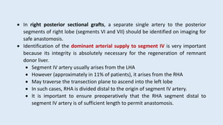
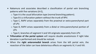
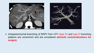
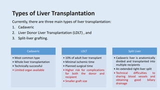
This document discusses imaging of the liver preoperatively for liver transplantation. Radiologists play a key role in evaluating patients' anatomy and suitability for transplantation. Preoperative imaging assesses liver parenchyma for tumors or other abnormalities, calculates liver volumes, and precisely maps the hepatic vasculature. Living donor liver transplantation requires imaging the donor's liver to ensure the safety of donation and adequate remnant liver volume. The document outlines various surgical techniques for cadaveric and living donor transplantation.
Liver transplant In India by Dr. Abhideep Chaudhary, Sir Ganga Ram Hospitaldrabhideep
This presentation is related to Liver Transplant, Liver Failure, It's causes and remedy.
Here we also talk about liver transplant scenario in india and success rate of liver transplant both cadaver or living donor.
We also give a brief about the cost of liver transplant.
Dr. Abhideep Chaudhary, is liver transplant consultant/surgeon at Sir Ganga Ram Hospital, New Delhi, India.
Email : drabhideep@yahoo.com , care@drabhideep.com
This presentation include biliary anatomy ,indication, contraindication post op care of percutaneus transhepatic biliary drainage with important technique. and advantage and disadvantage of different technique. This is important for radiologist, radiographers, intervention radiologist radiology resident. Thanks
Brief description on the benign tumors of liver that includes hemangioma, focal nodular hyperplasia, regenerative nodular hyperplasia, dysplastic foci, dysplastic nodules and focal fatty change.
This slide includes various CT protocol , liver ct triple phase protocol , with important findings, this power-point presentation help a lot for radiologist, radiology resident, radiographers, technician. Thanks.
Purpose of this presentation is to educate non radiologist about basic CT anatomy of abdominal viscera and basic interpretation of very common diseases
Liver transplantation current status, controversies and mythsAbhishek Yadav
Details the present status, indications, techniques about liver transplantation. Also dispels some common myths surrounding liver transplantation. #liver transplantation # living donor liver transplantation #liver cirrhosis #liver failure#transplantation#live donor#drabhishekyadav.com#liversurgeon#myths#livedonorlivertransplantation#organtransplantation#alcohololiverdisease
Hepatocellular carcinoma—role of interventional radiologist Dr. Muhammad Bin ...Dr. Muhammad Bin Zulfiqar
In these presentation we will discuss the merits, demrits and outcomes of various interventional radiology modalities for the treatment of hepatocellular carcinoma
Anaesthesia for liver transplantation.pptxKLahari7
Introduction
Liver transplantation (LT) is the treatment of choice for end-stage liver disease regardless of its aetiology. Ever since the first transplant interventions in the 1960s, mortality rates after LT have significantly improved and have led to an increase in the number of successful procedures and improved outcomes.
Significant challenges remain for the transplant team as the procedure is performed on high-risk patients with impaired cardiovascular, pulmonary, renal and coagulation systems. Recent publications have indicated that transplant candidates are older, sicker and with multiple associate co-morbidities and organ dysfunctions compared to those treated in the past. Adequate perioperative care is essential for a prompt graft function which will improve organ system recovery and recipient’s quality of life [1].
Though there is a potential worldwide liver graft shortage, the expansion of the donor pool using marginal donors and increasing donor age has resulted, never the less, in reduced waiting list mortality [2]. A successful LT requires teams with a particular set of skills and competences, including a complete and detailed understanding of the multi-organic pathophysiology of liver failure and its implications and management during the three stages of surgery.
There have been many innovations, updates and procedural changes in the anaesthetic management of patients during this time. This article gives an overview of the current literature regarding anaesthetic management during liver transplantation and its singularities during the three stages of surgery.
Go to:
Indications for liver transplantation
The indications for LT in patients with acute and chronic liver failure should be assessed independent of the aetiology and are listed in Table 1 [3].
Table 1
Indications for liver transplantation
Class Disease
Non-cholestatic liver disease HepatitisB
HepatitisC
HepatitisD
HepatitisA
Alcoholic liver disease
Autoimmune hepatitis
Cryptogenic cirrhosis
Non-alcoholic steatohepatitis
Other
Cholestatic liver disease Primary biliary cirrhosis
Secondary biliary cirrhosis (Caroli disease, choledochal cyst)
Primary sclerosing cholangitis
Other
Malignant disease Hepatocellular carcinoma
Cholangiocarcinoma
Hepatoblastoma
Other
Extrahepatic biliary atresia or hypoplasia Alagille syndrome
Other
Metabolic diseases Alpha-1 antitrypsin deficiency
Crigler-Najjar disease, Type I
Byler’s disease
Glycogen storage disease, Type I
Wilson’s disease
Hemochromatosis
Tyrosinemia
Wolman’s disease
Familial amyloidotic polyneuropathy
Primary hyperoxaluria type 1
Other
Hepatic vein thrombosis Budd-Chiari
Acute hepatic failure Hepatitis
Drugs
Unknown aetiology
Re-transplantation Primary non-function
Hepatic artery thrombosis
Acute/chronic rejection
Open in a separate window
The US and European countries have been using the Model for End-Stage Liver Disease (MELD) score for organ allocation since 2007. This is a grading system from 6 to 40 points, which depen
discusses in detail about approach and management of HCC. Other liver masses and abscesses including cholangiocarcinoma. liver abscess, Hydatid cyst, Hepatic adenoma, hemangioma, Focal Nodular Hyperplasia.
This is a general overview of options available to patients with liver dominant metastatic disease as well other focal areas of disease which may benefit from services provided by an interventional radiologist
Benign Biliary Stricture is a common condition which we encounter during gastro practice. Here we discuss in detail about its diagnosis and management.
Radiological and Clinical features of diffuse lung diseases.
Especially, HRCT features and some pathognomonic findings of diffuse lung disease.
Cystic lung diseases, Nodular lung diseases, Fibrotic lung diseases, Smoking related lung diseases,
Defecation
Normal defecation begins with movement in the left colon, moving stool toward the anus. When stool reaches the rectum, the distention causes relaxation of the internal sphincter and an awareness of the need to defecate. At the time of defecation, the external sphincter relaxes, and abdominal muscles contract, increasing intrarectal pressure and forcing the stool out
The Valsalva maneuver exerts pressure to expel faeces through a voluntary contraction of the abdominal muscles while maintaining forced expiration against a closed airway. Patients with cardiovascular disease, glaucoma, increased intracranial pressure, or a new surgical wound are at greater risk for cardiac dysrhythmias and elevated blood pressure with the Valsalva maneuver and need to avoid straining to pass the stool.
Normal defecation is painless, resulting in passage of soft, formed stool
CONSTIPATION
Constipation is a symptom, not a disease. Improper diet, reduced fluid intake, lack of exercise, and certain medications can cause constipation. For example, patients receiving opiates for pain after surgery often require a stool softener or laxative to prevent constipation. The signs of constipation include infrequent bowel movements (less than every 3 days), difficulty passing stools, excessive straining, inability to defecate at will, and hard feaces
IMPACTION
Fecal impaction results from unrelieved constipation. It is a collection of hardened feces wedged in the rectum that a person cannot expel. In cases of severe impaction the mass extends up into the sigmoid colon.
DIARRHEA
Diarrhea is an increase in the number of stools and the passage of liquid, unformed feces. It is associated with disorders affecting digestion, absorption, and secretion in the GI tract. Intestinal contents pass through the small and large intestine too quickly to allow for the usual absorption of fluid and nutrients. Irritation within the colon results in increased mucus secretion. As a result, feces become watery, and the patient is unable to control the urge to defecate. Normally an anal bag is safe and effective in long-term treatment of patients with fecal incontinence at home, in hospice, or in the hospital. Fecal incontinence is expensive and a potentially dangerous condition in terms of contamination and risk of skin ulceration
HEMORRHOIDS
Hemorrhoids are dilated, engorged veins in the lining of the rectum. They are either external or internal.
FLATULENCE
As gas accumulates in the lumen of the intestines, the bowel wall stretches and distends (flatulence). It is a common cause of abdominal fullness, pain, and cramping. Normally intestinal gas escapes through the mouth (belching) or the anus (passing of flatus)
FECAL INCONTINENCE
Fecal incontinence is the inability to control passage of feces and gas from the anus. Incontinence harms a patient’s body image
PREPARATION AND GIVING OF LAXATIVESACCORDING TO POTTER AND PERRY,
An enema is the instillation of a solution into the rectum and sig
CHAPTER 1 SEMESTER V PREVENTIVE-PEDIATRICS.pdfSachin Sharma
This content provides an overview of preventive pediatrics. It defines preventive pediatrics as preventing disease and promoting children's physical, mental, and social well-being to achieve positive health. It discusses antenatal, postnatal, and social preventive pediatrics. It also covers various child health programs like immunization, breastfeeding, ICDS, and the roles of organizations like WHO, UNICEF, and nurses in preventive pediatrics.
R3 Stem Cells and Kidney Repair A New Horizon in Nephrology.pptxR3 Stem Cell
R3 Stem Cells and Kidney Repair: A New Horizon in Nephrology" explores groundbreaking advancements in the use of R3 stem cells for kidney disease treatment. This insightful piece delves into the potential of these cells to regenerate damaged kidney tissue, offering new hope for patients and reshaping the future of nephrology.
CRISPR-Cas9, a revolutionary gene-editing tool, holds immense potential to reshape medicine, agriculture, and our understanding of life. But like any powerful tool, it comes with ethical considerations.
Unveiling CRISPR: This naturally occurring bacterial defense system (crRNA & Cas9 protein) fights viruses. Scientists repurposed it for precise gene editing (correction, deletion, insertion) by targeting specific DNA sequences.
The Promise: CRISPR offers exciting possibilities:
Gene Therapy: Correcting genetic diseases like cystic fibrosis.
Agriculture: Engineering crops resistant to pests and harsh environments.
Research: Studying gene function to unlock new knowledge.
The Peril: Ethical concerns demand attention:
Off-target Effects: Unintended DNA edits can have unforeseen consequences.
Eugenics: Misusing CRISPR for designer babies raises social and ethical questions.
Equity: High costs could limit access to this potentially life-saving technology.
The Path Forward: Responsible development is crucial:
International Collaboration: Clear guidelines are needed for research and human trials.
Public Education: Open discussions ensure informed decisions about CRISPR.
Prioritize Safety and Ethics: Safety and ethical principles must be paramount.
CRISPR offers a powerful tool for a better future, but responsible development and addressing ethical concerns are essential. By prioritizing safety, fostering open dialogue, and ensuring equitable access, we can harness CRISPR's power for the benefit of all. (2998 characters)
One of the most developed cities of India, the city of Chennai is the capital of Tamilnadu and many people from different parts of India come here to earn their bread and butter. Being a metropolitan, the city is filled with towering building and beaches but the sad part as with almost every Indian city
Medical Technology Tackles New Health Care Demand - Research Report - March 2...pchutichetpong
M Capital Group (“MCG”) predicts that with, against, despite, and even without the global pandemic, the medical technology (MedTech) industry shows signs of continuous healthy growth, driven by smaller, faster, and cheaper devices, growing demand for home-based applications, technological innovation, strategic acquisitions, investments, and SPAC listings. MCG predicts that this should reflects itself in annual growth of over 6%, well beyond 2028.
According to Chris Mouchabhani, Managing Partner at M Capital Group, “Despite all economic scenarios that one may consider, beyond overall economic shocks, medical technology should remain one of the most promising and robust sectors over the short to medium term and well beyond 2028.”
There is a movement towards home-based care for the elderly, next generation scanning and MRI devices, wearable technology, artificial intelligence incorporation, and online connectivity. Experts also see a focus on predictive, preventive, personalized, participatory, and precision medicine, with rising levels of integration of home care and technological innovation.
The average cost of treatment has been rising across the board, creating additional financial burdens to governments, healthcare providers and insurance companies. According to MCG, cost-per-inpatient-stay in the United States alone rose on average annually by over 13% between 2014 to 2021, leading MedTech to focus research efforts on optimized medical equipment at lower price points, whilst emphasizing portability and ease of use. Namely, 46% of the 1,008 medical technology companies in the 2021 MedTech Innovator (“MTI”) database are focusing on prevention, wellness, detection, or diagnosis, signaling a clear push for preventive care to also tackle costs.
In addition, there has also been a lasting impact on consumer and medical demand for home care, supported by the pandemic. Lockdowns, closure of care facilities, and healthcare systems subjected to capacity pressure, accelerated demand away from traditional inpatient care. Now, outpatient care solutions are driving industry production, with nearly 70% of recent diagnostics start-up companies producing products in areas such as ambulatory clinics, at-home care, and self-administered diagnostics.
Health Education on prevention of hypertensionRadhika kulvi
Hypertension is a chronic condition of concern due to its role in the causation of coronary heart diseases. Hypertension is a worldwide epidemic and important risk factor for coronary artery disease, stroke and renal diseases. Blood pressure is the force exerted by the blood against the walls of the blood vessels and is sufficient to maintain tissue perfusion during activity and rest. Hypertension is sustained elevation of BP. In adults, HTN exists when systolic blood pressure is equal to or greater than 140mmHg or diastolic BP is equal to or greater than 90mmHg. The
The Importance of Community Nursing Care.pdfAD Healthcare
NDIS and Community 24/7 Nursing Care is a specific type of support that may be provided under the NDIS for individuals with complex medical needs who require ongoing nursing care in a community setting, such as their home or a supported accommodation facility.
Global launch of the Healthy Ageing and Prevention Index 2nd wave – alongside...ILC- UK
The Healthy Ageing and Prevention Index is an online tool created by ILC that ranks countries on six metrics including, life span, health span, work span, income, environmental performance, and happiness. The Index helps us understand how well countries have adapted to longevity and inform decision makers on what must be done to maximise the economic benefits that comes with living well for longer.
Alongside the 77th World Health Assembly in Geneva on 28 May 2024, we launched the second version of our Index, allowing us to track progress and give new insights into what needs to be done to keep populations healthier for longer.
The speakers included:
Professor Orazio Schillaci, Minister of Health, Italy
Dr Hans Groth, Chairman of the Board, World Demographic & Ageing Forum
Professor Ilona Kickbusch, Founder and Chair, Global Health Centre, Geneva Graduate Institute and co-chair, World Health Summit Council
Dr Natasha Azzopardi Muscat, Director, Country Health Policies and Systems Division, World Health Organisation EURO
Dr Marta Lomazzi, Executive Manager, World Federation of Public Health Associations
Dr Shyam Bishen, Head, Centre for Health and Healthcare and Member of the Executive Committee, World Economic Forum
Dr Karin Tegmark Wisell, Director General, Public Health Agency of Sweden
CHAPTER 1 SEMESTER V - ROLE OF PEADIATRIC NURSE.pdfSachin Sharma
Pediatric nurses play a vital role in the health and well-being of children. Their responsibilities are wide-ranging, and their objectives can be categorized into several key areas:
1. Direct Patient Care:
Objective: Provide comprehensive and compassionate care to infants, children, and adolescents in various healthcare settings (hospitals, clinics, etc.).
This includes tasks like:
Monitoring vital signs and physical condition.
Administering medications and treatments.
Performing procedures as directed by doctors.
Assisting with daily living activities (bathing, feeding).
Providing emotional support and pain management.
2. Health Promotion and Education:
Objective: Promote healthy behaviors and educate children, families, and communities about preventive healthcare.
This includes tasks like:
Administering vaccinations.
Providing education on nutrition, hygiene, and development.
Offering breastfeeding and childbirth support.
Counseling families on safety and injury prevention.
3. Collaboration and Advocacy:
Objective: Collaborate effectively with doctors, social workers, therapists, and other healthcare professionals to ensure coordinated care for children.
Objective: Advocate for the rights and best interests of their patients, especially when children cannot speak for themselves.
This includes tasks like:
Communicating effectively with healthcare teams.
Identifying and addressing potential risks to child welfare.
Educating families about their child's condition and treatment options.
4. Professional Development and Research:
Objective: Stay up-to-date on the latest advancements in pediatric healthcare through continuing education and research.
Objective: Contribute to improving the quality of care for children by participating in research initiatives.
This includes tasks like:
Attending workshops and conferences on pediatric nursing.
Participating in clinical trials related to child health.
Implementing evidence-based practices into their daily routines.
By fulfilling these objectives, pediatric nurses play a crucial role in ensuring the optimal health and well-being of children throughout all stages of their development.
2. Introduction
• Liver transplantation is considered the only curative treatment for patients with
end stage liver disease
• Successful treatment for unresectable small hepatocellular carcinoma (HCC)
• Outcome had greatly improved during the last years giving the advance of
immunosuppressive treatments and surgical techniques
• Successful liver transplantation requires a multidisciplinary approach with careful
preprocedural preparation as well as long term follow up.
• Radiologists play a crucial role in the pre-procedural workup and the short/long
term follow up.
• Understanding the surgical procedure and its potential complications are
essential for an adequate radiological assessment
3. Indications for Liver Transplantation
Condition Indications
Alcoholic cirrhosis Patients must abstain from alcohol for 6 months, undergo treatment for
alcohol abuse, and have a strong psychosocial support system
Chronic hepatitis Hepatitis B, C, and D infection; autoimmune hepatitis; chronic drug use;
cryptogenic cirrhosis; chronic active hepatitis
Primary hepatic tumors Hepatocellular carcinoma (grade 1 or 2, <5 cm, and no extrahepatic
metastases or macrovascular invasion)
Cholestatic diseases Primary biliary cirrhosis, sclerosing cholangitis, cystic fibrosis, biliary atre-
sia, Caroli disease, Alagille syndrome, Byler disease, familial cholestatic
syndromes
Fulminant liver failure Drug toxicity, toxins, viral hepatitis infection
Metabolic disease a-1-antitrypsin deficiency, glycogen storage disease, Wilson disease, hemo-
chromatosis
Other conditions Budd-Chiari syndrome, parenteral nutrition–induced cirrhosis, trauma
4. Contraindications for Liver Transplantation
Absolute contraindications
• Active extrahepatic malignancy
• Diffuse hepatic tumor invasion
• Thrombosis of the entire portal and
superior mesenteric venous system
• Active or uncontrolled systemic
infection
• Active substance or alcohol abuse
• Severe cardiopulmonary disease
• Lack of social support
• Noncompliance
• Fatty infiltration >30%
Relative contraindications
• Age
• Cholangiocarcinoma
• Portal vein thrombosis
• Chronic or refractory infection
• HIV infection
• Previous malignancy
• Active psychiatric disorder
• Poor social support
5. Patient Selection
• Unfortunately, the disparity between the number of recipients and available
livers has been increasing.
• Thus, under the current guidelines, the sickest patients are given priority for
transplantation.
• Priority is given to
• patients with end-stage liver disease and who have the greatest risk for death
within 3 months due to progressive deterioration
• acute decompensation from chronic liver disease
• development of fulminant liver failure in those who were previously healthy
6. Model for End-Stage Liver Disease (MELD score)
• Used to assess the severity of chronic liver disease.
• Determined which patients need to do urgent liver transplantation.
• MELD scores range from 6 to 40
• 6 indicating the least sick patients
• 40 indicating the sickest patients.
• MELD scores are calculated on the basis of
• total bilirubin,
• international normalized ratio (INR)
• creatinine
• MELD scores Equation:
3.8 × log (e) (amount of bilirubin) + 11.2 × log (e) (INR) + 9.6 log (e) (amount of creatinine)
7. • According to the studies,
• 3-month mortality rate with a MELD score of 40 is 100%.
• For score 30–39 is 83%
• For score 20–29 is 76%
• For score 0–19 is 27% and
• For score 10 is 4%.
8. Hepatocellular Carcinoma
• Among patients with HCC, priority for liver transplantation should be given
to those with stage T2 disease
• Patients with T1 disease have high survival rates without transplantation
• T3 disease have a high rate of recurrence.
9. • T1 - A single tumor (of any size) without blood vessel invasion
• T2 - A single tumor >2 cm with vascular invasion or multiple tumors (none >5 cm)
• T3a - multiple tumors, with at least one is larger than 5 cm.
• T3b - extend into a major branch of the portal or hepatic vein.
• T4 - tumors extend beyond the liver, except gallbladder
HCC (T-staging)
10. • MELD scores are not a reliable indicator of mortality among patients who die
from HCC.
Milan criteria
• Eligible for liver transplantation in HCC patients;
• A single tumor that is smaller than 5 cm
• Up to three tumors that are 3 cm or smaller
• No vascular invasion
• No extrahepatic disease
University of California San Francisco (UCSF) criteria
• Eligible for transplantation in;
• single tumor that is 6.5 cm or smaller
• three or fewer tumor nodules are present, with the largest being 4.5 cm or
small
11. Tumor Volume
• When a cutoff of 115 𝒄𝒎𝟑 is used for selecting transplantation, post-
transplantation outcomes are no different from those achieved with the Milan
and UCSF classification systems.
High Recurrent Potential
• Tumor larger than 6 cm
• Disease that progresses after pre-transplantation treatment
• Tumor that is exposed to the liver surface.
12. Types of Liver Transplantation
Currently, there are three main types of liver transplantation:
1. Cadaveric
2. Liver Donor Liver Transplantation (LDLT) , and
3. Split-liver grafting.
Cadaveric
• Most common type
• Whole liver transplantation
• Technically successful
• Limited organ available
LDLT
• 10% of adult liver transplant
• Minimal ischemic time
• Planned surgical time
• Higher risk for complications
for both the donor and
recipient
• Smaller graft size
Split Liver
• Cadaveric liver is anatomically
divided and transplanted into
multiple recipients
• An extended right-liver split
• Technical difficulties to
sharing blood vessels and
obtaining good biliary
drainage
13. Living Donor Liver Transplantation
• The advantages of LDLT include a lack of preservation injuries and an ability to
perform the procedure on an elective basis.
• The disadvantages include a risk for complications in the donor.
• Hepatic segments cannot be transplanted separately because the transplanted
liver tissue requires arterial supply, venous drainage, and biliary outflow.
• The decision of whether to perform hepatectomy is determined on:
• Donor safety
• Size of the recipient
• Left lateral hepatectomy grafts are ideal for smaller recipients.
• Left or right hepatectomy grafts are more useful for larger patients.
15. Left Lateral Hepatectomy
• Most common LDLT technique
• Includes segments II and III of the resected liver
• Represents 20-25% of the whole liver volume
16. Left Lobectomy
• Includes segments 2, 3, and 4 together (known as the left lobe)
• Represents approximately 40% of the whole liver volume
• Harvesting the middle hepatic vein to obtain a reasonably large graft and
maintain high tissue viability for transplantation.
17. Right Hemihepatectomy
• Includes segments V, VI, VII and VIII (entire donor right lobe), Rt. hepatic artery,
Rt. hepatic vein, Rt. bile duct and Rt. portal vein
• Account for approximately 60% of the total liver volume
• Performed if the volume of the donor’s left lobe is more than 30% of the total
hepatic volume.
• Inclusion of the middle hepatic vein in the graft is controversial; however, it may
be included to avoid congestion of the right paramedian segment in the recipient.
18. • The caudate lobe usually remains in the donor because it is directly perfused by
the right or left portal vein branches.
• Middle hepatic vein remains with the donor when the right side of the liver is
being donated (Controversial)
• MHV is harvested when left hepatectomy is performed.
• The recipient left and middle hepatic veins are joined, followed by end-to-end
anastamosis with the donor hepatic vein.
• Thus, the venous ouflow is optimized by reconnecting the major veins and
anastomosing with a single reconstructed recipient vein.
19.
20.
21. Split-Liver Grafting Technique
• Two types of split-liver grafting
• Left lateral split
• Extended right split
• The classic left lateral split is performed at the falciform ligament
• Right extended graft (segments I and IV to VIII) used in adults
• Left lateral graft (segments II and III) used in children who are no larger than
30 kg.
• The extended right split, which results in right (segments V–VIII) and left
(segments II–IV) hemilivers, is performed in two adult recipients.
• Typically,
• common bile duct and inferior vena cava (IVC) are part of the right liver graft
• main arterial trunk and middle hepatic vein are part of the left liver graft.
22. Pre-operative Imaging
• Correct identification of donor anatomy is essential to determine whether the
donor is a suitable candidate and hepatectomy may be safely performed with no
risk to the donor.
• Preoperative evaluation of liver parenchyma, vascularization and biliary system in
both donors and recipients by using;
1. Magnetic resonance imaging (MRI),
2. Computed tomography (CT) or
3. Ultrasound (US) imaging with Doppler evaluation
23. CT Cholangiogram
• CT cholangiography is performed after intravenous infusion of cholangiographic
agent.
• To reduce the risk for allergic reactions:
• 20 mL of 52% iodipamide meglumine (cholangiographic contrast) is diluted in
80 mL of normal saline
• infused over a period of 30–60 minutes.
• Fifteen minutes after completion of the infusion, spiral CT of the liver is
performed.
24. • In some institution, contrast-enhanced MR imaging and MR
Cholangiopancreatography (MRCP) are the first choice for preoperative
evaluation,
1. radiation free,
2. ensure accurate parenchymal analysis
3. adequate evaluation of vasculature and biliary system
25. Living Donor
Cross sectional liver imaging of the donor is mandatory before liver donation to
detect any contraindication or finding that may modify the surgical approach.
Liver parenchymal evaluation, diffuse (mainly steatosis) or focal
(benign/malignant) conditions
Identification of anatomical variants
26. Liver Volume Calculation
• Very important to calculate graft and remnant liver volumes before hepatic
resection, in order to ensure adequate hepatic function and liver regeneration
after surgery, both in recipients and donors.
• A minimal residual volume of 30% is required to prevent small-for-size syndrome
on the assumption that the liver parenchyma is disease free (i.e. steatosis)
• Ideally, the recipient should receive a graft similar in size to the native organ.
• But a minimum corrected graft-to-recipient body weight ratio (GRBW) of 0.8% is
still acceptable.
27. Liver Parenchymal Evaluation
Look for presence of incidental hepatic focal lesions.
Majority are benign, presence of any large lesion or a malignant lesion is a
contraindication for organ donation.
Any diffuse liver disease, such as fatty infiltration should be identified.
Hepatic steatosis carries a high risk of postoperative liver dysfunction in donors
and graft non-function in recipients.
Each 1% increase in hepatic fat content, functional mass of donor liver reduces
by 1%.
In the donor liver, moderate to severe steatosis (≥30% fat) is considered
unacceptably high and should be rejected for donation.
28. Severe hepatic steatosis. Axial NECT
image shows a decreased hepatic
attenuation in comparison with spleen.
29. Vascular Anatomy
Anatomical variants of the hepatic vasculature are relatively frequent.
Adequate arterial inflow to graft liver is necessary for avoidance of biliary
necrosis.
Patency of the portal vein is crucial for graft survival and liver regeneration.
Patent hepatic vein outflow is needed to prevent hepatic congestion and graft
dysfunction.
MDCT is an excellent tool in providing a detailed road map of normal and variant
hepatic vascular anatomy in the donor, and helps in guiding the surgical
approach.
Any variant should be clearly described and reported for an appropriate pre-
procedural planification.
Some anatomical variants are contraindication to liver donation.
30. Hepatic arterial anatomy and variants
MDCT allows accurate delineation of the intrahepatic tertiary arterial branches
as small as 1 mm in size.
In normal hepatic arterial anatomy, common hepatic artery (CHA) arises from
the celiac axis.
It divides into gastro-duodenal artery (GDA) and proper hepatic artery.
Proper hepatic artery ascends toward the liver hilum and divides into left
hepatic artery (LHA) and right hepatic artery (RHA).
LHA supplies the entire left hemi-liver, including segment IV.
The RHA divides into anterior and posterior sectional branches which supply the
anterior (VIII and V) and posterior sections (VI and VII) of right hemi-liver,
respectively.
32. In right posterior sectional grafts, a separate single artery to the posterior
segments of right lobe (segments VI and VII) should be identified on imaging for
safe anastomosis.
Identification of the dominant arterial supply to segment IV is very important
because its integrity is absolutely necessary for the regeneration of remnant
donor liver.
Segment IV artery usually arises from the LHA
However (approximately in 11% of patients), it arises from the RHA
May traverse the transection plane to ascend into the left lobe
In such cases, RHA is divided distal to the origin of segment IV artery.
It is important to ensure preoperatively that the RHA segment distal to
segment IV artery is of sufficient length to permit anastomosis.
33. Presence of accessory arteries to a lobe requires two arterial anastomoses, and
therefore increases the surgical time and poses a higher risk of postoperative
hepatic arterial thrombosis.
Presence of multiple small vessels in a lobe precludes donation.
Other surgically important arterial variants include direct origin of hepatic artery
from the aorta or entire hepatic artery from the SMA, and separate origin of all
hepatic arterial branches from CHA.
34. Segment IV artery origin from RHA Intraoperative photograph of segment IV
artery (Black arrow) arising from
RHA(open arrow)
35. Portal vein anatomy and variants
Classically, the main portal vein trunk branches into right and left portal veins at
porta hepatis.
The right portal vein (RPV) subsequently divides into anterior and posterior
sectional branches at a variable length from the RPV origin.
The right anterior portal vein (RAPV) supplies segments VIII and V, while the
right posterior portal vein (RPPV) supplies segments VI and VII of the liver.
The left portal vein (LPV), on the other hand, ascends along the falciform
ligament and supplies the entire left hemi-liver (segments II, III, and IV)
This normal portal vein anatomy is most suitable for donation, as only one
anastomosis is required between the donor and recipient portal veins.
36. Nakamura and associates described a classification of portal vein branching
patterns with five variations (A-E),
o Type A is the usual bifurcation type (normal branching pattern).
o Type B is a trifurcation pattern without the trunk of RPV.
o Type C, RAPV arises separately from the proximal or extra-parenchymal part
of LPV.
o Type D, RAPV arises separately from a distal or intra-parenchymal portion of
LPV.
o Type E, branches of segment V and VIII originate separately from LPV.
Trifurcation of the portal system will require double anastomosis if right lobe
donation is performed and should be noticed.
When the right anteromedial branch arises from the left portal vein, the
resection of the latter can have deleterious effects on segments IV, V and VIII.
37. Intraparenchymal branching of RAPV from LPV (type D) and type E branching
patterns are uncommon and are considered absolute contraindications for
surgery.
38. Hepatic venous anatomy and variants
Three major hepatic veins which drain liver parenchyma into the inferior vena
cava.
Right hepatic vein (RHV) is the largest and drains a major part of right hemi-liver
into IVC.
Middle hepatic vein (MHV) drains central liver segments (i.e. IV, V, and VIII),
Left hepatic vein (LHV) predominantly drains from segments II and III
In 60-70% of cases, MHV and LHV join to form a common stump before entering
IVC, while RHV opens directly into IVC.
Even when MHV and LHV open separately into IVC, an intimate relationship
exists between the two in 100% of cases.
39. Drainage pattern of the MHV should be thoroughly evaluated, since it is an
important surgical landmark.
The usual position of the middle hepatic vein is between segments VIII and IV.
It is important to identify any variation in MHV as hepatectomy plane in right hemi-
liver donation is about 1 cm to the right of the middle hepatic vein, along the
gallbladder fossa.
Branches from the anterior segments of right lobe (V and VIII) draining into the MHV
run along the parenchymal resection plane.
These veins need to be preserved and re-anastomosed in the recipient to prevent
congestion and risk of graft failure in the transplanted right hemi-liver.
40. It is also important to determine if an accessory inferior right hepatic vein is
present.
They are seen in 40-50% of patients.
Majority of these veins drain segments VI and VII
Inferior RHVs more than 4 mm in diameter should be preserved and
anastomosed separately to recipient’s IVC to prevent hepatic congestion.
Accessory hepatic veins with a caliber of 5 mm or more require separate
anastomosis to the IVC.
41. 3D image showing drainage of right
anterior sector veins (arrows) into MHV
42. Biliary Anatomy
In routine clinical practice, preoperative assessment of biliary tree in potential
liver donors is done using MR cholangiography.
The common hepatic duct (CHD) emerges from the convergence of right and left
bile ducts.
Variant biliary anatomy has been observed in 30-35% patients.
With increasing surgical expertise, a bile duct variation rarely excludes a person
from being a liver donor.
However, it very important to preoperatively evaluate unusual patterns of bile
duct branching, in order to modify the cutting plane during graft retrieval and
the pattern of ductal anastomosis in the recipient.
This helps in reducing postoperative biliary complications.
43. Recipient
• Evaluation and description of the liver anatomy
• Size, number, and characteristics of the visualized lesions.
• Tumors should be localized according to the Couinaud classification system to
facilitate their identification during resection, intervention, and other treatment
procedures.
• Imaging of the recipient liver should be performed as close to the time of
transplantation as possible because tumors may grow rapidly, invade local
structures, or metastasize, which may affect the stage of disease, management
perspective, and surgical technique.
44. It is also critical to report the proximity of the tumor to important structures
such as major blood vessels, the gallbladder, and central bile ducts because the
outcome may be affected.
Identification of vascular invasion is important because it is a contraindication
for transplantation.
Varices develop as a result of sustained severe portal hypertension.
The peri-caval and peri-hepatic varices should be reported due to their
association with increased intraoperative bleeding.
To exclude extrahepatic malignancy, CT of the chest and abdomen should be
performed.
45. Conclusions
• Preoperative imaging in potential liver transplantation patients includes proper
evaluation of the liver parenchyma and vascular and biliary anatomy.
• Understanding the indications, contraindications, surgical techniques and
potential complications are important for a thorough assessment.
• A multidisciplinary approach is essential in liver transplantation in order to
achieve the best outcome, with the radiologist playing a crucial role during the
whole process.
Typically, venous branches from segments V and VIII which are larger than 4 mm in diameter are anastomosed in the back bench to a portal vein graft harvested from the explanted liver.