The document provides instructions for a plant tissue practical examination. Students will use a microscope to observe and identify parenchyma and sclerenchyma tissues from prepared slides. They will draw and label diagrams of the tissues and complete a table describing their structures and functions. Additionally, students will create their own slides of stomata from a leaf using nail polish and observe the epidermal tissue under the microscope.
![1
Practical 6 [25]
Examining Simple Plant tissue
Learning Outcome:
Afteryouhave completedthisinvestigationyoushouldbe able to handle andoperate amicroscope
inan efficientandsafe waytoinvestigateplanttissue.
At the endof the practical youshouldbe able to demonstrate the following assessmentcriteria:
Be confidentinfocusingonpreparedslidesusingamicroscope.
Identifyparenchymaandsclerenchymatissuesinplantsfrompreparedslides.
Draw labelleddiagrams of parenchymaandsclerenchyma.
Tabulate the characteristicsandfunctionsof parenchymaandsclerenchyma.
Prepare a slide of stomatausingthe correcttechniques.
Draw a labelleddiagramof sclerenchyma.
Introduction
A groupof cellsof the samesize andshape,orof amixedtype,havingacommonoriginand performing
an identical function is called tissue. Plant tissues are of two types—meristematic and permanent.
Meristematic tissue cellsare capable of dividing, while permanent tissue cells are not. Parenchyma,
collenchyma, and sclerenchyma are the three types of simple permanent tissues.
Instructions
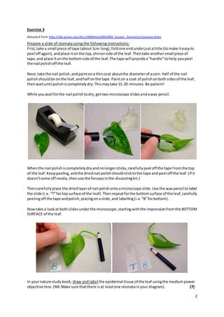
Exercise 1
Take the slide containing the prepared parenchyma tissue and place it on the stage of the
compound microscope.
Observe the features of the tissue starting at the low power lens, medium and high power
objective lens of the compound microscope.
In your nature study book, draw and label the parenchyma tissue using the medium power
objective lens.
Repeat the same procedure for the slides containing the sclerenchyma, collenchyma and
tissues of the leaf.
In yournature studybook, draw and label the sclerenchymatissueusing the mediumpower
objective lens.
(5x2=10)
Exercise 2
Describe your observation by redrawing and completing the table below:
Table 1: Structure of Parenchyma and sclerenchyma cells (8)
Type of tissue Structural Characteristics Function
Parenchyma
Sclerenchyma](https://image.slidesharecdn.com/practicalplanttissues-151012123222-lva1-app6891/85/Practical-plant-tissues-1-320.jpg)