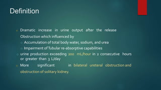
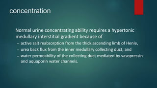
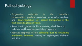
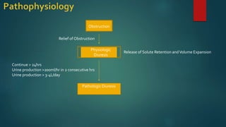
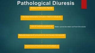
Post obstructive
Diuresis
DR.V.ARUL
Definition
o Dramatic increase in urine output after the release
Obstruction which influenced by
o Accumulation of total body water, sodium, and urea
o Impairment ofTubular re-absorptive capabilities
o urine production exceeding 200 mL/hour in 2 consecutive hours
or greater than 3 L/day
o More significant in bilateral ureteral obstruction and
obstruction of solitary kidney.
Risk Factors
 Edema
 Congestive Heart Failure
 Hypertension
 Azotemia (high levels of nitrogen-containing
compounds such as urea and creatinine)
Physiological Pathological
Urea
Diuresis
Sodium
Diuresis
Water
Diuresis
• Self-limiting
• Normal responseto volume expansion
and soluteaccumulationduring
obstruction
• ExcessNa and water retention
• Urea retention+ non-
reabsorbablesolutes
• Accumulationof ANP
• Once soluteand fluid homeostasisis
achieved,diuresisends – euvolumia
• Most common
• Self-limiting
• Lasts 24-28 hours
• Uosm > 250
• Fluid balance and
electrolytesshould
be monitores
• Unless
contraindicated,
fluid intake should
be increased and
suffices
• Secondmost
common
• Usuallyself-limiting
• Uosm > 250
• Longer duration(>
72 hours)
• Requires more
aggressive
monitoring for
larger diuresis
potential
• Rare
• Self limiting
• Uosm < 150
• Secondaryto
impaired renal
tubularresponse to
ADH
response to solute
and water overload.
Stops after return to
euvolumeic state.
Diuresis of water beyond euvolemic
state, due to insensitivity of
collecting tubule to ADH and other
defects in urinary concentrating
ability of the kidney and tubular
reabsorption of solutes
After the release of obstruction
Contributing factors are both physiological and pathological
➢ Physiological
1.Excess Na and water retention
2.Retention of urea and non reabsorbable solutes
3. Accumulation of ANP
➢ Pathological
1.Decreased tubular reabsorption of Na
2.Concentration defect
3.Increased tubular flow reducing equilibration time for reabsorption of Na and water
concentration
Normal urine concentrating ability requires a hypertonic
medullary interstitial gradient because of
– active salt reabsorption from the thick ascending limb of Henle,
– urea back flux from the inner medullary collecting duct, and
– water permeability of the collecting duct mediated by vasopressin
and aquaporin water channels.
Pathophysiology
 Progressive reduction in the medullary
concentration gradient secondary to vascular washout
and down-regulation of sodium transporters in the
thick ascending loop of Henle.
 Reduction in glomerular filtration rate, which leads to
ischemia and loss of juxtamedullary nephrons.
 Reduced response of the collecting duct to circulating
antidiuretic hormone, leading to nephrogenic diabetes
insipidus.
Obstruction
Relief of Obstruction
Physiologic
Diuresis
Release of Solute Retention andVolume Expansion
Pathologic Diuresis
Continue > 24hrs
Urine production >200ml/hr in 2 consecutive hrs
Urine production > 3-4L/day
Release of Natriuretic Peptide
Decrease reabsorption of Sodium (NaCl) in tubule (a)
Loss of medullary gradient (a)
Reduced response of cortical duct to Anti Diuretic Hormone (b)
Impairment ofTubuloGlomerularFeedback -c
Water cannot be taken out from the tubule
ADH mechanism
•AQUAPORIN 1 (AQP1) - renal proximal
tubules, the thin descending limb of Henle, and
the descending vasa recta in the kidney.
•It promotes urinary concentration through the
countercurrent multiplier
•by facilitating water transport from the
descending limb of Henle into the interstitium
Thus dysregulation of aquaporin water
channels in the proximal tubule, thin
descending loop, and collecting duct may
contribute to the long-term polyuria and
impaired concentrating capacity caused by
obstructive nephropathy.
Accumulation of ANP
• An accumulation of vasoactive substances in BUO that could contribute to
significant post obstructive natriuresis.
 ANP
– increases afferent arteriolar dilation
– efferent arteriolar vasoconstriction, thus increasing PGC
– decreases the sensitivity of tubuloglomerular feedback
– inhibits release of renin,
– secreted ANP contributes to a profound diuresis and
natriuresis.
Clinical features
 Massive sustained polyruria post-relief of obstruction
 Signs of dehydration
 Altered mental status
 Decompensated heart failure and arrhythmias
Obstruction Relieved
Instatement of Urine
Collection Instruments
Mental Status Impairment
Edema
Congestive Heart Failure
Hypertension
Azotemia
FolleyCatheter
Condom Catheter
Assess Hydration status and
Post-Obstructive Diuresis Risk
Post Obstructive Diuresis
POD Management
Discharge
Yes No
POD Management
Monitoring
Urine volume every hr in first 24 hr
Vital signs every 6 to 8 hr
Serum electrolyte levels every 12 to 24 hr
Urea and creatinine levels every 12 to 24 hr
Weight every 24hr
Physiologic Pathologic
Subside within 24 hrs
Persistent after 24 hrs
Fluid Management,Tight Monitoring,
and ReEvaluation
IV fluid replacement
Frequent monitoring of urine
and serum electrolyte levels
Repeat imaging to rule out
persistent obstruction
Discharge
HemodialysisIf fluid correction cannot be
achieved
complications
 Volume depletion
 Hyponatremia or hypernatremia
 Hypokalemia
 Hypomagenesemia
 Metabolicacidosis
 Shock
 Death
• Ureteral obstruction
– induces expression of COX-2 in collecting duct cells and
downregulation of AQP2 receptors is mediated by COX-2.
– COX-2 inhibitors prevented the downregulation of AQP2
and significantly diminished postobstructive diuresis in
rats.
• In addition, with ureteral obstruction,
– cGMP pathway has been demonstrated in both in vitro and in
vivo models to allow membrane insertion of AQP2.
– Sildenafil Citrate elevated intracellular cGMP and facilitate
collecting duct accumulation of AQP2.
• Pharmacological manipulation
–beneficial or harmful - unclear
Post obstructive-diuresis

Post obstructive-diuresis

  • 1.
  • 2.
    Definition o Dramatic increasein urine output after the release Obstruction which influenced by o Accumulation of total body water, sodium, and urea o Impairment ofTubular re-absorptive capabilities o urine production exceeding 200 mL/hour in 2 consecutive hours or greater than 3 L/day o More significant in bilateral ureteral obstruction and obstruction of solitary kidney.
  • 3.
    Risk Factors  Edema Congestive Heart Failure  Hypertension  Azotemia (high levels of nitrogen-containing compounds such as urea and creatinine)
  • 4.
    Physiological Pathological Urea Diuresis Sodium Diuresis Water Diuresis • Self-limiting •Normal responseto volume expansion and soluteaccumulationduring obstruction • ExcessNa and water retention • Urea retention+ non- reabsorbablesolutes • Accumulationof ANP • Once soluteand fluid homeostasisis achieved,diuresisends – euvolumia • Most common • Self-limiting • Lasts 24-28 hours • Uosm > 250 • Fluid balance and electrolytesshould be monitores • Unless contraindicated, fluid intake should be increased and suffices • Secondmost common • Usuallyself-limiting • Uosm > 250 • Longer duration(> 72 hours) • Requires more aggressive monitoring for larger diuresis potential • Rare • Self limiting • Uosm < 150 • Secondaryto impaired renal tubularresponse to ADH response to solute and water overload. Stops after return to euvolumeic state. Diuresis of water beyond euvolemic state, due to insensitivity of collecting tubule to ADH and other defects in urinary concentrating ability of the kidney and tubular reabsorption of solutes
  • 5.
    After the releaseof obstruction Contributing factors are both physiological and pathological ➢ Physiological 1.Excess Na and water retention 2.Retention of urea and non reabsorbable solutes 3. Accumulation of ANP ➢ Pathological 1.Decreased tubular reabsorption of Na 2.Concentration defect 3.Increased tubular flow reducing equilibration time for reabsorption of Na and water
  • 6.
    concentration Normal urine concentratingability requires a hypertonic medullary interstitial gradient because of – active salt reabsorption from the thick ascending limb of Henle, – urea back flux from the inner medullary collecting duct, and – water permeability of the collecting duct mediated by vasopressin and aquaporin water channels.
  • 7.
    Pathophysiology  Progressive reductionin the medullary concentration gradient secondary to vascular washout and down-regulation of sodium transporters in the thick ascending loop of Henle.  Reduction in glomerular filtration rate, which leads to ischemia and loss of juxtamedullary nephrons.  Reduced response of the collecting duct to circulating antidiuretic hormone, leading to nephrogenic diabetes insipidus.
  • 8.
    Obstruction Relief of Obstruction Physiologic Diuresis Releaseof Solute Retention andVolume Expansion Pathologic Diuresis Continue > 24hrs Urine production >200ml/hr in 2 consecutive hrs Urine production > 3-4L/day
  • 9.
    Release of NatriureticPeptide Decrease reabsorption of Sodium (NaCl) in tubule (a) Loss of medullary gradient (a) Reduced response of cortical duct to Anti Diuretic Hormone (b) Impairment ofTubuloGlomerularFeedback -c Water cannot be taken out from the tubule
  • 10.
  • 11.
    •AQUAPORIN 1 (AQP1)- renal proximal tubules, the thin descending limb of Henle, and the descending vasa recta in the kidney. •It promotes urinary concentration through the countercurrent multiplier •by facilitating water transport from the descending limb of Henle into the interstitium
  • 12.
    Thus dysregulation ofaquaporin water channels in the proximal tubule, thin descending loop, and collecting duct may contribute to the long-term polyuria and impaired concentrating capacity caused by obstructive nephropathy.
  • 13.
    Accumulation of ANP •An accumulation of vasoactive substances in BUO that could contribute to significant post obstructive natriuresis.  ANP – increases afferent arteriolar dilation – efferent arteriolar vasoconstriction, thus increasing PGC – decreases the sensitivity of tubuloglomerular feedback – inhibits release of renin, – secreted ANP contributes to a profound diuresis and natriuresis.
  • 14.
    Clinical features  Massivesustained polyruria post-relief of obstruction  Signs of dehydration  Altered mental status  Decompensated heart failure and arrhythmias
  • 15.
    Obstruction Relieved Instatement ofUrine Collection Instruments Mental Status Impairment Edema Congestive Heart Failure Hypertension Azotemia FolleyCatheter Condom Catheter Assess Hydration status and Post-Obstructive Diuresis Risk Post Obstructive Diuresis POD Management Discharge Yes No
  • 16.
    POD Management Monitoring Urine volumeevery hr in first 24 hr Vital signs every 6 to 8 hr Serum electrolyte levels every 12 to 24 hr Urea and creatinine levels every 12 to 24 hr Weight every 24hr Physiologic Pathologic Subside within 24 hrs Persistent after 24 hrs Fluid Management,Tight Monitoring, and ReEvaluation IV fluid replacement Frequent monitoring of urine and serum electrolyte levels Repeat imaging to rule out persistent obstruction Discharge HemodialysisIf fluid correction cannot be achieved
  • 17.
    complications  Volume depletion Hyponatremia or hypernatremia  Hypokalemia  Hypomagenesemia  Metabolicacidosis  Shock  Death
  • 18.
    • Ureteral obstruction –induces expression of COX-2 in collecting duct cells and downregulation of AQP2 receptors is mediated by COX-2. – COX-2 inhibitors prevented the downregulation of AQP2 and significantly diminished postobstructive diuresis in rats.
  • 19.
    • In addition,with ureteral obstruction, – cGMP pathway has been demonstrated in both in vitro and in vivo models to allow membrane insertion of AQP2. – Sildenafil Citrate elevated intracellular cGMP and facilitate collecting duct accumulation of AQP2. • Pharmacological manipulation –beneficial or harmful - unclear