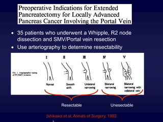
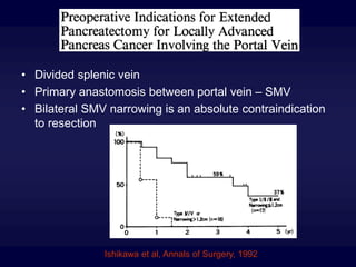
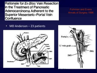
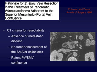
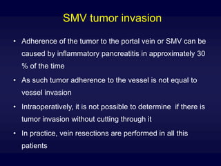
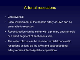
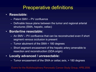
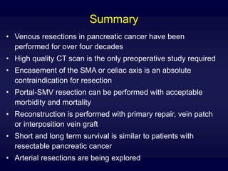
Venous resections in the surgical
treatment of pancreatic
adenocarcinoma
Eduardo A. Guzman MD
May 29, 2008
Outline
• History
• Indications
• Anatomy
• Technique
• Contemporary data
• Definitions
• Summary
• Conclusion
The 70’s
• Having recognized the poor survival associated with
pancreatic cancer, surgeons introduced extensive
pancreatic resections
• One of these, was the “regional pancreatectomy” or “en
bloc pancreatectomy”
• This operation required mesenteric vessel resections as
the standard approach regardless of invasion
• The goal was to enhance nodal harvest
• Did not impact survival but was associated with
increased morbidity and mortality
• Fortner et al. MSKCC
• “Regional pancreatectomy refers to an en bloc removal of a tumor in or
adjacent to the pancreas with an adequate soft tissue margin and with
its regional lymphatic drainage. The pancreatic segment of portal vein
is part of the en bloc resection with venous reconstruction by end-to-
end anastomosis without a graft. This operation, called a Type I
regional pancreatectomy, may utilize either a total or subtotal removal
of the pancreas. Localized arterial involvement by a neoplasm
necessitates adding a segmental resection of the artery with vascular
reconstruction, a Type II procedure”
• 1972-1982, 35 patients, 26% operative mortality
Fortner et al, Annals of Surgery, 1984
The 80’s
• Regional pancreatectomy was abandoned in favor of
extended nodal dissections without vessel resections
(R2 dissection)
• Refinements in surgical technique and postoperative
care made it possible to substantially improve outcomes
for pancreaticoduodenectomies
1969 - 1980 1981 - 1986
Morbidity 59 % 36 %
Mortality 24 % 2%
Johns Hopkins Hospital
Crist and Cameron, Annals of Surgery, 1987
The 90’s
• Improved perioperative outcomes brought a renewed
interest for vein resections in locally advanced
pancreatic cancer
• Specific indications were developed
 35 patients who underwent a Whipple, R2 node
dissection and SMV/Portal vein resection
 Use arteriography to determine resectability
I
Ishikawa et al, Annals of Surgery, 1992
Resectable Unesectable
Ishikawa et al, Annals of Surgery, 1992
• Divided splenic vein
• Primary anastomosis between portal vein – SMV
• Bilateral SMV narrowing is an absolute contraindication
to resection
• MD Anderson – 23 patients
Fuhrman and Evans
Annals of Surgery 1996
• CT criteria for resectability
– Absence of metastatic
disease
– No tumor encasement of
the SMA or celiac axis
– Patent PV/SMV
confluence
Fuhrman and Evans
Annals of Surgery 1996
• Outcomes
– No difference in morbidity, mortality, hospital stay,
– Increased surgical time, blood loss and blood transfusions
– Similar pathologic features than no vein resection patients
Fuhrman and Evans
Annals of Surgery 1996
The millenium
• Widespread acceptance of portal vein resections
• Performance of arterial resections in selected patients
• Precise preoperative definitions
• Importance of negative resection margins (R0)
• Introduction of neoadjuvant chemoradiotherapy
approaches in borderline resectable patients
• The majority of cases did not
require an interposition graft
• Splenic vein ligation was
performed for tumors at the
confluence
• Re-implantation was only done
in patients with poor splenic
hylar collaterals
• Arterial resections ocassionally
performed
Yakebas et al. Annals of Surgery 2008
Fukuda et al. Archives of Surgery 2007
Indications for portal vein resection
• A high quality pancreatic protocol CT scan is the only
preoperative study required to determine resectability
– Absence of metastatic disease
– No tumor encasement of the SMA or celiac axis
– Patent PV / SMV confluence
– Good performance status
• SMV encasement likely signifies SMA involvement
• Patients that are borderline resectable are sometimes
referred to neoadjuvant chemotherapy approaches
Anatomical considerations
• The splenic vein is anterior to the base of the SMA
• Splenic vein transection allows access to the proximal SMA
• Transection of the splenic vein frees the portal structures to
the right and allows for primary repair of the PV – SMV
following vein resections at the confluence
• The first jejunal branch courses underneath the SMA
• Bleeding from the first jejunal branch is difficult to control
with the standard exposure
• Ligation of the first jejunal branch can be performed after
transection of the root of the mesentery
Some technical aspects
• Appropriate patient selection
• Perform dissection of the SMA first leaving the tumor
attached to the SMV only
• Facilitated by division of the splenic vein yet not essential
• Evans leaves the splenic vein intact due to concerns for
venous hypertension if this vessel is ligated
• Systemic heparinization
• Inflow occlusion of the SMA to prevent intestinal edema
• Repair with interrupted 6-0 prolene sutures
• In distal SMV, either the jejunal or the iliac branch can be
safely ligated if the other branch remains patent
Some technical aspects
• Reconstruction is done
with either
– Primary anastomosis
– Vein patch
– Interposition vein graft
• Grafts used
– Saphenous vein
– Internal Jugular vein
– Left renal vein
SMV tumor invasion
• Adherence of the tumor to the portal vein or SMV can be
caused by inflammatory pancreatitis in approximately 30
% of the time
• As such tumor adherence to the vessel is not equal to
vessel invasion
• Intraoperatively, it is not possible to determine if there is
tumor invasion without cutting through it
• In practice, vein resections are performed in all this
patients
Arterial resections
• Controversial
• Focal involvement of the hepatic artery or SMA can be
amenable to resection
• Reconstruction can be either with a primary anastomosis
or a short segment of saphenous vein
• The celiac plexus can be resected in distal pancreatic
resections as long as the SMA and gastroduodenal
artery remain intact (Appleby’s operation)
Preoperative definitions
• Resectable:
– Patent SMV – PV confluence
– Definable tissue plane between the tumor and regional arterial
structures (SMA, hepatic, celiac)
• Borderline resectable:
– An SMV – PV confluence that can be reconstructed even if short
segment venous occlusion is present
– Tumor abutment of the SMA < 180 degrees
– Short segment encasement of the hepatic artery amenable to
resection and reconstruction (GDA origin)
• Locally advanced / unresectable:
– Tumor encasement of the SMA or celiac axis, > 180 degrees
Evans for the Multidisciplinary Pancreatic Cancer Study Group, HPB 2006
Summary
• Venous resections in pancreatic cancer have been
performed for over four decades
• High quality CT scan is the only preoperative study required
• Encasement of the SMA or celiac axis is an absolute
contraindication for resection
• Portal-SMV resection can be performed with acceptable
morbidity and mortality
• Reconstruction is performed with primary repair, vein patch
or interposition vein graft
• Short and long term survival is similar to patients with
resectable pancreatic cancer
• Arterial resections are being explored
Conclusion
• Vessel resections have expanded the pool of patients
with pancreatic adenocarcinoma that are able to undergo
an R0 resection
But
• Many more patients with resectable disease are denied
surgery secondary to nihilistic views present in the
medical community
• 71 % of patients with Stage 1 pancreatic cancer do not
undergo surgery
Bilimoria et al.
National failure to operate on early stage pancreatic cancer
Annals of Surgery 2007

Portal vein resection

  • 1.
    Venous resections inthe surgical treatment of pancreatic adenocarcinoma Eduardo A. Guzman MD May 29, 2008
  • 2.
    Outline • History • Indications •Anatomy • Technique • Contemporary data • Definitions • Summary • Conclusion
  • 3.
    The 70’s • Havingrecognized the poor survival associated with pancreatic cancer, surgeons introduced extensive pancreatic resections • One of these, was the “regional pancreatectomy” or “en bloc pancreatectomy” • This operation required mesenteric vessel resections as the standard approach regardless of invasion • The goal was to enhance nodal harvest • Did not impact survival but was associated with increased morbidity and mortality
  • 4.
    • Fortner etal. MSKCC • “Regional pancreatectomy refers to an en bloc removal of a tumor in or adjacent to the pancreas with an adequate soft tissue margin and with its regional lymphatic drainage. The pancreatic segment of portal vein is part of the en bloc resection with venous reconstruction by end-to- end anastomosis without a graft. This operation, called a Type I regional pancreatectomy, may utilize either a total or subtotal removal of the pancreas. Localized arterial involvement by a neoplasm necessitates adding a segmental resection of the artery with vascular reconstruction, a Type II procedure” • 1972-1982, 35 patients, 26% operative mortality Fortner et al, Annals of Surgery, 1984
  • 5.
    The 80’s • Regionalpancreatectomy was abandoned in favor of extended nodal dissections without vessel resections (R2 dissection) • Refinements in surgical technique and postoperative care made it possible to substantially improve outcomes for pancreaticoduodenectomies
  • 6.
    1969 - 19801981 - 1986 Morbidity 59 % 36 % Mortality 24 % 2% Johns Hopkins Hospital Crist and Cameron, Annals of Surgery, 1987
  • 7.
    The 90’s • Improvedperioperative outcomes brought a renewed interest for vein resections in locally advanced pancreatic cancer • Specific indications were developed
  • 8.
     35 patientswho underwent a Whipple, R2 node dissection and SMV/Portal vein resection  Use arteriography to determine resectability I Ishikawa et al, Annals of Surgery, 1992 Resectable Unesectable
  • 9.
    Ishikawa et al,Annals of Surgery, 1992 • Divided splenic vein • Primary anastomosis between portal vein – SMV • Bilateral SMV narrowing is an absolute contraindication to resection
  • 10.
    • MD Anderson– 23 patients Fuhrman and Evans Annals of Surgery 1996
  • 11.
    • CT criteriafor resectability – Absence of metastatic disease – No tumor encasement of the SMA or celiac axis – Patent PV/SMV confluence Fuhrman and Evans Annals of Surgery 1996
  • 12.
    • Outcomes – Nodifference in morbidity, mortality, hospital stay, – Increased surgical time, blood loss and blood transfusions – Similar pathologic features than no vein resection patients Fuhrman and Evans Annals of Surgery 1996
  • 13.
    The millenium • Widespreadacceptance of portal vein resections • Performance of arterial resections in selected patients • Precise preoperative definitions • Importance of negative resection margins (R0) • Introduction of neoadjuvant chemoradiotherapy approaches in borderline resectable patients
  • 14.
    • The majorityof cases did not require an interposition graft • Splenic vein ligation was performed for tumors at the confluence • Re-implantation was only done in patients with poor splenic hylar collaterals • Arterial resections ocassionally performed Yakebas et al. Annals of Surgery 2008
  • 16.
    Fukuda et al.Archives of Surgery 2007
  • 17.
    Indications for portalvein resection • A high quality pancreatic protocol CT scan is the only preoperative study required to determine resectability – Absence of metastatic disease – No tumor encasement of the SMA or celiac axis – Patent PV / SMV confluence – Good performance status • SMV encasement likely signifies SMA involvement • Patients that are borderline resectable are sometimes referred to neoadjuvant chemotherapy approaches
  • 18.
    Anatomical considerations • Thesplenic vein is anterior to the base of the SMA • Splenic vein transection allows access to the proximal SMA • Transection of the splenic vein frees the portal structures to the right and allows for primary repair of the PV – SMV following vein resections at the confluence • The first jejunal branch courses underneath the SMA • Bleeding from the first jejunal branch is difficult to control with the standard exposure • Ligation of the first jejunal branch can be performed after transection of the root of the mesentery
  • 19.
    Some technical aspects •Appropriate patient selection • Perform dissection of the SMA first leaving the tumor attached to the SMV only • Facilitated by division of the splenic vein yet not essential • Evans leaves the splenic vein intact due to concerns for venous hypertension if this vessel is ligated • Systemic heparinization • Inflow occlusion of the SMA to prevent intestinal edema • Repair with interrupted 6-0 prolene sutures • In distal SMV, either the jejunal or the iliac branch can be safely ligated if the other branch remains patent
  • 20.
    Some technical aspects •Reconstruction is done with either – Primary anastomosis – Vein patch – Interposition vein graft • Grafts used – Saphenous vein – Internal Jugular vein – Left renal vein
  • 21.
    SMV tumor invasion •Adherence of the tumor to the portal vein or SMV can be caused by inflammatory pancreatitis in approximately 30 % of the time • As such tumor adherence to the vessel is not equal to vessel invasion • Intraoperatively, it is not possible to determine if there is tumor invasion without cutting through it • In practice, vein resections are performed in all this patients
  • 22.
    Arterial resections • Controversial •Focal involvement of the hepatic artery or SMA can be amenable to resection • Reconstruction can be either with a primary anastomosis or a short segment of saphenous vein • The celiac plexus can be resected in distal pancreatic resections as long as the SMA and gastroduodenal artery remain intact (Appleby’s operation)
  • 23.
    Preoperative definitions • Resectable: –Patent SMV – PV confluence – Definable tissue plane between the tumor and regional arterial structures (SMA, hepatic, celiac) • Borderline resectable: – An SMV – PV confluence that can be reconstructed even if short segment venous occlusion is present – Tumor abutment of the SMA < 180 degrees – Short segment encasement of the hepatic artery amenable to resection and reconstruction (GDA origin) • Locally advanced / unresectable: – Tumor encasement of the SMA or celiac axis, > 180 degrees Evans for the Multidisciplinary Pancreatic Cancer Study Group, HPB 2006
  • 24.
    Summary • Venous resectionsin pancreatic cancer have been performed for over four decades • High quality CT scan is the only preoperative study required • Encasement of the SMA or celiac axis is an absolute contraindication for resection • Portal-SMV resection can be performed with acceptable morbidity and mortality • Reconstruction is performed with primary repair, vein patch or interposition vein graft • Short and long term survival is similar to patients with resectable pancreatic cancer • Arterial resections are being explored
  • 25.
    Conclusion • Vessel resectionshave expanded the pool of patients with pancreatic adenocarcinoma that are able to undergo an R0 resection
  • 26.
    But • Many morepatients with resectable disease are denied surgery secondary to nihilistic views present in the medical community • 71 % of patients with Stage 1 pancreatic cancer do not undergo surgery Bilimoria et al. National failure to operate on early stage pancreatic cancer Annals of Surgery 2007