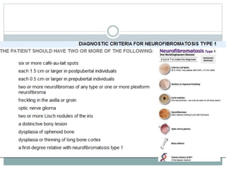
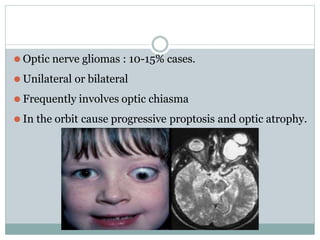
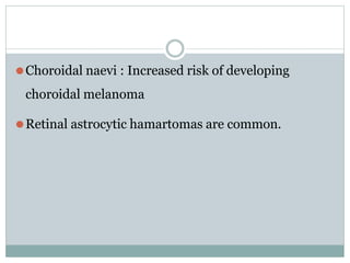
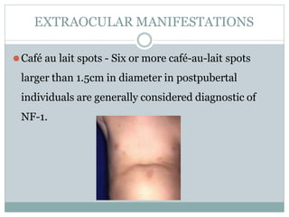
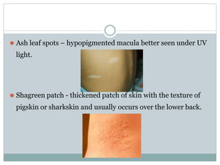
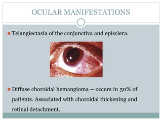
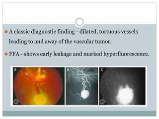
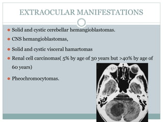
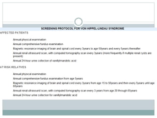
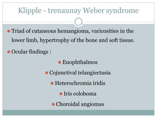
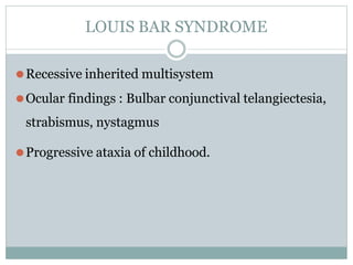
DavudAhmadzada220I1-a
PHAKOMATOSES
DEFINITION
⚫ Coined by van der Hoeve.
⚫ No satisfactory definition present.
⚫ Phakomatoses (or neuro-oculo-cutaneous
syndromes, neurocutaneous disorders) are multisystem
disorders that have characteristic CNS, ocular, and cutaneous
lesions/hamartomas of variable severity.
Common syndromes
⚫Neurofibromatosis type 1
⚫Neurofibromatosis type 2
⚫Tuberous sclerosis
⚫Von Hippel-Lindau syndrome
⚫Sturge Weber syndrome
⚫Wyburn-Mason syndrome
Uncommon syndromes
⚫Klipple trenaunay Weber syndrome
⚫Louis bar syndrome
⚫Diffuse congenital hemangiomatosis
⚫Oculodermal melanocytosis
⚫Basal cell naevus syndrome
NEUROFIBROMATOSIS TYPE 1
⚫ Also known as peripheral NF, von Recklinghausen’s disease.
⚫ Neuroectodermal tumors with autosomal dominant
inheritance.
⚫ 1 person per 3500–4000 persons in the general population.
⚫ Men and women equally affected.
⚫ No racial predilection.
⚫ The gene for NF-1 has been localized to chromosome 17q11.
OCULAR MANIFESTATIONS
⚫ Lignes grises – intrastromal hyperplastic nerves.
⚫ Subcutaneous pedunculated and plexiform neurofibromas
of the eyelids
⚫ Lisch nodules : melanocytic hamartomas of the iris stroma.
⚫ Tan to light brown nodules that stud the iris surface.
⚫ Histopathologically - closely packed dendritic or spindle-
shaped melanocytes within the anterior layers of iris stroma.
These cells are normal uveal melanocytes and not nevus cells.
⚫ Optic nerve gliomas : 10-15% cases.
⚫ Unilateral or bilateral
⚫ Frequently involves optic chiasma
⚫ In the orbit cause progressive proptosis and optic atrophy.
⚫Choroidal naevi : Increased risk of developing
choroidal melanoma
⚫Retinal astrocytic hamartomas are common.
⚫Glaucoma :
EXTRAOCULAR MANIFESTATIONS
⚫Café au lait spots - Six or more café-au-lait spots
larger than 1.5cm in diameter in postpubertal
individuals are generally considered diagnostic of
NF-1.
⚫Axillary or inguinal freckling – 90-95% of the cases.
⚫Subcutaneous or neurological plexiform
neurofibromas.
⚫Sphenoid wing dysplasia, Lamboid suture defects.
NEUROFIBROMATOSIS TYPE 2
⚫ 1 person per 40000–50000 persons
⚫ localized to chromosome 22q12.
⚫ bilateral vestibular schwannomas (acoustic neuromas) and
widely scattered neurofibromas, meningiomas, gliomas, and
schwannomas.
⚫Ophthalmologic findings in NF-2 are relatively
uncommon.
⚫Combined hamartomas of the retina and juvenile
posterior subcapsular or cortical lens opacities.
TUBEROUS SCLEROSIS
⚫Multiorgan tumor syndrome
⚫Multifocal, bilateral retinal astrocytic hamartomas,
astrocytic tumors of the CNS, several unusual
cutaneous lesions, mental retardation, seizures, and
a variety of cysts and tumors of other organs.
⚫ 1 case per 10000 persons
⚫ one third of cases are familial and two thirds are sporadic.
⚫ No racial predilection
⚫ Sexes are affected equally.
⚫ Signs and symptoms begin by the time the patient is 6 years of
age.
⚫ Loci on the long arm of chromosome 9 (9q32-34), on the long
arm of chromosome 11, on the short arm of chromosome 16
(16p13), and on the long arm of chromosome 12 (12q22-24).
OCULAR MANIFESTATIONS
⚫ Astrocytic hamartomas : 50% of the patients develop retinal
astrocytoma in atleast one eye.
⚫ Histologically – composed of felt-like network of atypical
astrocytes and small blood vessels located in the superficial layers.
⚫ Vision loss occurs when the papillomacular bundle is affected.
CUTANEOUS LESIONS
⚫ Adenoma sebaceum : unusual facial dermatological
eruption characterized by pinhead to pea-sized yellowish
to reddish-brown papules distributed in a butterfly
fashion over the nose, cheeks, and nasolabial folds.
⚫ Ash leaf spots – hypopigmented macula better seen under UV
light.
⚫ Shagreen patch - thickened patch of skin with the texture of
pigskin or sharkskin and usually occurs over the lower back.
⚫Common visceral tumor in TS appears to be the
angiomyolipoma of the kidney.
⚫Probably the most distinctive visceral tumor-
rhabdomyoma.
STURGE WEBER SYNDROME
⚫ Dermato-oculo-neural syndrome.
⚫ Cutaneous facial nevus flammeus in the distribution of
the branches of the trigeminal nerve
⚫ Ipsilateral diffuse cavernous hemangioma of the choroid
⚫ Ipsilateral meningeal hemangiomatosis.
⚫ The lesions in the eye, skin, and brain are always present
at birth
⚫Sporadic nonfamilial disease.
⚫No racial prediliction
⚫Men and female affected equally.
OCULAR MANIFESTATIONS
⚫ Telangiectasia of the conjunctiva and episclera.
⚫ Diffuse choroidal hemangioma – occurs in 50% of
patients. Associated with choroidal thickening and
retinal detachment.
⚫ Glaucoma : Occurs in 30 to 70 % Bilateral glaucoma can occur in the
presence of bilateral facial hemangiomas.
⚫ Mechanisms of glaucoma - Developmental anomaly of the
anterior chamber angle and elevated episcleral venous pressure,
each of which leads to aqueous outflow obstruction.
⚫ Clinical and histopathological features of the drainage angle in SWS
are similar to those seen in primary congenital glaucoma.
⚫ On gonioscopy, the angle structures appear indistinct, with a high
iris insertion. An anteriorly displaced iris root, poorly developed
scleral spur, and thickened uveal meshwork have been observed.
CUTANEOUS MANIESTATIONS
⚫ Facial nevus flammeus, a flat to moderately thick zone of dilated
telangiectatic cutaneous capillaries lined by a single layer of endothelial
cells in the dermis.
⚫ Unilateral.
⚫ Involves the regions of the face innervated by the first branch of the
trigeminal nerve.
CNS MANIFESTATTIONS
⚫Ipsilateral leptomeningeal hemangiomatosis, which
causes atrophy of the cortical parenchyma of the
brain, seizures, and frequently mental retardation.
⚫Present at birth and are detectable by MRI or CT.
⚫Progressive throughout life.
VON HIPPLE LINDAU DISEASE
⚫Characterized by :
⚫Retinal capillary hemangiomas,
⚫CNS hemangioblastomas,
⚫Solid and cystic visceral hamartomas
⚫Renal cell carcinomas
⚫Pheochromocytomas.
⚫ Capillary hemangiomas of the retina-earliest detected
manifestation
⚫ The cumulative probability of developing retinal capillary
hemangiomas and CNS hemangioblastomas in a patient who
has VHLS is >80%, and the probability of developing renal
cell carcinoma is >60%.
⚫ Autosomal dominant inheritance pattern
⚫ The median age at detection is 20–25 years
⚫ VHLS gene to chromosome 3p25-26
OCULAR MANIFESTATTIONS
⚫ Retinal capillary hemangioblastoma
commonly seen in 60% patients
⚫ Peripheral lesions hav subtle red hue
and are no larger than a few hundred
microns.
⚫ As the proliferation continues, acquire
a more nodular appearance with
marked dilated and engorged afferent
and efferent blood vessels.
⚫ Retinal edema and hard exudates.
⚫ Juxtapapillary lesions,11-15% of cases, can cause
pseudopapilledema from elevation and exudation around
the optic nerve .
⚫ A classic diagnostic finding - dilated, tortuous vessels
leading to and away of the vascular tumor.
⚫ FFA - shows early leakage and marked hyperfluorescence.
EXTRAOCULAR MANIFESTATIONS
⚫ Solid and cystic cerebellar hemangioblastomas.
⚫ CNS hemangioblastomas,
⚫ Solid and cystic visceral hamartomas
⚫ Renal cell carcinomas( 5% by age of 30 years but >40% by age of
60 years)
⚫ Pheochromocytomas.
⚫Differential diagnosis for retinal capillary
hemangioblastomas :
⚫Coat's disease
⚫Retinal Cavernous Hemangioma
⚫Retinal Macroaneurysm
⚫ Visual loss due to RCH:
⚫ Exudation : increase in capillary tumor vasopermeablity leading to
macular edema or exudative retinal detachment.
⚫ Tractional effects : glial proliferation on the surface of the
tumor may induce retinal striae & distortion or even tractional
retinal
detachment
⚫ Vitreous Hemorrhage : from rupture and bleeding of the RCH into
the vitreous cavity
⚫Neovascular glaucoma : leaking of angiogenic factors, such as
VEGF, to the anterior chamber causing neovascularization of the angle.
WYBURN MASON SYNDROME
⚫ Arteriovenous malformations (AVMs) of the retina and
ipsilateral CNS.
⚫ Abnormal lesions are not distinct tumors but anomalous
arteriovenous communications, hence not a true
phakomatoses.
⚫ The retinal and intracranial AVMs are congenital.
⚫ Incompletely developed at birth but progress during growth
and aging.
⚫ Occur in the orbit, in the periorbital soft tissues and
bones, and in the midbrain ipsilateral to the retinal AVM.
⚫ More complex the retinal vascular anomalies, the higher
the likelihood of associated CNS AVMs.
Klipple - trenaunay Weber syndrome
⚫ Triad of cutaneous hemangioma, varicosities in the
lower limb, hypertrophy of the bone and soft tissue.
⚫ Ocular findings :
⚫ Enophthalmos
⚫ Cojunctival telangiectasia
⚫ Heterochromia iridis
⚫ Iris coloboma
⚫ Choroidal angiomas
LOUIS BAR SYNDROME
⚫Recessive inherited multisystem
⚫Ocular findings : Bulbar conjunctival telangiectesia,
strabismus, nystagmus
⚫Progressive ataxia of childhood.
OCULODERMAL MELANOCYTOSIS
phakomatosesppt-170818165457.pptx

phakomatosesppt-170818165457.pptx

  • 1.
  • 2.
    DEFINITION ⚫ Coined byvan der Hoeve. ⚫ No satisfactory definition present. ⚫ Phakomatoses (or neuro-oculo-cutaneous syndromes, neurocutaneous disorders) are multisystem disorders that have characteristic CNS, ocular, and cutaneous lesions/hamartomas of variable severity.
  • 3.
    Common syndromes ⚫Neurofibromatosis type1 ⚫Neurofibromatosis type 2 ⚫Tuberous sclerosis ⚫Von Hippel-Lindau syndrome ⚫Sturge Weber syndrome ⚫Wyburn-Mason syndrome
  • 4.
    Uncommon syndromes ⚫Klipple trenaunayWeber syndrome ⚫Louis bar syndrome ⚫Diffuse congenital hemangiomatosis ⚫Oculodermal melanocytosis ⚫Basal cell naevus syndrome
  • 5.
    NEUROFIBROMATOSIS TYPE 1 ⚫Also known as peripheral NF, von Recklinghausen’s disease. ⚫ Neuroectodermal tumors with autosomal dominant inheritance. ⚫ 1 person per 3500–4000 persons in the general population. ⚫ Men and women equally affected. ⚫ No racial predilection. ⚫ The gene for NF-1 has been localized to chromosome 17q11.
  • 7.
    OCULAR MANIFESTATIONS ⚫ Lignesgrises – intrastromal hyperplastic nerves. ⚫ Subcutaneous pedunculated and plexiform neurofibromas of the eyelids
  • 8.
    ⚫ Lisch nodules: melanocytic hamartomas of the iris stroma. ⚫ Tan to light brown nodules that stud the iris surface. ⚫ Histopathologically - closely packed dendritic or spindle- shaped melanocytes within the anterior layers of iris stroma. These cells are normal uveal melanocytes and not nevus cells.
  • 9.
    ⚫ Optic nervegliomas : 10-15% cases. ⚫ Unilateral or bilateral ⚫ Frequently involves optic chiasma ⚫ In the orbit cause progressive proptosis and optic atrophy.
  • 10.
    ⚫Choroidal naevi :Increased risk of developing choroidal melanoma ⚫Retinal astrocytic hamartomas are common.
  • 11.
  • 12.
    EXTRAOCULAR MANIFESTATIONS ⚫Café aulait spots - Six or more café-au-lait spots larger than 1.5cm in diameter in postpubertal individuals are generally considered diagnostic of NF-1.
  • 13.
    ⚫Axillary or inguinalfreckling – 90-95% of the cases. ⚫Subcutaneous or neurological plexiform neurofibromas. ⚫Sphenoid wing dysplasia, Lamboid suture defects.
  • 14.
    NEUROFIBROMATOSIS TYPE 2 ⚫1 person per 40000–50000 persons ⚫ localized to chromosome 22q12. ⚫ bilateral vestibular schwannomas (acoustic neuromas) and widely scattered neurofibromas, meningiomas, gliomas, and schwannomas.
  • 15.
    ⚫Ophthalmologic findings inNF-2 are relatively uncommon. ⚫Combined hamartomas of the retina and juvenile posterior subcapsular or cortical lens opacities.
  • 17.
    TUBEROUS SCLEROSIS ⚫Multiorgan tumorsyndrome ⚫Multifocal, bilateral retinal astrocytic hamartomas, astrocytic tumors of the CNS, several unusual cutaneous lesions, mental retardation, seizures, and a variety of cysts and tumors of other organs.
  • 18.
    ⚫ 1 caseper 10000 persons ⚫ one third of cases are familial and two thirds are sporadic. ⚫ No racial predilection ⚫ Sexes are affected equally. ⚫ Signs and symptoms begin by the time the patient is 6 years of age. ⚫ Loci on the long arm of chromosome 9 (9q32-34), on the long arm of chromosome 11, on the short arm of chromosome 16 (16p13), and on the long arm of chromosome 12 (12q22-24).
  • 19.
    OCULAR MANIFESTATIONS ⚫ Astrocytichamartomas : 50% of the patients develop retinal astrocytoma in atleast one eye. ⚫ Histologically – composed of felt-like network of atypical astrocytes and small blood vessels located in the superficial layers. ⚫ Vision loss occurs when the papillomacular bundle is affected.
  • 20.
    CUTANEOUS LESIONS ⚫ Adenomasebaceum : unusual facial dermatological eruption characterized by pinhead to pea-sized yellowish to reddish-brown papules distributed in a butterfly fashion over the nose, cheeks, and nasolabial folds.
  • 21.
    ⚫ Ash leafspots – hypopigmented macula better seen under UV light. ⚫ Shagreen patch - thickened patch of skin with the texture of pigskin or sharkskin and usually occurs over the lower back.
  • 22.
    ⚫Common visceral tumorin TS appears to be the angiomyolipoma of the kidney. ⚫Probably the most distinctive visceral tumor- rhabdomyoma.
  • 23.
    STURGE WEBER SYNDROME ⚫Dermato-oculo-neural syndrome. ⚫ Cutaneous facial nevus flammeus in the distribution of the branches of the trigeminal nerve ⚫ Ipsilateral diffuse cavernous hemangioma of the choroid ⚫ Ipsilateral meningeal hemangiomatosis. ⚫ The lesions in the eye, skin, and brain are always present at birth
  • 24.
    ⚫Sporadic nonfamilial disease. ⚫Noracial prediliction ⚫Men and female affected equally.
  • 25.
    OCULAR MANIFESTATIONS ⚫ Telangiectasiaof the conjunctiva and episclera. ⚫ Diffuse choroidal hemangioma – occurs in 50% of patients. Associated with choroidal thickening and retinal detachment.
  • 26.
    ⚫ Glaucoma :Occurs in 30 to 70 % Bilateral glaucoma can occur in the presence of bilateral facial hemangiomas. ⚫ Mechanisms of glaucoma - Developmental anomaly of the anterior chamber angle and elevated episcleral venous pressure, each of which leads to aqueous outflow obstruction. ⚫ Clinical and histopathological features of the drainage angle in SWS are similar to those seen in primary congenital glaucoma. ⚫ On gonioscopy, the angle structures appear indistinct, with a high iris insertion. An anteriorly displaced iris root, poorly developed scleral spur, and thickened uveal meshwork have been observed.
  • 27.
    CUTANEOUS MANIESTATIONS ⚫ Facialnevus flammeus, a flat to moderately thick zone of dilated telangiectatic cutaneous capillaries lined by a single layer of endothelial cells in the dermis. ⚫ Unilateral. ⚫ Involves the regions of the face innervated by the first branch of the trigeminal nerve.
  • 28.
    CNS MANIFESTATTIONS ⚫Ipsilateral leptomeningealhemangiomatosis, which causes atrophy of the cortical parenchyma of the brain, seizures, and frequently mental retardation. ⚫Present at birth and are detectable by MRI or CT. ⚫Progressive throughout life.
  • 29.
    VON HIPPLE LINDAUDISEASE ⚫Characterized by : ⚫Retinal capillary hemangiomas, ⚫CNS hemangioblastomas, ⚫Solid and cystic visceral hamartomas ⚫Renal cell carcinomas ⚫Pheochromocytomas.
  • 30.
    ⚫ Capillary hemangiomasof the retina-earliest detected manifestation ⚫ The cumulative probability of developing retinal capillary hemangiomas and CNS hemangioblastomas in a patient who has VHLS is >80%, and the probability of developing renal cell carcinoma is >60%. ⚫ Autosomal dominant inheritance pattern ⚫ The median age at detection is 20–25 years ⚫ VHLS gene to chromosome 3p25-26
  • 31.
    OCULAR MANIFESTATTIONS ⚫ Retinalcapillary hemangioblastoma commonly seen in 60% patients ⚫ Peripheral lesions hav subtle red hue and are no larger than a few hundred microns. ⚫ As the proliferation continues, acquire a more nodular appearance with marked dilated and engorged afferent and efferent blood vessels. ⚫ Retinal edema and hard exudates.
  • 32.
    ⚫ Juxtapapillary lesions,11-15%of cases, can cause pseudopapilledema from elevation and exudation around the optic nerve .
  • 33.
    ⚫ A classicdiagnostic finding - dilated, tortuous vessels leading to and away of the vascular tumor. ⚫ FFA - shows early leakage and marked hyperfluorescence.
  • 34.
    EXTRAOCULAR MANIFESTATIONS ⚫ Solidand cystic cerebellar hemangioblastomas. ⚫ CNS hemangioblastomas, ⚫ Solid and cystic visceral hamartomas ⚫ Renal cell carcinomas( 5% by age of 30 years but >40% by age of 60 years) ⚫ Pheochromocytomas.
  • 36.
    ⚫Differential diagnosis forretinal capillary hemangioblastomas : ⚫Coat's disease ⚫Retinal Cavernous Hemangioma ⚫Retinal Macroaneurysm
  • 37.
    ⚫ Visual lossdue to RCH: ⚫ Exudation : increase in capillary tumor vasopermeablity leading to macular edema or exudative retinal detachment. ⚫ Tractional effects : glial proliferation on the surface of the tumor may induce retinal striae & distortion or even tractional retinal detachment ⚫ Vitreous Hemorrhage : from rupture and bleeding of the RCH into the vitreous cavity ⚫Neovascular glaucoma : leaking of angiogenic factors, such as VEGF, to the anterior chamber causing neovascularization of the angle.
  • 38.
    WYBURN MASON SYNDROME ⚫Arteriovenous malformations (AVMs) of the retina and ipsilateral CNS. ⚫ Abnormal lesions are not distinct tumors but anomalous arteriovenous communications, hence not a true phakomatoses. ⚫ The retinal and intracranial AVMs are congenital. ⚫ Incompletely developed at birth but progress during growth and aging.
  • 40.
    ⚫ Occur inthe orbit, in the periorbital soft tissues and bones, and in the midbrain ipsilateral to the retinal AVM. ⚫ More complex the retinal vascular anomalies, the higher the likelihood of associated CNS AVMs.
  • 41.
    Klipple - trenaunayWeber syndrome ⚫ Triad of cutaneous hemangioma, varicosities in the lower limb, hypertrophy of the bone and soft tissue. ⚫ Ocular findings : ⚫ Enophthalmos ⚫ Cojunctival telangiectasia ⚫ Heterochromia iridis ⚫ Iris coloboma ⚫ Choroidal angiomas
  • 42.
    LOUIS BAR SYNDROME ⚫Recessiveinherited multisystem ⚫Ocular findings : Bulbar conjunctival telangiectesia, strabismus, nystagmus ⚫Progressive ataxia of childhood.
  • 43.