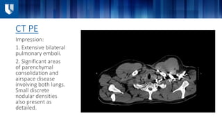
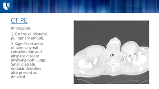
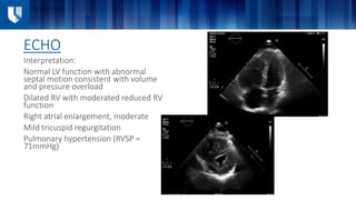
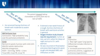
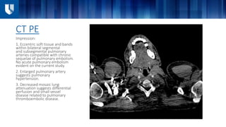
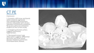
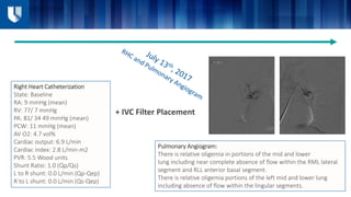
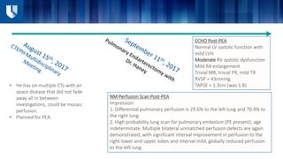
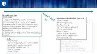
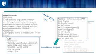
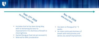
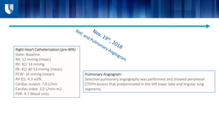
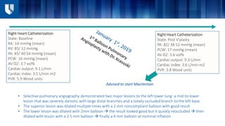
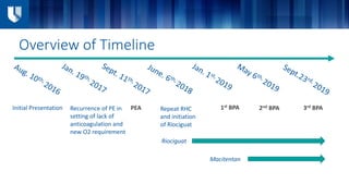
Clinical Case from DUH
Emory Buck, MD
Internal Medicine, PGY-3
November 4th, 2022
Annual North Carolina Research Triangle
Pulmonary Hypertension Symposium
• 45-year-old male presented with subacute shortness of breath and chest pain x 2 days
• He described the pain as sharp, located over center of chest, worse with exertion and
relieved by rest.
• He noted associated dyspnea on exertion. The day prior to presentation, he developed
hemoptysis.
• He denied any history of blood clots
• ROS otherwise negative
• PMH: Notable for asthma, uses an albuterol inhaler
• PSH: Negative
• FH: HTN in mother and father, negative for blood clots.
• Social History: The patient worked as truck driver and he had just returned from a long
trip to Baltimore last night. He endorsed occasional marijuana use, but no other illicit
drugs and never smoker.
Case Presentation (Aug. 10th, 2016)
Initial Exam
VS: Temp 98.6F, HR 77, BP 121/82, RR 20, and SpO2 89% on room air  5L NC 92%
• GENERAL: Well-developed well-nourished in mild respiratory distress.
• HEENT: Pupils equally round and reactive to light. Anicteric sclera. Moist mucous membranes.
• NECK: Supple no masses. Elevated jugular venous pressure with mild abdominal jugular reflux.
• PULM: Tachypneic. Bilateral chest expansion. Bronchial breath sounds in the right upper lobe. No
other major adventitious breath sounds.
• CV: Tachycardic. Fixed S-2 verses an S3 summation gallop difficult to differentiate. Nondisplaced
PMI.
• ABDOMEN: Distended with good bowel sounds. Overall soft with no masses. Nontender.
• EXTREMITIES: Warm and well perfused. No clubbing cyanosis or edema.
• MSK: No evidence of active synovitis.
• NEURO: Alert and oriented x3. Nonfocal exam.
• SKIN/WOUND: No rash lesions or ulcers.
12.6 (H)
15.9
269
46.4
138
3.9
107
21
14
1.5
113
Troponin I: 0.03
ESR: 10
ANA: Positive, 1:40
ANCA: Negative
RF: Negative
Anti GBM Ab: Negative
AST: 22
ALT: 22
Alkaline phosphatase: 46
T. bilirubin: 1.0
Total Protein: 7.8
Albumin: 4.2
1.2
13.8 27.2
Basic Labs
CXR
Impression:
1. Right upper lobe pneumonia. This
process should be followed to
resolution.
2. Possible left perihilar opacity which
could also represent pneumonia
or mass. This finding should be
followed to resolution.
CT PE
Impression:
1. Extensive bilateral
pulmonary emboli.
2. Significant areas
of parenchymal
consolidation and
airspace disease
involving both lungs.
Small discrete
nodular densities
also present as
detailed.
CT PE
Impression:
1. Extensive bilateral
pulmonary emboli.
2. Significant areas
of parenchymal
consolidation and
airspace disease
involving both lungs.
Small discrete
nodular densities
also present as
detailed.
ECHO
Interpretation:
Normal LV function with abnormal
septal motion consistent with volume
and pressure overload
Dilated RV with moderated reduced RV
function
Right atrial enlargement, moderate
Mild tricuspid regurgitation
Pulmonary hypertension (RVSP =
71mmHg)
Hospital Course Aug. 10th – Aug. 18th 2016
He was
started on
heparin 
hemoptysis
 concern
for DAH 
additional
A/C was
held
Bronchoscopy
 negative for
DAH 
resumed on
heparin
Treated with
azithromycin
and
Ceftriaxone
for CAP
High oxygen
requirement,
up to 10L NC
Heparin 
transitioned
to
Rivaroxaban
On discharge,
he was able
to be weaned
off oxygen
and
ambulated
well on room
air.
NM Perfusion Scan
Conclusions:​ High probability lung
scan for pulmonary embolism.
• He remained having shortness of
breath with moderate to severe
exertion.
ECHO
Normal LV systolic function with mild cLVH.
Mild RV systolic dysfunction
Mild RA enlargement
RVSP = 32mmHg; TAPSE = 1.9cm
Trivial MR, trivial PR, trivial TR
• Endorsed a two-week history of
a cough, but less shortness of
breath with exertional activities
in general.
• Oxygen titration study showed
new O2 requirement: 2 L/min at
rest, up to 6 L when he sleeps at
night and up to 15 L with
exertion.
• He was resumed on Rivaroxaban
and prescribed Augmentin given
cough and elevated WBC and
infiltration on CXR.
The patient stopped taking
rivaroxaban at some point due to
lack of refills
CXR
Impression:
1. New right upper lung opacity.
Differential includes infection or
hemorrhage.
2. Increased prominence of the
pulmonary arteries, suggestive of
pulmonary hypertension.
CT PE
Impression:
1. Eccentric soft tissue and bands
within bilateral segmental
and subsegmental pulmonary
arteries compatible with chronic
sequelae of pulmonary embolism.
No acute pulmonary embolism
evident on the current study.
2. Enlarged pulmonary artery
suggests pulmonary
hypertension.
3. Decreased mosaic lung
attenuation suggests differential
perfusion and small vessel
disease related to pulmonary
thromboembolic disease.
Right Heart Catheterization
State: Baseline
RA: 9 mmHg (mean)
RV: 77/ 7 mmHg
PA: 81/ 34 49 mmHg (mean)
PCW: 11 mmHg (mean)
AV O2: 4.7 vol%
Cardiac output: 6.9 L/min
Cardiac index: 2.8 L/min-m2
PVR: 5.5 Wood units
Shunt Ratio: 1.0 (Qp/Qs)
L to R shunt: 0.0 L/min (Qp-Qep)
R to L shunt: 0.0 L/min (Qs-Qep)
+ IVC Filter Placement
Pulmonary Angiogram:
There is relative oligemia in portions of the mid and lower
lung including near complete absence of flow within the RML lateral
segment and RLL anterior basal segment.
There is relative oligemia portions of the left mid and lower lung
including absence of flow within the lingular segments.
In a patient with these findings on RHC and PA
angiogram, what should be the next step in
management?
PEA Outcomes
• By ESC 2022 guidelines, PEA is the treatment of choice for
accessible lesions since surgery may normalize pulmonary
hemodynamics and functional capacity
• Long term outcomes after PEA surgery are excellent
regarding survival (averaging 90% at 3 years) and quality of
life, even in patients with distal PA obstructions.
• Delcroix et al (2016) prospective trial found that in
operated patients, the preoperative characteristics
increasing mortality included bridging therapy with
PAH drugs
• Quadry et al (2018) found that in patients with
proximal operable disease declining surgery have a
poor long-term outcome, with a 5-year survival of
53% compared with 83% in patients undergoing PEA
6MWD
• 6MWD increased by an average of 95.7 m
after PEA
• (n = 11 studies)
mPAP
• mPAP reduced by an average of 21.42 mm Hg
after PEA
• (n = 23 studies)
PVR
• PVR decreased by an average of
560.3 dyns/cm5 after PEA
• (n= 19 studies)
•RPH
• RPH in 25% of patients
Meta-analysis by Hsieh et al (2018) found that PEA:
Back to the Case
• He has on multiple CTs with air
space disease that did not fade
away all in between
investigations, could be mosaic
perfusion.
• Planned for PEA
NM Perfusion Scan Post-PEA
Impression:
1. Differential pulmonary perfusion is 29.6% to the left lung and 70.4% to
the right lung.
2. High probability lung scan for pulmonary embolism (PE present), age
indeterminate. Multiple bilateral unmatched perfusion defects are again
demonstrated, with significant interval improvement in perfusion to the
right lower and upper lobes and interval mild, globally reduced perfusion
to the left lung.
ECHO Post-PEA
Normal LV systolic function with
mild LVH
Moderate RV systolic dysfunction
Mild RA enlargement
Trivial MR, trivial PR, mild TR
RVSP = 43mmHg
TAPSE = 1.3cm (was 1.8)
• He has had no increasing shortness of
breath or chest pain or tightness, does
not have a cough either.
• He says that he is able now to do
workouts, but not able to walk for
longer distances or tolerate inclines.
• He slowly getting better.
• He was able to return to work
• 6MWT performed off of O2
• Since discharge, he continues to
progress well
• He reports activity level and tolerance
since discharge has been continuing to
improve.
Right Heart Catheterization (7/13/2017,
pre-PEA)
State: Baseline
RA: 9 mmHg (mean)
RV: 77/ 7 mmHg
PA: 81/ 34 49 mmHg (mean)
PCW: 11 mmHg (mean)
AV O2: 4.7 vol%
Cardiac output: 6.9 L/min
Cardiac index: 2.8 L/min-m2
PVR: 5.5 Wood units
Shunt Ratio: 1.0 (Qp/Qs)
L to R shunt: 0.0 L/min (Qp-Qep)
R to L shunt: 0.0 L/min (Qs-Qep)
Right Heart Catheterization (post-PEA)
State: Baseline
RA: 1 mmHg (mean)
RV: 71/ 1 mmHg
PA: 71/ 26 40 mmHg (mean)
PCW: 6 mmHg (mean)
AV O2: 5.5 vol%
Cardiac output: 6.1 L/min
Cardiac index: 2.4 L/min-m2
PVR: 5.6 Wood units
NM Perfusion Scan
Conclusions:
1. High probability lung scan for pulmonary
embolism, age indeterminate, which appears
relatively similar to prior with no new perfusion
defects. Findings may be seen in the setting of
chronic thromboembolic pulmonary
hypertension.
2. Scintigraphic findings of mild obstructive airways
disease.
ECHO
Interpretation:
Normal LV systolic function with mild LVH
Moderate RV systolic dysfunction
Trivial MR, trivial PR, mild TR
RVSP = 87mmHg
TAPSE = 1.7cm
In a patient with this RHC post-PEA, what
should be the next step in management?
Back to the Case
Right Heart Catheterization (7/13/2017,
pre-PEA)
State: Baseline
RA: 9 mmHg (mean)
RV: 77/ 7 mmHg
PA: 81/ 34 49 mmHg (mean)
PCW: 11 mmHg (mean)
AV O2: 4.7 vol%
Cardiac output: 6.9 L/min
Cardiac index: 2.8 L/min-m2
PVR: 5.5 Wood units
Shunt Ratio: 1.0 (Qp/Qs)
L to R shunt: 0.0 L/min (Qp-Qep)
R to L shunt: 0.0 L/min (Qs-Qep)
Right Heart Catheterization (post-PEA)
State: Baseline
RA: 1 mmHg (mean)
RV: 71/ 1 mmHg
PA: 71/ 26 40 mmHg (mean)
PCW: 6 mmHg (mean)
AV O2: 5.5 vol%
Cardiac output: 6.1 L/min
Cardiac index: 2.4 L/min-m2
PVR: 5.6 Wood units
NM Perfusion Scan
Conclusions:
1. High probability lung scan for pulmonary
embolism, age indeterminate, which appears
relatively similar to prior with no new perfusion
defects. Findings may be seen in the setting of
chronic thromboembolic pulmonary
hypertension.
2. Scintigraphic findings of mild obstructive airways
disease.
ECHO
Interpretation:
Normal LV systolic function with mild LVH
Moderate RV systolic dysfunction
Trivial MR, trivial PR, mild TR
RVSP = 87mmHg
TAPSE = 1.7cm
Advised to start Riociguat
• Has been on Riociguat for ~3
weeks.
• He notes continued shortness of
breath with mild activities and
denies any overall improvement.
• He states that he has been doing okay,
with no worsening but also no
improvement in his shortness of breath or
chest tightness.
• Started Riociguat (had not yet received it)
• Referred for BPA consideration
Right Heart Catheterization (pre-BPA)
State: Baseline
RA: 12 mmHg (mean)
RV: 82/ 14 mmHg
PA: 82/ 40 53 mmHg (mean)
PCW: 16 mmHg (mean)
AV O2: 4.3 vol%
Cardiac output: 7.8 L/min
Cardiac index: 3.0 L/min-m2
PVR: 4.7 Wood units
Pulmonary Angiogram:
Selective pulmonary angiography was performed and showed peripheral
CTEPH lesions that predominated in the left lower lobe and lingular lung
segments.
Right Heart Catheterization
State: Baseline
RA: 14 mmHg (mean)
RV: 85/ 12 mmHg
PA: 85/ 40 54 mmHg (mean)
PCW: 16 mmHg (mean)
AV O2: 3.7 vol%
Cardiac output: 9.1 L/min
Cardiac index: 3.5 L/min-m2
PVR: 5.9 Wood units
Right Heart Catheterization
State: Post V'plasty
PA: 82/ 38 52 mmHg (mean)
PCW: 17 mmHg (mean)
AV O2: 3.6 vol%
Cardiac output: 9.3 L/min
Cardiac index: 3.6 L/min-m2
PVR: 3.8 Wood units
• Selective pulmonary angiography demonstrated two major lesions to the left lower lung: a mid-to-lower
lesion that was severely stenotic with large distal branches and a totally occluded branch to the left base.
• The superior lesion was dilated multiple times with a 2 mm noncompliant balloon with good result
• The lower lesion was dilated with 2mm balloon  the result looked good but it quickly reoccluded  then
dilated with lesion with a 2.5 mm balloon  finally a 4 mm balloon at nominal inflation
Advised to start Macitentan
Overview of Timeline
Initial Presentation Recurrence of PE in
setting of lack of
anticoagulation and
new O2 requirement
PEA Repeat RHC
and initiation
of Riociguat
1st BPA 2nd BPA 3rd BPA
Riociguat
Macitentan
• He doesn't have to stop while going for
walk with his wife anymore
• He does get tired and short of breath
after walking few flights of stairs
• He does have significant bendopnea
• Unfortunately, he had a lapse in Riociguat;
restarted but had not titrated up
ECHO
Interpretation:
Normal LV systolic function with
mild LVH
Mild RV systolic dysfunction, mild
enlargement
Normal RA size
Trivial MR, mild PR, mild TR
RVSP = 58mmHg
TAPSE = 1.5cm
NM Perfusion Scan
Conclusions:
Multiple perfusion defects. No new
perfusion defect identified compared to
June 6, 2018 consistent with chronic PE.
• Overall feeling better, able to
walk 2.5 miles
ECHO
Interpretation:
Normal LV systolic function with
mild LVH
Mild RV systolic dysfunction, normal
size
Normal RA size
Trivial MR, trivial PR, mild TR
RVSP = 34mmHg
Discussion
PEA versus BPA
• No head-to-head comparison of PEA versus BPA
• A systematic review and meta-analysis by Zhang et al in 2021
compared the safety and efficacy of BPA and PEA in the treatment
of CTEPH
• The survival rates at perioperative/in-hospital period, 2 years,
and 3 years were 100%, 99%, and 97%, in BPA group and 93%,
90%, and 88%, respectively, in PEA group.
• The variation of 6-min walk distance was 141.80 m in BPA and
100.73 m in PEA
• At < 1-month, 1–6-month, and > 12-month follow-up, the
changed results of mean pulmonary arterial pressure were −
18.31, − 17.00, and − 12.97 mmHg in BPA group and − 18.93, −
21.21, and − 21.35 mmHg in PEA group.
• At < 1-month and 1–6-month follow-up, the changed values of
pulmonary vascular resistance were − 542.24 and − 599.77
dyne•s•cm−5 in PEA group and − 443.49 and − 280.00
dyne•s•cm−5 in BPA group.
BPA might have higher survival
rate and fewer types of
complications compared with PEA.
The improvement in exercise
capacity in the BPA group might
be more pronounced than in PEA
group.
PEA might be superior in
improvement of hemodynamic
parameters
BPA versus Medical Therapy
• A systematic review and meta-analysis
by Wang et al in 2019 compared the
efficacy and safety of BPA with riociguat
therapy in inoperable CTEPH patients.
As compared with Riociguat, BPA was
associated with a greater improvement
in:
• RAP (MD = -3.53 mmHg vs = -1.05
mmHg)
• mPAP (MD = -15.02 mmHg vs MD
= -4.19 mmHg)
• PVR (MD = -1.32 woods vs
standard MD = -0.65 woods)
• NYHA functional class (RR = 6.78 vs
RR = 1.49)
• 6MWD (MD = 71.66 m vs MD =
45.25 m)
Medical Options:
• Riociguat (CHEST-1 Study: improved
6MWD and reduced PVR by 31% compared
with placebo in patients with inoperable
CTEPH or those with persistent/recurrent
PH after PEA)
• Treprostinil SC (CTREPH Study: improved
6MWD at week 24 in patients with
inoperable CTEPH or those with
persistent/recurrent PH after PEA receiving
a high dose compared with a low dose)
• Macitentan (MERIT-1 Study: improved PVR
and 6MWD vs. placebo at 16 and 24 weeks
including only patients with inoperable
CTEPH)
Questions?
References
1. Ghofrani HA, D’Armini AM, Grimminger F, Hoeper MM, Jansa P, Kim NH, et al. Riociguat for the treatment of chronic
thromboembolic pulmonary hypertension. N Engl J Med 2013;369:319–329.
2. Hsieh WC, Jansa P, Huang WC, Niznansky M, Omara M, Lindner J. Residual pul- monary hypertension after pulmonary
endarterectomy: a meta-analysis. J Thorac Cardiovasc Surg 2018;156:1275–1287.
3. Delcroix M, Lang I, Pepke-Zaba J, Jansa P, D’Armini AM, Snijder R, et al. Long-term outcome of patients with chronic
thromboembolic pulmonary hypertension: re- sults from an international prospective registry. Circulation 2016;133:859–
871.
4. Quadery SR, Swift AJ, Billings CG, Thompson AAR, Elliot CA, Hurdman J, et al. The impact of patient choice on survival in
chronic thromboembolic pulmonary hyper- tension. Eur Respir J 2018;52:1800589.
5. Zhang L, Bai Y, Yan P, He T, Liu B, Wu S, Qian Z, Li C, Cao Y, Zhang M. Balloon pulmonary angioplasty vs. pulmonary
endarterectomy in patients with chronic thromboembolic pulmonary hypertension: a systematic review and meta-analysis.
Heart Fail Rev. 2021 Jul;26(4):897-917. doi: 10.1007/s10741-020-10070-w. Epub 2021 Feb 5. PMID: 33544306.
6. Jaïs X, Brenot P, Bouvaist H, Jevnikar M, Canuet M, Chabanne C, et al. Balloon pul- monary angioplasty versus riociguat for
the treatment of inoperable chronic thromboembolic pulmonary hypertension (RACE): a multicentre, phase 3, open-la- bel,
randomised controlled trial and ancillary follow-up study. Lancet Respir Med 2022. doi:10.1016/S2213-2600(22)00214-4.
7. Wang W, Wen L, Song Z, Shi W, Wang K, Huang W. Balloon pulmonary angioplasty vs riociguat in patients with inoperable
chronic thromboembolic pulmonary hypertension: A systematic review and meta-analysis. Clin Cardiol. 2019 Aug;42(8):741-
752. doi: 10.1002/clc.23212. Epub 2019 Jun 12. Erratum in: Clin Cardiol. 2020 Mar;43(3):315-316. PMID: 31188483; PMCID:
PMC6671827.

Perplexing PH Case 2

  • 1.
    Clinical Case fromDUH Emory Buck, MD Internal Medicine, PGY-3 November 4th, 2022 Annual North Carolina Research Triangle Pulmonary Hypertension Symposium
  • 2.
    • 45-year-old malepresented with subacute shortness of breath and chest pain x 2 days • He described the pain as sharp, located over center of chest, worse with exertion and relieved by rest. • He noted associated dyspnea on exertion. The day prior to presentation, he developed hemoptysis. • He denied any history of blood clots • ROS otherwise negative • PMH: Notable for asthma, uses an albuterol inhaler • PSH: Negative • FH: HTN in mother and father, negative for blood clots. • Social History: The patient worked as truck driver and he had just returned from a long trip to Baltimore last night. He endorsed occasional marijuana use, but no other illicit drugs and never smoker. Case Presentation (Aug. 10th, 2016)
  • 3.
    Initial Exam VS: Temp98.6F, HR 77, BP 121/82, RR 20, and SpO2 89% on room air  5L NC 92% • GENERAL: Well-developed well-nourished in mild respiratory distress. • HEENT: Pupils equally round and reactive to light. Anicteric sclera. Moist mucous membranes. • NECK: Supple no masses. Elevated jugular venous pressure with mild abdominal jugular reflux. • PULM: Tachypneic. Bilateral chest expansion. Bronchial breath sounds in the right upper lobe. No other major adventitious breath sounds. • CV: Tachycardic. Fixed S-2 verses an S3 summation gallop difficult to differentiate. Nondisplaced PMI. • ABDOMEN: Distended with good bowel sounds. Overall soft with no masses. Nontender. • EXTREMITIES: Warm and well perfused. No clubbing cyanosis or edema. • MSK: No evidence of active synovitis. • NEURO: Alert and oriented x3. Nonfocal exam. • SKIN/WOUND: No rash lesions or ulcers.
  • 4.
    12.6 (H) 15.9 269 46.4 138 3.9 107 21 14 1.5 113 Troponin I:0.03 ESR: 10 ANA: Positive, 1:40 ANCA: Negative RF: Negative Anti GBM Ab: Negative AST: 22 ALT: 22 Alkaline phosphatase: 46 T. bilirubin: 1.0 Total Protein: 7.8 Albumin: 4.2 1.2 13.8 27.2 Basic Labs
  • 5.
    CXR Impression: 1. Right upperlobe pneumonia. This process should be followed to resolution. 2. Possible left perihilar opacity which could also represent pneumonia or mass. This finding should be followed to resolution.
  • 6.
    CT PE Impression: 1. Extensivebilateral pulmonary emboli. 2. Significant areas of parenchymal consolidation and airspace disease involving both lungs. Small discrete nodular densities also present as detailed.
  • 7.
    CT PE Impression: 1. Extensivebilateral pulmonary emboli. 2. Significant areas of parenchymal consolidation and airspace disease involving both lungs. Small discrete nodular densities also present as detailed.
  • 8.
    ECHO Interpretation: Normal LV functionwith abnormal septal motion consistent with volume and pressure overload Dilated RV with moderated reduced RV function Right atrial enlargement, moderate Mild tricuspid regurgitation Pulmonary hypertension (RVSP = 71mmHg)
  • 9.
    Hospital Course Aug.10th – Aug. 18th 2016 He was started on heparin  hemoptysis  concern for DAH  additional A/C was held Bronchoscopy  negative for DAH  resumed on heparin Treated with azithromycin and Ceftriaxone for CAP High oxygen requirement, up to 10L NC Heparin  transitioned to Rivaroxaban On discharge, he was able to be weaned off oxygen and ambulated well on room air.
  • 10.
    NM Perfusion Scan Conclusions:​High probability lung scan for pulmonary embolism. • He remained having shortness of breath with moderate to severe exertion. ECHO Normal LV systolic function with mild cLVH. Mild RV systolic dysfunction Mild RA enlargement RVSP = 32mmHg; TAPSE = 1.9cm Trivial MR, trivial PR, trivial TR • Endorsed a two-week history of a cough, but less shortness of breath with exertional activities in general. • Oxygen titration study showed new O2 requirement: 2 L/min at rest, up to 6 L when he sleeps at night and up to 15 L with exertion. • He was resumed on Rivaroxaban and prescribed Augmentin given cough and elevated WBC and infiltration on CXR. The patient stopped taking rivaroxaban at some point due to lack of refills CXR Impression: 1. New right upper lung opacity. Differential includes infection or hemorrhage. 2. Increased prominence of the pulmonary arteries, suggestive of pulmonary hypertension.
  • 11.
    CT PE Impression: 1. Eccentricsoft tissue and bands within bilateral segmental and subsegmental pulmonary arteries compatible with chronic sequelae of pulmonary embolism. No acute pulmonary embolism evident on the current study. 2. Enlarged pulmonary artery suggests pulmonary hypertension. 3. Decreased mosaic lung attenuation suggests differential perfusion and small vessel disease related to pulmonary thromboembolic disease.
  • 12.
    CT PE Impression: 1. Eccentricsoft tissue and bands within bilateral segmental and subsegmental pulmonary arteries compatible with chronic sequelae of pulmonary embolism. No acute pulmonary embolism evident on the current study. 2. Enlarged pulmonary artery suggests pulmonary hypertension. 3. Decreased mosaic lung attenuation suggests differential perfusion and small vessel disease related to pulmonary thromboembolic disease.
  • 13.
    Right Heart Catheterization State:Baseline RA: 9 mmHg (mean) RV: 77/ 7 mmHg PA: 81/ 34 49 mmHg (mean) PCW: 11 mmHg (mean) AV O2: 4.7 vol% Cardiac output: 6.9 L/min Cardiac index: 2.8 L/min-m2 PVR: 5.5 Wood units Shunt Ratio: 1.0 (Qp/Qs) L to R shunt: 0.0 L/min (Qp-Qep) R to L shunt: 0.0 L/min (Qs-Qep) + IVC Filter Placement Pulmonary Angiogram: There is relative oligemia in portions of the mid and lower lung including near complete absence of flow within the RML lateral segment and RLL anterior basal segment. There is relative oligemia portions of the left mid and lower lung including absence of flow within the lingular segments.
  • 14.
    In a patientwith these findings on RHC and PA angiogram, what should be the next step in management?
  • 15.
    PEA Outcomes • ByESC 2022 guidelines, PEA is the treatment of choice for accessible lesions since surgery may normalize pulmonary hemodynamics and functional capacity • Long term outcomes after PEA surgery are excellent regarding survival (averaging 90% at 3 years) and quality of life, even in patients with distal PA obstructions. • Delcroix et al (2016) prospective trial found that in operated patients, the preoperative characteristics increasing mortality included bridging therapy with PAH drugs • Quadry et al (2018) found that in patients with proximal operable disease declining surgery have a poor long-term outcome, with a 5-year survival of 53% compared with 83% in patients undergoing PEA 6MWD • 6MWD increased by an average of 95.7 m after PEA • (n = 11 studies) mPAP • mPAP reduced by an average of 21.42 mm Hg after PEA • (n = 23 studies) PVR • PVR decreased by an average of 560.3 dyns/cm5 after PEA • (n= 19 studies) •RPH • RPH in 25% of patients Meta-analysis by Hsieh et al (2018) found that PEA:
  • 16.
  • 17.
    • He hason multiple CTs with air space disease that did not fade away all in between investigations, could be mosaic perfusion. • Planned for PEA NM Perfusion Scan Post-PEA Impression: 1. Differential pulmonary perfusion is 29.6% to the left lung and 70.4% to the right lung. 2. High probability lung scan for pulmonary embolism (PE present), age indeterminate. Multiple bilateral unmatched perfusion defects are again demonstrated, with significant interval improvement in perfusion to the right lower and upper lobes and interval mild, globally reduced perfusion to the left lung. ECHO Post-PEA Normal LV systolic function with mild LVH Moderate RV systolic dysfunction Mild RA enlargement Trivial MR, trivial PR, mild TR RVSP = 43mmHg TAPSE = 1.3cm (was 1.8)
  • 18.
    • He hashad no increasing shortness of breath or chest pain or tightness, does not have a cough either. • He says that he is able now to do workouts, but not able to walk for longer distances or tolerate inclines. • He slowly getting better. • He was able to return to work • 6MWT performed off of O2 • Since discharge, he continues to progress well • He reports activity level and tolerance since discharge has been continuing to improve.
  • 19.
    Right Heart Catheterization(7/13/2017, pre-PEA) State: Baseline RA: 9 mmHg (mean) RV: 77/ 7 mmHg PA: 81/ 34 49 mmHg (mean) PCW: 11 mmHg (mean) AV O2: 4.7 vol% Cardiac output: 6.9 L/min Cardiac index: 2.8 L/min-m2 PVR: 5.5 Wood units Shunt Ratio: 1.0 (Qp/Qs) L to R shunt: 0.0 L/min (Qp-Qep) R to L shunt: 0.0 L/min (Qs-Qep) Right Heart Catheterization (post-PEA) State: Baseline RA: 1 mmHg (mean) RV: 71/ 1 mmHg PA: 71/ 26 40 mmHg (mean) PCW: 6 mmHg (mean) AV O2: 5.5 vol% Cardiac output: 6.1 L/min Cardiac index: 2.4 L/min-m2 PVR: 5.6 Wood units NM Perfusion Scan Conclusions: 1. High probability lung scan for pulmonary embolism, age indeterminate, which appears relatively similar to prior with no new perfusion defects. Findings may be seen in the setting of chronic thromboembolic pulmonary hypertension. 2. Scintigraphic findings of mild obstructive airways disease. ECHO Interpretation: Normal LV systolic function with mild LVH Moderate RV systolic dysfunction Trivial MR, trivial PR, mild TR RVSP = 87mmHg TAPSE = 1.7cm
  • 20.
    In a patientwith this RHC post-PEA, what should be the next step in management?
  • 21.
  • 22.
    Right Heart Catheterization(7/13/2017, pre-PEA) State: Baseline RA: 9 mmHg (mean) RV: 77/ 7 mmHg PA: 81/ 34 49 mmHg (mean) PCW: 11 mmHg (mean) AV O2: 4.7 vol% Cardiac output: 6.9 L/min Cardiac index: 2.8 L/min-m2 PVR: 5.5 Wood units Shunt Ratio: 1.0 (Qp/Qs) L to R shunt: 0.0 L/min (Qp-Qep) R to L shunt: 0.0 L/min (Qs-Qep) Right Heart Catheterization (post-PEA) State: Baseline RA: 1 mmHg (mean) RV: 71/ 1 mmHg PA: 71/ 26 40 mmHg (mean) PCW: 6 mmHg (mean) AV O2: 5.5 vol% Cardiac output: 6.1 L/min Cardiac index: 2.4 L/min-m2 PVR: 5.6 Wood units NM Perfusion Scan Conclusions: 1. High probability lung scan for pulmonary embolism, age indeterminate, which appears relatively similar to prior with no new perfusion defects. Findings may be seen in the setting of chronic thromboembolic pulmonary hypertension. 2. Scintigraphic findings of mild obstructive airways disease. ECHO Interpretation: Normal LV systolic function with mild LVH Moderate RV systolic dysfunction Trivial MR, trivial PR, mild TR RVSP = 87mmHg TAPSE = 1.7cm Advised to start Riociguat
  • 23.
    • Has beenon Riociguat for ~3 weeks. • He notes continued shortness of breath with mild activities and denies any overall improvement. • He states that he has been doing okay, with no worsening but also no improvement in his shortness of breath or chest tightness. • Started Riociguat (had not yet received it) • Referred for BPA consideration
  • 24.
    Right Heart Catheterization(pre-BPA) State: Baseline RA: 12 mmHg (mean) RV: 82/ 14 mmHg PA: 82/ 40 53 mmHg (mean) PCW: 16 mmHg (mean) AV O2: 4.3 vol% Cardiac output: 7.8 L/min Cardiac index: 3.0 L/min-m2 PVR: 4.7 Wood units Pulmonary Angiogram: Selective pulmonary angiography was performed and showed peripheral CTEPH lesions that predominated in the left lower lobe and lingular lung segments.
  • 25.
    Right Heart Catheterization State:Baseline RA: 14 mmHg (mean) RV: 85/ 12 mmHg PA: 85/ 40 54 mmHg (mean) PCW: 16 mmHg (mean) AV O2: 3.7 vol% Cardiac output: 9.1 L/min Cardiac index: 3.5 L/min-m2 PVR: 5.9 Wood units Right Heart Catheterization State: Post V'plasty PA: 82/ 38 52 mmHg (mean) PCW: 17 mmHg (mean) AV O2: 3.6 vol% Cardiac output: 9.3 L/min Cardiac index: 3.6 L/min-m2 PVR: 3.8 Wood units • Selective pulmonary angiography demonstrated two major lesions to the left lower lung: a mid-to-lower lesion that was severely stenotic with large distal branches and a totally occluded branch to the left base. • The superior lesion was dilated multiple times with a 2 mm noncompliant balloon with good result • The lower lesion was dilated with 2mm balloon  the result looked good but it quickly reoccluded  then dilated with lesion with a 2.5 mm balloon  finally a 4 mm balloon at nominal inflation Advised to start Macitentan
  • 26.
    Overview of Timeline InitialPresentation Recurrence of PE in setting of lack of anticoagulation and new O2 requirement PEA Repeat RHC and initiation of Riociguat 1st BPA 2nd BPA 3rd BPA Riociguat Macitentan
  • 27.
    • He doesn'thave to stop while going for walk with his wife anymore • He does get tired and short of breath after walking few flights of stairs • He does have significant bendopnea • Unfortunately, he had a lapse in Riociguat; restarted but had not titrated up ECHO Interpretation: Normal LV systolic function with mild LVH Mild RV systolic dysfunction, mild enlargement Normal RA size Trivial MR, mild PR, mild TR RVSP = 58mmHg TAPSE = 1.5cm NM Perfusion Scan Conclusions: Multiple perfusion defects. No new perfusion defect identified compared to June 6, 2018 consistent with chronic PE. • Overall feeling better, able to walk 2.5 miles ECHO Interpretation: Normal LV systolic function with mild LVH Mild RV systolic dysfunction, normal size Normal RA size Trivial MR, trivial PR, mild TR RVSP = 34mmHg
  • 28.
  • 29.
    PEA versus BPA •No head-to-head comparison of PEA versus BPA • A systematic review and meta-analysis by Zhang et al in 2021 compared the safety and efficacy of BPA and PEA in the treatment of CTEPH • The survival rates at perioperative/in-hospital period, 2 years, and 3 years were 100%, 99%, and 97%, in BPA group and 93%, 90%, and 88%, respectively, in PEA group. • The variation of 6-min walk distance was 141.80 m in BPA and 100.73 m in PEA • At < 1-month, 1–6-month, and > 12-month follow-up, the changed results of mean pulmonary arterial pressure were − 18.31, − 17.00, and − 12.97 mmHg in BPA group and − 18.93, − 21.21, and − 21.35 mmHg in PEA group. • At < 1-month and 1–6-month follow-up, the changed values of pulmonary vascular resistance were − 542.24 and − 599.77 dyne•s•cm−5 in PEA group and − 443.49 and − 280.00 dyne•s•cm−5 in BPA group. BPA might have higher survival rate and fewer types of complications compared with PEA. The improvement in exercise capacity in the BPA group might be more pronounced than in PEA group. PEA might be superior in improvement of hemodynamic parameters
  • 30.
    BPA versus MedicalTherapy • A systematic review and meta-analysis by Wang et al in 2019 compared the efficacy and safety of BPA with riociguat therapy in inoperable CTEPH patients. As compared with Riociguat, BPA was associated with a greater improvement in: • RAP (MD = -3.53 mmHg vs = -1.05 mmHg) • mPAP (MD = -15.02 mmHg vs MD = -4.19 mmHg) • PVR (MD = -1.32 woods vs standard MD = -0.65 woods) • NYHA functional class (RR = 6.78 vs RR = 1.49) • 6MWD (MD = 71.66 m vs MD = 45.25 m) Medical Options: • Riociguat (CHEST-1 Study: improved 6MWD and reduced PVR by 31% compared with placebo in patients with inoperable CTEPH or those with persistent/recurrent PH after PEA) • Treprostinil SC (CTREPH Study: improved 6MWD at week 24 in patients with inoperable CTEPH or those with persistent/recurrent PH after PEA receiving a high dose compared with a low dose) • Macitentan (MERIT-1 Study: improved PVR and 6MWD vs. placebo at 16 and 24 weeks including only patients with inoperable CTEPH)
  • 31.
  • 32.
    References 1. Ghofrani HA,D’Armini AM, Grimminger F, Hoeper MM, Jansa P, Kim NH, et al. Riociguat for the treatment of chronic thromboembolic pulmonary hypertension. N Engl J Med 2013;369:319–329. 2. Hsieh WC, Jansa P, Huang WC, Niznansky M, Omara M, Lindner J. Residual pul- monary hypertension after pulmonary endarterectomy: a meta-analysis. J Thorac Cardiovasc Surg 2018;156:1275–1287. 3. Delcroix M, Lang I, Pepke-Zaba J, Jansa P, D’Armini AM, Snijder R, et al. Long-term outcome of patients with chronic thromboembolic pulmonary hypertension: re- sults from an international prospective registry. Circulation 2016;133:859– 871. 4. Quadery SR, Swift AJ, Billings CG, Thompson AAR, Elliot CA, Hurdman J, et al. The impact of patient choice on survival in chronic thromboembolic pulmonary hyper- tension. Eur Respir J 2018;52:1800589. 5. Zhang L, Bai Y, Yan P, He T, Liu B, Wu S, Qian Z, Li C, Cao Y, Zhang M. Balloon pulmonary angioplasty vs. pulmonary endarterectomy in patients with chronic thromboembolic pulmonary hypertension: a systematic review and meta-analysis. Heart Fail Rev. 2021 Jul;26(4):897-917. doi: 10.1007/s10741-020-10070-w. Epub 2021 Feb 5. PMID: 33544306. 6. Jaïs X, Brenot P, Bouvaist H, Jevnikar M, Canuet M, Chabanne C, et al. Balloon pul- monary angioplasty versus riociguat for the treatment of inoperable chronic thromboembolic pulmonary hypertension (RACE): a multicentre, phase 3, open-la- bel, randomised controlled trial and ancillary follow-up study. Lancet Respir Med 2022. doi:10.1016/S2213-2600(22)00214-4. 7. Wang W, Wen L, Song Z, Shi W, Wang K, Huang W. Balloon pulmonary angioplasty vs riociguat in patients with inoperable chronic thromboembolic pulmonary hypertension: A systematic review and meta-analysis. Clin Cardiol. 2019 Aug;42(8):741- 752. doi: 10.1002/clc.23212. Epub 2019 Jun 12. Erratum in: Clin Cardiol. 2020 Mar;43(3):315-316. PMID: 31188483; PMCID: PMC6671827.

Editor's Notes

  • #5 ALSO had Factor V leiden Prothrombin gene mutation Anticardiolipin Glycoprotein 2 Lupus AC negative No D-dimer or BNP obtained during admission
  • #6 CXR on presentation
  • #7 DVT U/S negative in bilateral lower extremities
  • #8 DVT U/S negative in bilateral lower extremities
  • #11 His V/Q scan two months later showed high probability for PE still and his ECHo was notable for mile RV systolic dysfunction with RVSP = 32
  • #12 BNP: 692 on 5/2017 The patient stopped using oxygen since his lack of insurance for a time caused him to have his O2 equipment taken away… ECHO Normal LV systolic function with mild LVH Mild RV systolic dysfunction Mild RA enlargement Trivial MR, trivial PR, mild TR RVSP = 41mmHg although the velocity from the trivial TR may underestimate RVSP TAPSE = 1.8cm
  • #13 DVT U/S negative in bilateral lower extremities
  • #14 There is relative oligemia in portions of the mid and lower lung including near complete absence of flow within the right middle lobe lateral segment and right lower lobe anterior basal segment. In addition, there is a suggestion of blebs and linear filling defects within several segmental branches of the right lower lobe.   There is relative oligemia portions of the left mid and lower lung including absence of flow within the lingular segments.
  • #16 Meta-analysis by Hsieh et al (2018) found that PEA: PVR decreased by an average of 560.3 dyn.s/cm5 after PEA (n= 19 studies) mPAP reduced by an average of 21.42 mm Hg after PEA (n = 23 studies) 6MWD increased by an average of 95.7 m after PEA (n = 11 studies) Incidence of patients with New York Heart Association/WHO III-IV decreased by an average of 41.2 after PEA (n = 6 studies) In Delcroix et al (2016) prospective trial found that in operated patients, the preoperative characteristics increasing mortality included bridging therapy with PAH drugs (HR, 2.62; 95% CI, 1.30–5.28; P=0.0072) – although notably before Riociguat
  • #18 Ventilation scan: On the breath-hold image, there is delayed yet homogeneous wash-in. On the wash-out images, there is no xenon retention in both lungs.   Perfusion scan: There are multiple unmatched segmental defects involving the upper and lower lobes of the bilateral lungs. Unmatched segmental defects include the apical posterior, superior lingual, and inferior lingual segments of the left upper lobe, as well as the left latero-basal segment, similar to the previous exam but with interval improved perfusion to the right lower lobe and right upper lobe when compared to prior. Unmatched segmental defects in the right lung include the posterior segment of the right upper lobe, the lateral and medial segments of the right middle lobe, as well as the superior and latero-basal segments of the right lower lobe. Differential pulmonary perfusion is as follows:   Left upper zone: 6.5%     Right upper zone: 20.5% Left mid zone: 18.9%         Right mid zone: 36.7% Left lower zone: 4.1%      Right lower zone: 13.2% Total left lung: 29.6%         Total right lung: 70.4%   Hospitalized until 9/22, had delayed weaning from the ventilator but otherwise did fine
  • #26 PH Follow up clinic on April 4th, 2019 Started on Macitentan 10mg daily after 1st BPA Continued on Riociguat 2.5mg TID Started on Lasix by PCP for pedal edema   Impressions Right heart pressures were severely elevated consistent with severe pulmonary hypertension.
  • #30 54 studies, mostly retrospective or prospective BPA might have higher survival rate (perioperative/in-hospital period, 2-year and 3-year follow-up) and fewer types of complications compared with PEA. The improvement in exercise capacity (1–6-month follow-up) in the BPA group might be more pronounced than in PEA group. BUT, PEA might be superior in improvement of hemodynamic parameters
  • #31 RACE Trial A phase 3, multicenter, open-label, parallel-group, randomized controlled trial of treatment-naive patients aged 18–80 years with newly diagnosed, inoperable CTEPH and pulmonary vascular resistance of more than 320 dyn·s/cm5 At week 26, the mean pulmonary vascular resistance decreased to 39·9% (95% CI 36·2–44·0) of baseline pulmonary vascular resistance in the BPA group and 66·7% (60·5–73·5) of baseline pulmonary vascular resistance in the riociguat group (ratio of geometric means 0·60, 95% CI 0·52–0·69; p<0·0001). Treatment-related serious adverse events occurred in 22 (42%) of 52 patients in the BPA group and five (9%) of 53 patients in the riociguat group. At week 26, pulmonary vascular resistance reduction was more pronounced with BPA than with riociguat, but treatment-related serious adverse events were more common with BPA.  Fewer BPA-related serious adverse events among patients who were pretreated with riociguat in the follow-up study compared with those who received BPA as first-line treatment