Download to read offline


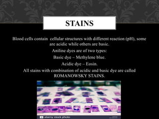
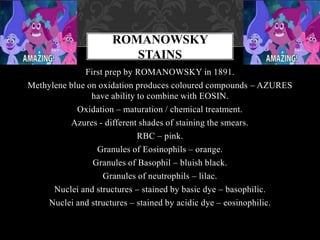


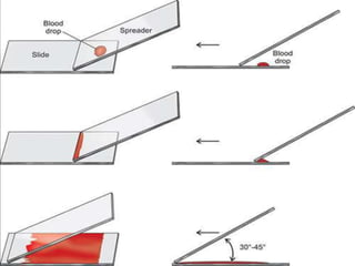


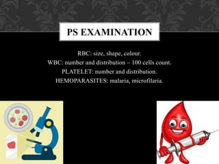


- A peripheral smear (PS) provides important diagnostic information about red blood cells, white blood cells, platelets, and any hemoparasites present. - Romanowsky stains like Leishman and Giemsa stains are commonly used because they differentially stain cellular components through a combination of basic and acidic dyes. - To perform a peripheral smear, a small blood sample is spread in a thin layer on a slide and allowed to dry before being stained using a Romanowsky stain and examined under a microscope.


![Peripheral blood smear [autosaved]](https://cdn.slidesharecdn.com/ss_thumbnails/peripheralbloodsmearautosaved-201029200454-thumbnail.jpg?width=640&height=640&fit=bounds)


