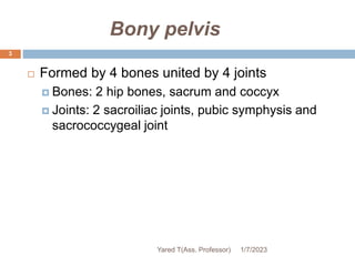
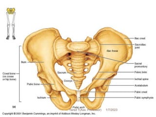
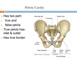
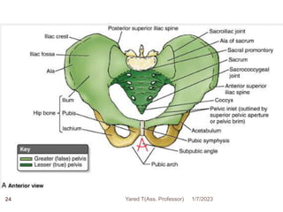
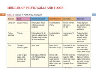
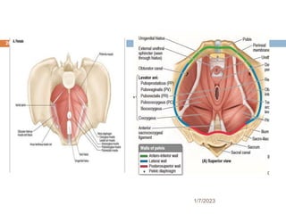
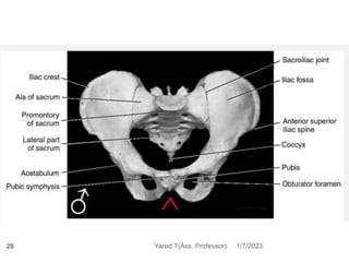
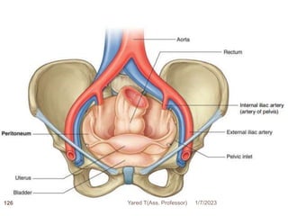
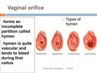
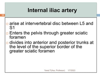
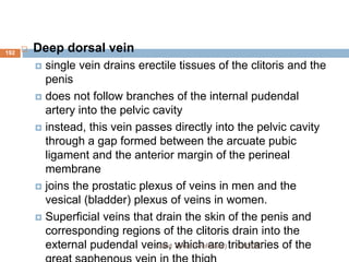
Pelvis perineum
1/7/2023
Yared T(Ass. Professor)
1
Pelvis perineum
 Formed by:
Skin
Subcutaneous tissue
Muscles
Bones
Fascia
Visceral organs(urinary, digestive and
reproductive)
1/7/2023
Yared T(Ass. Professor)
2
Bony pelvis
 Formed by 4 bones united by 4 joints
 Bones: 2 hip bones, sacrum and coccyx
 Joints: 2 sacroiliac joints, pubic symphysis and
sacrococcygeal joint
1/7/2023
Yared T(Ass. Professor)
3
4 1/7/2023
Yared T(Ass. Professor)
5 1/7/2023
Yared T(Ass. Professor)
Ilium
6
 Located superior to the acetabulum
Parts of ilium
 Body
 joins ischium and pubis to acetabulum
 Ala (wing)
 bordered superiorly by iliac crest
 Posteriorly it has; anterior, posterior and inferior
gluteal lines (origins of gluteus minimus,
medius and maximus muscles)
1/7/2023
Yared T(Ass. Professor)
…cnt
 Iliac crest
 a line joining the most superior point of the
bilateral iliac spine.
 cross the 4th lumbar spine (L4)(iliac
tuberosity)
 this is a guide to lumbar spinal tap because
the spinal cord ends around L1/L2
1/7/2023
Yared T(Ass. Professor)
7
Landmarks:
 ASIS
 AIIS
 PSIS
 PIIS
 Greater
sciatic notch
1/7/2023
Yared T(Ass. Professor)
8
Ischium
 Forms posteroinferior part of the acetabulum and
hip bone
 Body
 joins ilium and superior ramus of pubis to form
acetabulum
 Ramus
 fuses with the inferior ramus of pubis
 The pubic arch or sub pubic angle is formed by
joined rami of ischium and pubis, bilaterally
9
1/7/2023
Yared T(Ass. Professor)
Landmarks
 ischial spine
 lesser sciatic
notch
 ischial tuberosity
that supports
body during
sitting
1/7/2023
Yared T(Ass. Professor)
10
Pubis
11
 Forms anterior part of the acetabulum and
anteromedial part of hip bone
 Body
 has a symphyseal surface for articulation with
the contralateral pubis
 Rami
 two (inf. & sup.) which supports its body
1/7/2023
Yared T(Ass. Professor)
Landmarks
 pubic crest (medial) - ends at pubic tubercle (lateral)
 superior ramus - has a ridge called pecten pubis or
pectineal line
1/7/2023
Yared T(Ass. Professor)
12
1/7/2023
Yared T(Ass. Professor)
13
Sacrum
 Triangular shaped,
formed by five fused
vertebrae
 Forms the posterior wall
of the pelvis
 Articulates with L5 and
iliac bone
 Transfers the weight of
the body to the lower
extremities
. 1/7/2023
Yared T(Ass. Professor)
Coccyx
 tail bone
 composed
of 4 fused
vertebrae
 articulates
with
sacrum
15
1/7/2023
Yared T(Ass. Professor)
Pelvic Cavity
 Has two part;
1) true and
2) false pelvis
 True pelvis has-
inlet & outlet
 Has true border.
1/7/2023
Yared T(Ass. Professor)
16
Pelvic Cavity
 Is divided by pelvic brim into 2 parts:
1.pelvis major (false pelvis)
 Also called greater pelvis
 lies between iliac fossa; is part of abdominal wall
 Superior to the pelvic inlet
 Bounded by the abdominal wall anteriorly, the iliac alae
laterally, and the L5 and S1 vertebrae posteriorly
1/7/2023
Yared T(Ass. Professor)
17
1/7/2023
Yared T(Ass. Professor)
18
2. pelvis minor (true pelvis)
 Is also called lesser pelvis
 actual pelvis; contains pelvic viscera(rectum,
bladder, and reproductive organs)
 Between the pelvic inlet and the pelvic outlet
 Bounded by the pelvic surfaces of the hip bones,
sacrum, and coccyx
 Limited inferiorly by the musculofascial pelvic
diaphragm (levator ani).
1/7/2023
Yared T(Ass. Professor)
19
Pelvic inlet
20
 Bounded by:
 Superior margin of the pubic symphysis
 Posterior border of the pubic crest
 Pecten pubis
 Arcuate line of the ilium
 Anterior border of the ala of the sacrum
 Sacral promontory
1/7/2023
Yared T(Ass. Professor)
1/7/2023
Yared T(Ass. Professor)
21
Pelvic outlet
 bounded by:
 Inferior margin of the pubic symphysis
anteriorly
 Inferior rami of the pubis and ischial
tuberosities anterolaterally
 Sacrotuberous ligaments posterolaterally
 Tip of the coccyx posteriorly
1/7/2023
Yared T(Ass. Professor)
22
1/7/2023
Yared T(Ass. Professor)
23
24 1/7/2023
Yared T(Ass. Professor)
MUSCLES OF PELVIC WALLS AND FLOOR
25
1/7/2023
26
1/7/2023
Sexual differences in bony pelvis
27
 sexual differences are related to:
 heavier build and larger muscles of men
 adaptation in women for childbearing
 The male pelvis is heavier ,thicker, & has
more prominent bone markings
 The female pelvis is wider, shallower, lighter,
and rounder than the male and has a larger
pelvic inlet and outlet
1/7/2023
Yared T(Ass. Professor)
28 1/7/2023
Yared T(Ass. Professor)
29 1/7/2023
Yared T(Ass. Professor)
30 1/7/2023
Yared T(Ass. Professor)
Types of female pelvis
 Gynecoid
 Android
 Anthropoid
 platypelloid
1/7/2023
Yared T(Ass. Professor)
31
32
A and C are most common in males
B and A in white females
B and C in black females
D is uncommon in both sexes 1/7/2023
Yared T(Ass. Professor)
Pelvic diameters (Conjugates)
 shape and size of the pelvic inlet are significant
during labor
 are noted radiographically or manually
 pelvic diameters are :
-Inlet diameter, pelvic cavity & outlet
diameter
1/7/2023
Yared T(Ass. Professor)
33
Inlet Diameters
34
 oval (female) or heart shaped (male) opening
 larger in females than in males
 is demarcated by linea terminalis which
separates the false from the true pelvis.
1/7/2023
Yared T(Ass. Professor)
The diameters of the inlet are:
35
1.The anterioposterior (true conjugate) diameter
 is measured between the midpoint of sacral
promontory and the upper border of the pubic
symphysis
 is 10 cm in male and 11cm in female
1/7/2023
Yared T(Ass. Professor)
…cnt
36
2.The transverse diameter
 maximum distance that can be measured
between similar points on the opposite sides
of pelvic brim
 is 12.5 cm in male and 13 cm in female.
1/7/2023
Yared T(Ass. Professor)
…cnt
37
3. The oblique diameter
 is measured from one iliopubic eminence to
the opposite sacroiliac joint
 is 12cm in male and 12.5 cm in female
1/7/2023
Yared T(Ass. Professor)
1/7/2023
Yared T(Ass. Professor)
38
Pelvic outlet
39
 diamond shaped opening in both
 much wider in females than males
 Bounded:
 anteriorly by pubic symphysis
 posteriorly by sacrum and coccyx
 laterally by ischial tuberosities
 is partially closed because of coccyx is curved
in.
1/7/2023
Yared T(Ass. Professor)
Diameters of the outlet are:
40
1.The anterioposterior diameter
 is usually measured from apex(tip) of the
coccyx to the midpoint of the lower rim of
pubic symphysis
 is 8cm in male and 12.5 in female
1/7/2023
Yared T(Ass. Professor)
…cnt
41
2. The transverse diameter
 between ischial tuberosities, from the lower
border of their medial surfaces
 is 8.5 cm in male and 11.8 cm in female.
1/7/2023
Yared T(Ass. Professor)
1/7/2023
Yared T(Ass. Professor)
42
…cnt
43
3. The oblique diameter
 from midpoint of the sacrotuberous ligament
on one side to the junction of ischial and
pubic ramus on the other
 is 10 cm in male and 11.8cm in female.
1/7/2023
Yared T(Ass. Professor)
Nerves of pelvis
44
 Pelvis is innervated mainly by sacral and
coccygeal nerves and pelvic part of ANS
 This nerves includes
-sacral plexus
- coxygeal plexus
1/7/2023
Yared T(Ass. Professor)
Sacral plexus
45
 Origin anterior primary rami of L4–S4
 L4 is shared by both lumbar & sacral plexus
 sacral nerves emerge from the anterior sacral
foramina and unite in front of piriformis where
they are joined by the lumbosacral trunk
 Branches from the plexus supply:
 the pelvic muscles
 the muscles of the hip
 the skin of the buttock and the back of the thigh
1/7/2023
Yared T(Ass. Professor)
1/7/2023
Yared T(Ass. Professor)
46
Sacral Plexus
47
1/7/2023
Yared T(Ass. Professor)
48
1/7/2023
Yared T(Ass. Professor)
Branches
 Superior gluteal nerve - gluteus medius, minimus,
tensor fascia lata
 Inferior gluteal nerve – gluteus maximus
 Nerve to quadratus femoris
 Nerve to piriformis
 Nerve to obturator internus
 Posterior cutaneous nerve of the thigh
 The plexus itself terminates as the pudendal
nerve and the sciatic nerve
49
1/7/2023
Yared T(Ass. Professor)
50
Pudendal nerve
 forms from ventral divisions of S2 to S4
 It leaves the pelvic cavity through the greater
sciatic foramen, inferior to the piriformis muscle,
and enters the gluteal region;
 innervates skin and skeletal muscles of the
perineum, including the external anal and external
urethral sphincters
 Mediates the act of erection
 Voluntary control of urination
1/7/2023
Yared T(Ass. Professor)
51 1/7/2023
Yared T(Ass. Professor)
1/7/2023
Yared T(Ass. Professor)
52
53
Sciatic nerve
 The largest nerve in the body
 Formed by the anterior rami of spinal nerves L4 -S3
 Passes through the greater sciatic foramen inferior
to the piriformis to enter the gluteal (buttock) region
 It then descends along the posterior aspect of the
thigh to supply the posterior aspect of the lower
limb
1/7/2023
Yared T(Ass. Professor)
54
Coccygeal plexus
 Formed from S4 ,S5 and coccygeal nerve
 Supply part of levator ani, coccygeus and anal
sphincter
1/7/2023
Yared T(Ass. Professor)
55 1/7/2023
Yared T(Ass. Professor)
56 1/7/2023
Yared T(Ass. Professor)
57 1/7/2023
Yared T(Ass. Professor)
Pelvic Viscera
58
The organization of pelvic viscera from anterior to
posterior:
 In the male: bladder, prostate and male genital
organs and rectum
 In the female: bladder, uterus and female genital
organs and rectum
 The pelvic viscera are partly covered by
peritoneum in their upper aspects but are
extraperitoneal inferiorly
1/7/2023
Yared T(Ass. Professor)
1/7/2023
Yared T(Ass. Professor)
59
Urinary bladder
60
 Smooth, collapsible, muscular sac that stores
urine
 Position
 shape, size and position vary with amount of urine
and age
 In adults: empty bladder lies in pelvis minor
posterior and slightly superior to pubic bones
 In infants: found in abdomen
 Enters pelvic major at about 6 years of age and
pelvic minor at puberty
 When filled ascends to pelvic major and abdomen
1/7/2023
Yared T(Ass. Professor)
61
 Parts
 empty bladder has the form of pyramid
 4 surfaces: superior, 2 inferolateral and posterior
(fundus).
 anterior end is termed as apex
 inferior end is termed as neck
1/7/2023
Yared T(Ass. Professor)
1/7/2023
Yared T(Ass. Professor)
62
63
Internal structure
 The interior of the bladder has openings for both
ureters and the urethra
 The triangular region of the bladder base outlined by
these openings is called the trigone
 The ureters open at superior angle of trigone and the
urethra open at inferior angle.
1/7/2023
Yared T(Ass. Professor)
64
Arterial supply
 Branches of internal iliac arteries
 Superior vesical artery: supply anterosuperior parts
 Inferior vesical artery and Vaginal artery: supply fundus in
male and female, respectively
 Obturator and inferior gluteal arteries: small branches
 Venous drainage
 Correspond to arteries and drain into internal iliac vein
 Lymphatic drainage
 Superior part: to external iliac lymph nodes
 Inferior part: to internal iliac lymph nodes
 Innervation
 Parasympathetic fibers from pelvic splanchnic nerves
(S2,S3,S4)
 Motor to muscle and inhibitory to internal sphincter
 Sympathetic fibers from T11,T12,L1 & L2 through
vesical plexus
 inhibitory
1/7/2023
Yared T(Ass. Professor)
Urethra
65
Male urethra
 long, about 15-
20cm
 has 3 portions:
1.prostatic
urethra
 about 3cm long
 passes through
prostate
 widest and most
dilatable part
 ejaculatory
ducts open on
each side of
prostatic utricle
1/7/2023
Yared T(Ass. Professor)
1/7/2023
Yared T(Ass. Professor)
66
67
2.membranous urethra
 shortest (1-2cm), thinnest and narrowest
 traverse urogenital diaphragm
 surrounded by external urethrae sphincter muscle
and perineal membrane
 posterolateral to it on each side is bulbourethral
gland and duct
3.spongy urethra
 longest, about 15 cm
 passes through bulb of penis and corpus
spongiosum and ends at external urethral orifice
 ducts of bulbourethral glands opens into proximal
part
 expanded in bulb and in glans penis
1/7/2023
Yared T(Ass. Professor)
68
Blood supply
 arteries: branches to prostate, bulb and urethral
artery
 veins: accompany arteries
 Innervation
 branches of pudendal nerve
 pelvic splanchnic nerves
1/7/2023
Yared T(Ass. Professor)
69
Female urethra
shorter (4cm)
 It opens 2 cm posterior to the clitoris in vestibule of vagina
 Its inferior end is surrounded by sphincter urethrae muscle
 Urethral glands are found in superior part called
paraurethral glands
 Blood supply: internal pudendal and vaginal vessels
 Innervation: pudendal nerve and pelvic splanchnic nerves
 Lymphatic drainage: sacral and internal iliac lymph nodes
1/7/2023
Yared T(Ass. Professor)
70 1/7/2023
Yared T(Ass. Professor)
Male internal genital organs
Include :
 The testes (male gonads),
 System of ducts
 Epididymis,
 Ductus deferens,
 Ejaculatory ducts, and
 Urethra,
 Accessory sex glands
 seminal vesicles,
 prostate, and
 bulbourethral glands,
 Supporting structures
 the scrotum and
 the penis
1/7/2023
71
The Testes
 Small, oval shaped organ
 Housed in the scrotum
 Produces:
 Sperm
 androgens.
 Coverings
 tunica vaginalis:- is serous membrane which has
 Parietal layer
 Visceral layer.
 Tunica albuginea
250 lobules per testis
 Each lobule has up to 4 seminiferous tubules
72
1/7/2023
73
1/7/2023
The Fetus: Formation of Testes
 Testes formed inside body cavity (abdominal cavity).
 Descent of testis: During seventh month.
• Each testis:
– moves through abdominal musculature
– are accompanied by pockets of peritoneal cavity
 Accessary structures accompany testis during descent
 Form body of spermatic cord:
 ductus deferens
 testicular blood vessels, nerves, and lymphatic vessels
74
1/7/2023
Structure of testes
 seminiferous tubules: site of spermatogenesis
contain :- germ cell; Sperm forming cells
- Sertoli cells (supporting cells)
- form blood-testis barrier
-support developing sperm cells
- produce fluid & control release of sperm into lumen
- secrete inhibin which slows sperm production
 Interstitial spaces: surround the seminiferous tubules.
 Contain interstitial (Leydig) cells
 produce hormones called androgens.
75
1/7/2023
Structure of testes….
 Rete testis
= network of canals in the mediastinum testis
 Efferent Ductules
 15–20 large efferent ductules:
 connect rete testis to epididymis
76
1/7/2023
77
1/7/2023
Testes cont…
 Series of tubes (path of sperm )
Seminiferous tubules (site of sperm formation)
Straight ducts
Rete testis
Efferent ductule
Epididymis (where sperm matures & temporarily stored)
Ductus deferens
Ejaculatory duct
urethra
78
1/7/2023
The epididymis
 The epididymis is a comma-shaped structure composed of
an internal duct and an external covering of connective
tissue.
 Has three parts:-head, body and tail.
 Its head lies on the superior surface of the testis, while the
body and tail are posterior to the testis.
 Internally, the epididymis contains a long, convoluted duct
of the epididymis, which is approximately 7m.
 Sperm must reside in the epididymis for a period of time to
become mature and fully motile.
 If sperm are not ejected from the male reproductive
system in a timely manner, the old sperm degenerate in
the epididymis.
Yared T
79
1/7/2023
Yared T
80
1/7/2023
Epididymis cont…
 Testes produce physically immature spermatozoa that can
not fertilize an oocyte
 Other parts of reproductive system are responsible for:
 functional maturation, nourishment, storage, and transport
 3 Functions of the Epididymis
1. Monitors and adjusts fluid produced by seminiferous
tubules
2. Recycles damaged spermatozoa
3. Stores and protects spermatozoa:
– facilitates functional maturation of sperm
81
1/7/2023
Male internal genital organs
82
.Ductus (vas) deferens
 Thick walled muscular tube, about 45cm long.
 Continuation of ductus epididymis
 Course
 Begins in tail of epididymis and ends by joining duct of
seminal vesicle
 Ascends in spermatic cord, passes through inguinal
canal and cross over external iliac vessels to enter
pelvis minor
 Enlarges to form ampulla posterior to bladder
 Finally, it narrows and joins duct of seminal vesicle to
form ejaculatory duct.
1/7/2023
Yared T(Ass. Professor)
1/7/2023
Yared T(Ass. Professor)
83
Blood supply to ductus deference
 Usually from-superior vesical artery
 Some times-from inferior vesical artery
 Vein –to testicular vein
1/7/2023
Yared T(Ass. Professor)
84
Clinical Correlation
Male Sterilization
 The common method of sterilizing males is a
deferentectomy, popularly called a vasectomy.
 During this procedure, part of the ductus deferens is
ligated and/or excised through an incision in the
superior part of the scrotum .
 Hence, the subsequent ejaculated fluid from the
seminal glands, prostate, and bulbourethral glands
contains no sperms.
 The unexpelled sperms degenerate in the
epididymis and the proximal part of the ductus
deferens.
1/7/2023
Yared T(Ass. Professor)
85
Ejaculatory ducts
2.5cm long.
Formed by
union of ducts
of seminal
vesicle and
ductus
deferens.
Traverse
prostate.
Open into
posterior wall
of prostatic
1/7/2023
Yared T(Ass. Professor)
86
Blood supply to ejaculatory duct
 From superior or inferior vesical artery
 Vein-to prostatic & vesical venous plexus
1/7/2023
Yared T(Ass. Professor)
87
88
Seminal vesicles
Thin-walled and pear-shaped
 About 5 cm long
 Lies between fundus of bladder and rectum
 Secrete a fluid which ;
 accounts for about 60% of the volume of the semen
-It is a yellowish, viscous, alkaline fluid
 The ducts of the seminal vesicles join with the vas
deferens to form the ejaculatory ducts.
 Sperm and seminal fluid mix in the ejaculatory
duct to enter the prostatic urethra together during
ejaculation
1/7/2023
Yared T(Ass. Professor)
1/7/2023
Yared T(Ass. Professor)
89
1/7/2023
Yared T(Ass. Professor)
90
Contents Of Seminal Secretion
 Fructose
 Monosaccharides
 Citric Acid
 Coagulation Proteins
 Prostaglandins
 Fibrinogen
Arterial Supply and Venous
Drainage
 The arteries derive from the
inferior vesical and middle
rectal arteries.
 The veins accompany the
arteries and have similar
names
1/7/2023
Yared T(Ass. Professor)
91
92 1/7/2023
Yared T(Ass. Professor)
Prostate
93
 Surrounds prostatic
urethra
 Parts: base, apex, 4
surfaces (posterior,
anterior and 2
inferolateral)
 has 15-20 ducts
 It is 3 cm long, 4 cm wide,
and 2 cm in AP depth)
 about 20 g.
 largest accessory gland
1/7/2023
Yared T(Ass. Professor)
1/7/2023
Yared T(Ass. Professor)
94
95
 is divided into 2 lateral lobes (site of carcinoma -
may be felt on palpation through the rectum) and a
middle lobe (the upper part is the site of benign
hypertrophy)
 Secrete milky, alkaline fluid
 prostate progressively enlarges after 50 years of
age.
1/7/2023
Yared T(Ass. Professor)
Parts prostate gland
 Apex:-directed downwards; 1.5 cm below the margin of
symphysis pubis
 Base:-continuous with neck of bladder.
 Surfaces:-
 The anterior surface is separated from the pubic
symphysis by retroperitoneal fat in the retropubic space.
 A posterior surface that is related to the ampulla of the
rectum.
 Inferolateral surfaces that are related to the levator ani.
1/7/2023
Yared T(Ass. Professor)
96
Blood supply prostate
 Inferior vesical-major
 Middle rectal & internal pudendal artery
 Vein-prostatic plexus-drain to internal iliac
1/7/2023
Yared T(Ass. Professor)
97
Functions of the Prostate Gland
 Produce milky secretions
 Prostatic secretion makes from 20 -40% of volume of
the semen.
 Their secretion has the following advantages
1. assist motility of spermatozoa
2. help to nourish the spermatozoa
3. makes the semen more liquefied
4. its alkalinity protects sperm as it passes through the
acidic vagina
1/7/2023
Yared T(Ass. Professor)
98
Clinical Condition
Hypertrophy of Prostate
 The prostate is of considerable medical interest
because enlargement or benign hypertrophy of the
prostate (BHP) is common after middle age, affecting
virtually every male who lives long enough.
 An enlarged prostate projects into the urinary bladder
and impedes urination by distorting the prostatic
urethra.
 The middle lobule usually enlarges the most and
obstructs the internal urethral orifice.
 The more the person strains, the more the valve-like
prostatic mass occludes the urethra.
1/7/2023
Yared T(Ass. Professor)
99
Innervation to internal genital organs
 All internal genital structures such as;
 vas deference
 ejaculatory duct
 seminal vesicle and
 prostate are innervated;
 Symphatatic-T12-L3(abdominopelvic splanchnic)
 Parasympha-S2 &S3(pelvic splanchnic)
1/7/2023
Yared T(Ass. Professor)
100
Bulbourethral gland/Cowper
glands/
101
 Pea size
 lie posterolateral to membranous urethra,
largely embedded in External urethral sphincter
muscle
 duct passes through perineal membrane and
open in proximal spongy urethra in bulb of
penis
 Their mucus-like secretion enters the urethra
during sexual arousal, before ejaculation.
1/7/2023
Yared T(Ass. Professor)
1/7/2023
Yared T(Ass. Professor)
102
Female internal genital
organs
103
1.Primary reproductive organs- ovaries
 Used to:
 Produce gametes
 Produce female sex hormones (estogen and
progesterone)
2. The accessory ducts (uterine tubes, uterus,
and vagina)
1/7/2023
Yared T(Ass. Professor)
1/7/2023
Yared T(Ass. Professor)
104
Uterus
105
 hollow, thick walled, pear-shaped muscular
organ
 functions to receive, retain, and nourish the ovum
 located between bladder and rectum
 For non pregnant, uterus is:
 7-8cm long, 5-7cm wide, 3cm thick
 It is larger in woman who have borne children
 projects superoanteriorly over bladder
1/7/2023
Yared T(Ass. Professor)
Parts:
body: expanded superior 2/3
fundus: superior part of body
cervix: cylindrical inferior third
isthmus: junction between body and cervix
106
1/7/2023 Yared T(Ass. Professor)
Cervix
 Narrow part
 Characterized by internal and external os.
 Its mucosa contains cervical glands that secrete a mucus
 Mucus fills the cervical canal and covers the external os,
presumably to block the spread of bacteria from the vagina into
the uterus
 Cervical mucus also blocks the entry of sperm, except at
midcycle, when it becomes less viscous and allows sperm to pass
through.
107
1/7/2023
Yared T(Ass. Professor)
108
Position of uterus:
 usually bent anteriorly (anteflexed) between
cervix and body and the whole uterus is inclined
anterioly (anteverted)
 It is less anteverted when the bladder is full
 the uterus is often turned backward, or retroverted
in older woman
1/7/2023
Yared T(Ass. Professor)
109 1/7/2023
Yared T(Ass. Professor)
The Uterine Wall
110
 3 layers:
1.Perimetrium
 outermost serous layer composed of visceral
peritoneum
2.Myometrium
 bulky middle layer, composed of interlacing
bundles of smooth muscle
 contracts during childbirth to expel the baby
3.Endometrium
 simple columnar epithelium underlain by a
thick lamina propria of highly cellular
connective tissue
1/7/2023
Yared T(Ass. Professor)
111
Endometrium
 For Implantation of embryo
 has two strata/layer/
 stratum functionalis (functional layer)
 undergoes cyclic changes in response to blood levels of
ovarian hormones and is shed during menstruation (28
day cycle)
 stratum basale
 thinner deeper layer which forms a new functionalis layer
after menstruation ends
 unresponsive to ovarian hormones
 The endometrium has numerous uterine glands that
changes with the endometrium 1/7/2023
Yared T(Ass. Professor)
112
Blood supply
 Internal iliac Artery-uterine artery- arcuate
arteries within the myometrium-radial
branches into the endometrium-straight
arteries to the stratum basalis- spiral arteries
to the stratum functionalis
 spiral arteries undergo repeated degeneration and
regeneration, and it is their spasms that actually
cause the functionalis layer to be shed during
menstruation
1/7/2023
Yared T(Ass. Professor)
1/7/2023
Yared T(Ass. Professor)
113
114 1/7/2023
Yared T(Ass. Professor)
115
 Vein-uterine vein
 Lymphatics
 aortic, external iliac, internal iliac and sacral lymph
nodes
 Innervation
 from inferior hypogastric plexus
 parasympathetic - pelvic splanchnic
 sympathetic – symphatetic trunk
1/7/2023
Yared T(Ass. Professor)
Uterine tubes
116
 10-12cm long
 carry oocytes from ovaries and sperms from uterus to
site of fertilization (ampulla)
 opens at its proximal end to horn of uterus and distal
end into peritoneal cavity
 Parts:
 infundibulum: funnel shaped, margin have finger like
processes called fimbriae which spread over ovary
 ampulla: middle wide part, site of fertilization
 isthmus: narrow part which enters uterus
 uterine (intramural) portion: passes through
myometrium
1/7/2023
Yared T(Ass. Professor)
1/7/2023
Yared T(Ass. Professor)
117
118
 Arterial supply: from uterine and ovarian
arteries
 Venous drainage: to uterine and ovarian veins
 Lymphatics: to aortic lymph nodes
 Innervation: from ovarian plexus and uterine
plexus
1/7/2023
Yared T(Ass. Professor)
1/7/2023
Yared T(Ass. Professor)
119
Ovaries
120
 Oval shaped gland, about 3cm long
 Lies on the lateral wall of the pelvis major/in
ovarian fossa/
 The ovarian fossa is framed by the external iliac
vessels superiorly, the obliterated umbilical artery
anteriorly, the ureter and internal iliac vessels
posteriorly. Obturator nerve and vessels are lateral
to the ovarian fossa.
 Each ovary is held in place within the peritoneal
cavity by several ligaments
 ovarian ligament
 suspensory ligament
 mesovarium
1/7/2023
Yared T(Ass. Professor)
…cnt
 ovarian ligament
 anchors the ovary medially to the uterus
 suspensory ligament
 anchors the ovaries laterally to the pelvic wall
 mesovarium
 suspends the ovaries between the fallopian tubes and the
uterus
 Both the suspensory ligament and the
mesovarium are part of the broad ligament
121
1/7/2023
Yared T(Ass. Professor)
1/7/2023
Yared T(Ass. Professor)
122
123
 Arterial supply: ovarian arteries
 Venous drainage: ovarian veins; right drains
into inferior vena cava and left into left renal
vein
The ovarian blood vessels reach the
ovaries by traveling through the
suspensory ligaments and mesovaria
 Lymphatics: to aortic lymph nodes
 Innervation: ovarian plexus and vagus
1/7/2023
Yared T(Ass. Professor)
1/7/2023
Yared T(Ass. Professor)
124
Rectum
125
 Fixed terminal part of large intestine
 Begins at the level of S3
 about 12-15cm long, follows curves of sacrum
and coccyx
 Ends 3-4cm anteroinferior to tip of coccyx by
turning posteroinferiorly to become anal canal
 Terminal part dilate called rectal ampulla
1/7/2023
Yared T(Ass. Professor)
1/7/2023
Yared T(Ass. Professor)
126
127
Arterial supply
To rectum
 Superior rectal artery
 Middle rectal arteries
 Inferior rectal arteries
1/7/2023
Yared T(Ass. Professor)
PERINEUM
128
 diamond shaped inferior to pelvic diaphragm between
thighs
 Boundaries:
 anteriorly by the symphysis, the inferior ramus of the pubis and
the ramus of the ischium
 Posteriorly it is bound by the lower part of the sacrum and the
coccyx connected to the ischial tuberosity by the sacrotuberous
ligament
 The superficial part of this region is called the perineum
including the external genitalia (in the urogenital triangle)
and the anus (in the anal triangle)
1/7/2023
Yared T(Ass. Professor)
1/7/2023
Yared T(Ass. Professor)
129
1/7/2023
Yared T(Ass. Professor)
130
131
…cnt
 Divided into 2 triangles by line joining ischial
tuberosities
 Anal triangle: contains anus and ischioanal fossa
 Urogenital triangle: contains external genitalia
 Inferior pelvic aperture (pelvic outlet) is closed by
 Urogenital diaphragm: anterior part
 Pelvic diaphragm: posterior part
1/7/2023
Yared T(Ass. Professor)
132 1/7/2023
Yared T(Ass. Professor)
Pelvic diaphragm
133
 Levator ani and coccygeus form the pelvic
diaphragm, separating the pelvis from the
ischiorectal fossa
 urethra, vagina and rectum pass through the pelvic
diaphragm
 pudendal nerve and vessels go around it
1/7/2023
Yared T(Ass. Professor)
Levator ani
134
 Muscular floor of the pelvis and supports the
viscera
 Has 3 parts:
1. puborectalis
2. pubococcygeus
3. iliococcygeus muscle
1/7/2023
Yared T(Ass. Professor)
1/7/2023
Yared T(Ass. Professor)
135
136
Coccygeus
 deep to the sacrospinous ligament
 has similar attachments with levator ani
 forms posterior part of pelvic floor
 Nerve supply: branches of S4 & S5
 Action: assist levator ani
1/7/2023
Yared T(Ass. Professor)
137 1/7/2023
Yared T(Ass. Professor)
138
 Urogenital diaphragm
 strong triangular membrane (thin sheet of muscle)
 Anterior and posterior fibers called deep
transverse perenial muscles
 Middle fibers called sphincter urethrae muscle
 A small gap at the apex of the triangle, posterior to
the pubic symphysis, exists for passage of the
deep dorsal vein and the dorsal nerve of the
penis or clitoris
1/7/2023
Yared T(Ass. Professor)
139 1/7/2023
Yared T(Ass. Professor)
…cnt
 The perineal membrane is pierced by:
 the urethra
 vagina
 ducts of the bulbourethral glands
 deep and dorsal arteries of the penis or
clitoris
1/7/2023
Yared T(Ass. Professor)
140
Anal triangle
141
 Boundaries
 tip of coccyx, line joining ischial tuberosities,
sacrotuberous ligament
 Contents
 anal canal, external anal sphincter and ischioanal
fossae
1/7/2023
Yared T(Ass. Professor)
142 1/7/2023
Yared T(Ass. Professor)
Anal canal
 Terminal about 4 cm part of large intestine
 Begins at u-shaped sling formed by puborectalis
muscle and ends at anus
 Descends posterioinferiorly
1/7/2023
Yared T(Ass. Professor)
143
1/7/2023
Yared T(Ass. Professor)
144
145
 Arterial supply
 superior rectal artery: superior to pectinate line
 Inferior rectal arterie: inferior to pectinate line
 Middle rectal arteries: anastomose with both
 Venous drainage
 Similar to artery
 Lymphatic drainage
 Superior part: to internal iliac lymph nodes
 Inferior part: to superficial inguinal lymph nodes
 Innervation
 Superior part: from inferior hypogastric and pelvic splanchnic
 Inferior part: from inferior rectal nerves
1/7/2023
Yared T(Ass. Professor)
Anal sphincter
 Smooth muscle
 On each side of superior
2/3rd
 contracts involuntarily
 prevent feces from leaking
 Not trained
 Skeletal muscle
 On each side of inferior 2/3rd
 contracts voluntarily
 prevent feces from leaking
 children learn to control during
toilet training
Internal External
1/7/2023
Yared T(Ass. Professor)
146
Urogenital triangle
147
 Consists of male and
 female external genital,urinary
structure
1/7/2023
Yared T(Ass. Professor)
Male external genitalia
 cutaneous sac ( fold of skin) that envelops the
testis
 Double layered
1. outer: highly pigmented skin
2. inner: Subcutaneous tissue (dartos fascia)
and dartos muscle which is responsible for
wrinkling of the skin.
1/7/2023
Yared T(Ass. Professor)
148
 scrotal raphe-externally
 Scrotal septum-internally
1/7/2023
Yared T(Ass. Professor)
149
 Innervations is:
 Genital branch of the genitofemoral nerve:
anterolateral surface.
 Anterior scrotal nerves, branches of the
ilioinguinal nerve anterior surface.
 Posterior scrotal nerves, branches of the
perineal branch of the pudendal nerve:
posterior surface.
 Perineal branches of the posterior femoral
cutaneous nerve: inferior surface.
1/7/2023
Yared T(Ass. Professor)
150
 Posterior scrotal branches of the perineal
artery from internal pudendal artery.
 Anterior scrotal branches of the deep external
pudendal artery, from femoral artery.
 Cremasteric artery from inferior epigastric
artery
1/7/2023
Yared T(Ass. Professor)
151
Veins and lymphatic's
 Scrotal veins accompany the arteries
corresponding veins
Lymphatic drainage
superficial inguinal lymph nodes
1/7/2023
Yared T(Ass. Professor)
152
Penis
153
 Organ of copulation and common outlet for urine
and semen
 Composed of 3 cylindrical bodies of erectile
tissue
1.Two Corpora cavernosa penis
 fused in median plane anteriorly
2.Corpus spongiosum penis
 urethra runs through it
 enlarged at the end to form the glans of the penis
1/7/2023
Yared T(Ass. Professor)
154 1/7/2023
Yared T(Ass. Professor)
155 1/7/2023
Yared T(Ass. Professor)
156
parts of penis
 Root
 attached portion
 consists of crura, bulb and muscles
 crura are posterior continuations of the corpora
cavernosa
 bulb is between crura in superficial perineal space
and is a continuation of the corpus spongiosum
 The crura are attached to the inferior ramus of the
pubis and the ramus of the ischium
1/7/2023
Yared T(Ass. Professor)
…cnt
 the bulb is attached to the undersurface of the
perineal membrane
 The urethra pierces the membrane in order to
enter the bulb
 The crura are surrounded by the
ischiocavernosus muscle and the bulb by the
bulbospongiosus, which expels the last drop of
urine
1/7/2023
Yared T(Ass. Professor)
157
158
 Body
 free part
 consists of corpora cavernosa and corpus
spongiosum
 distal end - glans penis, formed by corpus
spongiosum
 near tip is external urethral orifice
 The skin of the penis is thin and loose and the fold
over the glans with fasciae to form prepuce
1/7/2023
Yared T(Ass. Professor)
159 1/7/2023
Yared T(Ass. Professor)
160 1/7/2023
Yared T(Ass. Professor)
161 1/7/2023
Yared T(Ass. Professor)
162
 Arterial supply: dorsal arteries, deep arteries,
artery to the bulb & Superficial external pudendal artery
 Venous drainage:
 The superficial dorsal vein is in the midline and drains
skin, into a tributary of the saphenous vein
 The deep dorsal vein is in the fascia penis, in the
midline and drains the deep structures. It passes
through the gap of the perineal membrane and joins
the prostatic venous plexus.
 Lymphatics: superficial and deep inguinal lymph
nodes
 Innervation: dorsal nerve of penis, cavernous
nerves, ilioinguinal nerves 1/7/2023
Yared T(Ass. Professor)
Female external genital organs
 The external genitalia also called vulva
include
 Mons pubis
 Labia majora & labia minora
 Clitoris
 Vestibule structures
163
1/7/2023
Yared T(Ass. Professor)
164
…cnt
1/7/2023
Yared T(Ass. Professor)
165 1/7/2023
Yared T(Ass. Professor)
Vagina & Vaginal orifice
166
 Female organ of copulation
 Often called the birth canal
 provides a passageway for infant & menstrual flow
 Musculomembranous tube
 Anterior wall is shorter (7 cm) than posterior wall (9
cm)
 passes between levator ani muscles, pierces
urogenital diaphragm
 The upper end of the vaginal canal loosely surrounds the
cervix of the uterus, producing vaginal fornix
 posterior fornix, is much deeper
 2 lateral
 anterior fornices
1/7/2023
Yared T(Ass. Professor)
1/7/2023
Yared T(Ass. Professor)
167
168
1/7/2023
Yared T(Ass. Professor)
169
 Arteries: vaginal, internal pudendal, vaginal
branch of middle rectal
 Veins: vaginal venous plexus drain into internal
iliac veins
 Lymphatic drainage: internal and external iliac,
superficial inguinal lymph nodes
 Innervation: from uterovaginal plexus (in broad
ligament) derived from inferior hypogastric
plexus and pelvic splanchnic nerves
1/7/2023
Yared T(Ass. Professor)
Vaginal orifice
forms an
incomplete
partition called
hymen
hymen is quite
vascular and
tends to bleed
during first
coitus
 Types of
hymen
1/7/2023
Yared T(Ass. Professor)
170
171
 Mons pubis
 Rounded fatty elevation anterior to pubic
symphysis
 Consists of pad of fatty tissue
 After puberty this area is covered with pubic
hair
 Labia majora
 Two symmetrical hair covered, fatty skin folds
 Each passes posteriorly from mons pubis to
anterior of anus
 the female counterpart of the male scrotum
1/7/2023
Yared T(Ass. Professor)
…cnt
 Labia minora
 Thin, hair free folds of skin between labia
majora
 Enclose a recess called vestibule of vagina
and lie on each side of orifices of urethra and
vagina
 Meet superior to clitoris to form a fold of skin
called prepuce
 Posteriorly united by fold of skin called
frenulum of labia minora
 homologous to the ventral penis
1/7/2023
Yared T(Ass. Professor)
172
173 1/7/2023
Yared T(Ass. Professor)
174
 Vestibule of vagina
Space between labia minora
Urethra, vagina and ducts of vestibular
glands open into it.
External urethral orifice is posterior to
clitoris and anterior to vaginal orifice
Vaginal orifice lies posterior to urethral
orifice
Surrounded by thin fold of mucous
membrane called hymen
1/7/2023
Yared T(Ass. Professor)
…cnt
 Vestibular glands
The greater vestibular (Bartholin's)
glands
Flanking/on the side/ the vaginal opening
pea sized
release mucus into the vestibule to keep it
moist and lubricated, facilitating intercourse
 homologous to the bulbourethral glands of
males
open into the vestibule just below
the hymen. 1/7/2023
Yared T(Ass. Professor)
175
176
Clitoris
 composed of erectile tissue
 homologous to the penis
 not traversed by urethra
 located posterior to anterior labial commissure
 parts: root ,body & glans
 Has corpora cavernosa but it lacks corpus spongiosum
 parts of labia minora anterior to clitoris form prepuce
and posterior form frenulum
 richly innervated with sensory nerve endings sensitive
to touch, and it becomes swollen with blood and erect
during tactile stimulation,
contributing to a female’s sexual arousal
1/7/2023
Yared T(Ass. Professor)
177 1/7/2023
Yared T(Ass. Professor)
178 1/7/2023
Yared T(Ass. Professor)
Arteries of pelvis and perineum
179
 The major artery of the pelvis and perineum is
the internal iliac artery on each side
 Other vessels that originate in the abdomen and
contribute to the supply of pelvic structures
include
 median sacral artery
 ovarian arteries in women
1/7/2023
Yared T(Ass. Professor)
Internal iliac artery
180
 arise at intervertebral disc between L5 and
S1
 Enters the pelvis through greater sciatic
foramen
 divides into anterior and posterior trunks at
the level of the superior border of the
greater sciatic foramen
1/7/2023
Yared T(Ass. Professor)
181
Branches of the posterior trunk
 iliolumbar artery
 lateral sacral artery
 superior gluteal artery
 Iliolumbar artery
 climbing over the brim it ascends in front of the sacrum and
anastomoses with the lumbar arteries
 divides into lumbar branch and iliac branch
 lumbar branch
 contributes to the supply of the posterior abdominal wall, psoas,
quadratus lumborum muscles, and cauda equina via a small spinal
branch that passes through the intervertebral foramen between L5 and
S1
 iliac branch
 passes laterally into the iliac fossa to supply muscle and bone
1/7/2023
Yared T(Ass. Professor)
182
 Lateral sacral arteries
 usually two
 bone and soft tissues, structures in the
vertebral (sacral) canal, and skin and muscle
posterior to the sacrum
 Superior gluteal artery
 the largest branch
 muscles and skin in the gluteal region and also
supplies branches to adjacent muscles and
bones of the pelvic walls
1/7/2023
Yared T(Ass. Professor)
183 1/7/2023
Yared T(Ass. Professor)
184
Branches of the anterior trunk
 umbilical artery - gives origin to the superior
vesical artery
 superior vesical artery -
 obturator artery
 inferior vesical artery
 middle rectal artery
 uterine artery (in female)
 2 terminal branches
 internal pudendal artery
 inferior gluteal artery
1/7/2023
Yared T(Ass. Professor)
185
 Superior vesical artery
 superior aspect of the bladder and distal parts of the ureter
 ductus deferens
 Inferior vesical artery
 occurs in men and supplies branches to the bladder, ureter,
seminal vesicle, and prostate
 Vaginal artery
 adjacent parts of the bladder and rectum
 Middle rectal artery
 courses medially to supply the rectum
 anastomoses with the superior rectal artery, which originates from
the inferior mesenteric artery in the abdomen, and the inferior
rectal artery, which originates from the internal pudendal artery in
the perineum
1/7/2023
Yared T(Ass. Professor)
186
 Obturator artery
 leaves the pelvic cavity via the obturator canal
 together with the obturator nerve, above, and
obturator vein, below, it enters and supplies the
adductor region of the thigh
 Internal pudendal artery
 the main artery of the perineum
 among the structures it supplies are the erectile
tissues of the clitoris and the penis.
1/7/2023
Yared T(Ass. Professor)
187
 Inferior gluteal artery
 a large terminal branch
 supply of the gluteal region and anastomoses with a network of
vessels around the hip joint
 Uterine artery
1/7/2023
Yared T(Ass. Professor)
188 1/7/2023
Yared T(Ass. Professor)
189 1/7/2023
Yared T(Ass. Professor)
Veins
190
 Within the pelvic cavity, extensive interconnected
venous plexuses are associated with the surfaces
of the viscera (bladder, rectum, prostate, uterus,
and vagina)
 together, these plexuses form the pelvic plexus of
veins
1/7/2023
Yared T(Ass. Professor)
191
 part of venous plexus surrounding the rectum
and anal canal drains via superior rectal veins
(tributaries of inferior mesenteric veins) into the
hepatic portal system, and via middle and
inferior rectal veins into the caval system
 this pelvic plexus is an important portacaval shunt
when the hepatic portal system is blocked
1/7/2023
Yared T(Ass. Professor)
192  Deep dorsal vein
 single vein drains erectile tissues of the clitoris and the
penis
 does not follow branches of the internal pudendal
artery into the pelvic cavity
 instead, this vein passes directly into the pelvic cavity
through a gap formed between the arcuate pubic
ligament and the anterior margin of the perineal
membrane
 joins the prostatic plexus of veins in men and the
vesical (bladder) plexus of veins in women.
 Superficial veins that drain the skin of the penis and
corresponding regions of the clitoris drain into the
external pudendal veins, which are tributaries of the
1/7/2023
Yared T(Ass. Professor)
193
 Median sacral veins
 coalesce to form a single vein that joins either the
left common iliac vein or the junction of the two
common iliac veins to form the inferior vena cava
 Ovarian veins
 follow the course of the corresponding arteries
 on the left, they join the left renal vein and, on the
right, they join the inferior vena cava in the
abdomen
1/7/2023
Yared T(Ass. Professor)
194 1/7/2023
Yared T(Ass. Professor)
195 1/7/2023
Yared T(Ass. Professor)
vessels & nerves of
perineum
196
 pudendal nerve, internal pudendal artery and nerve to
obturator internus
 They cross the spine of ischium
 enter the perineum through the lesser sciatic foramen
 Internal pudendal artery and pudendal nerve
 They both give an inferior rectal branch
 Pass through ischiorectal fossa
 supplies the lower part of the anal canal and its muscles
 The inferior rectal nerve is sensory to the lower half of
the anal canal, motor to the sphincter ani externus and
levator ani. These muscles are also innervated by a
separate branch from S4.
1/7/2023
Yared T(Ass. Professor)
197
Pudendal nerve
 divides into a dorsal nerve of the penis (clitoris)
and the perineal nerve
 perineal artery
 gives rise to scrotal or labial branches;
 It supplies the bulb of the penis (clitoris) and the
urethra and ends by dividing into dorsal and deep
arteries of the penis
1/7/2023
Yared T(Ass. Professor)
Lymphatics of the abdomen and pelvis
198
 Lymph nodes are closely related to the main arteries
 Preaortic nodes
 related to the 3 ventral branches of the aorta
 All lymph vessels converge to form the intestinal
trunks leading to the cisterna chyli
 The preaortic nodes include the celiac, gastric,
pancreaticosplenic, hepatic, pyloric and gastroepiploic
(gastro-omental) nodes
 The liver drains mainly into the hepatic group. Part of
the upper surface drains into nodes around the inferior
vena cava and the left lobe drains into the left gastric
group.
1/7/2023
Yared T(Ass. Professor)
…cnt
 The pancreas drains into the pancreaticosplenic
nodes but also into the superior mesenteric nodes.
 The superior mesenteric and inferior mesenteric
nodes drain along the major arteries
 The lower part of the rectum and the upper part of the
anal canal drain along the middle rectal artery to
internal iliac nodes. The lower part of the anal canal
drains to inguinal nodes.
1/7/2023
Yared T(Ass. Professor)
199
200
Lateral aortic (Para aortic) nodes
 related to the lateral aortic branches
 The iliac nodes are classified into common, internal and
external
 Kidneys and suprarenal glands drain into the local lateral
aortic nodes
 The bladder, lower end of the ureter, ductus deferens
and seminal vesicles drain mainly to external iliac nodes
and partly to the internal iliac nodes.
 The prostate gland drains to internal iliac and sacral
nodes.
 The cervix of the uterus drains to external, internal iliac
and sacral nodes.
 The body of the uterus drains into external iliac nodes
and superiorly to lateral aortic and preaortic nodes.
 Retroaortic nodes lie posterior to the aorta
1/7/2023
Yared T(Ass. Professor)
201 1/7/2023
Yared T(Ass. Professor)
202
1/7/2023
Yared T(Ass. Professor)

Pelvis perineum.pptx

  • 1.
  • 2.
    Pelvis perineum  Formedby: Skin Subcutaneous tissue Muscles Bones Fascia Visceral organs(urinary, digestive and reproductive) 1/7/2023 Yared T(Ass. Professor) 2
  • 3.
    Bony pelvis  Formedby 4 bones united by 4 joints  Bones: 2 hip bones, sacrum and coccyx  Joints: 2 sacroiliac joints, pubic symphysis and sacrococcygeal joint 1/7/2023 Yared T(Ass. Professor) 3
  • 4.
  • 5.
  • 6.
    Ilium 6  Located superiorto the acetabulum Parts of ilium  Body  joins ischium and pubis to acetabulum  Ala (wing)  bordered superiorly by iliac crest  Posteriorly it has; anterior, posterior and inferior gluteal lines (origins of gluteus minimus, medius and maximus muscles) 1/7/2023 Yared T(Ass. Professor)
  • 7.
    …cnt  Iliac crest a line joining the most superior point of the bilateral iliac spine.  cross the 4th lumbar spine (L4)(iliac tuberosity)  this is a guide to lumbar spinal tap because the spinal cord ends around L1/L2 1/7/2023 Yared T(Ass. Professor) 7
  • 8.
    Landmarks:  ASIS  AIIS PSIS  PIIS  Greater sciatic notch 1/7/2023 Yared T(Ass. Professor) 8
  • 9.
    Ischium  Forms posteroinferiorpart of the acetabulum and hip bone  Body  joins ilium and superior ramus of pubis to form acetabulum  Ramus  fuses with the inferior ramus of pubis  The pubic arch or sub pubic angle is formed by joined rami of ischium and pubis, bilaterally 9 1/7/2023 Yared T(Ass. Professor)
  • 10.
    Landmarks  ischial spine lesser sciatic notch  ischial tuberosity that supports body during sitting 1/7/2023 Yared T(Ass. Professor) 10
  • 11.
    Pubis 11  Forms anteriorpart of the acetabulum and anteromedial part of hip bone  Body  has a symphyseal surface for articulation with the contralateral pubis  Rami  two (inf. & sup.) which supports its body 1/7/2023 Yared T(Ass. Professor)
  • 12.
    Landmarks  pubic crest(medial) - ends at pubic tubercle (lateral)  superior ramus - has a ridge called pecten pubis or pectineal line 1/7/2023 Yared T(Ass. Professor) 12
  • 13.
  • 14.
    Sacrum  Triangular shaped, formedby five fused vertebrae  Forms the posterior wall of the pelvis  Articulates with L5 and iliac bone  Transfers the weight of the body to the lower extremities . 1/7/2023 Yared T(Ass. Professor)
  • 15.
    Coccyx  tail bone composed of 4 fused vertebrae  articulates with sacrum 15 1/7/2023 Yared T(Ass. Professor)
  • 16.
    Pelvic Cavity  Hastwo part; 1) true and 2) false pelvis  True pelvis has- inlet & outlet  Has true border. 1/7/2023 Yared T(Ass. Professor) 16
  • 17.
    Pelvic Cavity  Isdivided by pelvic brim into 2 parts: 1.pelvis major (false pelvis)  Also called greater pelvis  lies between iliac fossa; is part of abdominal wall  Superior to the pelvic inlet  Bounded by the abdominal wall anteriorly, the iliac alae laterally, and the L5 and S1 vertebrae posteriorly 1/7/2023 Yared T(Ass. Professor) 17
  • 18.
  • 19.
    2. pelvis minor(true pelvis)  Is also called lesser pelvis  actual pelvis; contains pelvic viscera(rectum, bladder, and reproductive organs)  Between the pelvic inlet and the pelvic outlet  Bounded by the pelvic surfaces of the hip bones, sacrum, and coccyx  Limited inferiorly by the musculofascial pelvic diaphragm (levator ani). 1/7/2023 Yared T(Ass. Professor) 19
  • 20.
    Pelvic inlet 20  Boundedby:  Superior margin of the pubic symphysis  Posterior border of the pubic crest  Pecten pubis  Arcuate line of the ilium  Anterior border of the ala of the sacrum  Sacral promontory 1/7/2023 Yared T(Ass. Professor)
  • 21.
  • 22.
    Pelvic outlet  boundedby:  Inferior margin of the pubic symphysis anteriorly  Inferior rami of the pubis and ischial tuberosities anterolaterally  Sacrotuberous ligaments posterolaterally  Tip of the coccyx posteriorly 1/7/2023 Yared T(Ass. Professor) 22
  • 23.
  • 24.
  • 25.
    MUSCLES OF PELVICWALLS AND FLOOR 25 1/7/2023
  • 26.
  • 27.
    Sexual differences inbony pelvis 27  sexual differences are related to:  heavier build and larger muscles of men  adaptation in women for childbearing  The male pelvis is heavier ,thicker, & has more prominent bone markings  The female pelvis is wider, shallower, lighter, and rounder than the male and has a larger pelvic inlet and outlet 1/7/2023 Yared T(Ass. Professor)
  • 28.
  • 29.
  • 30.
  • 31.
    Types of femalepelvis  Gynecoid  Android  Anthropoid  platypelloid 1/7/2023 Yared T(Ass. Professor) 31
  • 32.
    32 A and Care most common in males B and A in white females B and C in black females D is uncommon in both sexes 1/7/2023 Yared T(Ass. Professor)
  • 33.
    Pelvic diameters (Conjugates) shape and size of the pelvic inlet are significant during labor  are noted radiographically or manually  pelvic diameters are : -Inlet diameter, pelvic cavity & outlet diameter 1/7/2023 Yared T(Ass. Professor) 33
  • 34.
    Inlet Diameters 34  oval(female) or heart shaped (male) opening  larger in females than in males  is demarcated by linea terminalis which separates the false from the true pelvis. 1/7/2023 Yared T(Ass. Professor)
  • 35.
    The diameters ofthe inlet are: 35 1.The anterioposterior (true conjugate) diameter  is measured between the midpoint of sacral promontory and the upper border of the pubic symphysis  is 10 cm in male and 11cm in female 1/7/2023 Yared T(Ass. Professor)
  • 36.
    …cnt 36 2.The transverse diameter maximum distance that can be measured between similar points on the opposite sides of pelvic brim  is 12.5 cm in male and 13 cm in female. 1/7/2023 Yared T(Ass. Professor)
  • 37.
    …cnt 37 3. The obliquediameter  is measured from one iliopubic eminence to the opposite sacroiliac joint  is 12cm in male and 12.5 cm in female 1/7/2023 Yared T(Ass. Professor)
  • 38.
  • 39.
    Pelvic outlet 39  diamondshaped opening in both  much wider in females than males  Bounded:  anteriorly by pubic symphysis  posteriorly by sacrum and coccyx  laterally by ischial tuberosities  is partially closed because of coccyx is curved in. 1/7/2023 Yared T(Ass. Professor)
  • 40.
    Diameters of theoutlet are: 40 1.The anterioposterior diameter  is usually measured from apex(tip) of the coccyx to the midpoint of the lower rim of pubic symphysis  is 8cm in male and 12.5 in female 1/7/2023 Yared T(Ass. Professor)
  • 41.
    …cnt 41 2. The transversediameter  between ischial tuberosities, from the lower border of their medial surfaces  is 8.5 cm in male and 11.8 cm in female. 1/7/2023 Yared T(Ass. Professor)
  • 42.
  • 43.
    …cnt 43 3. The obliquediameter  from midpoint of the sacrotuberous ligament on one side to the junction of ischial and pubic ramus on the other  is 10 cm in male and 11.8cm in female. 1/7/2023 Yared T(Ass. Professor)
  • 44.
    Nerves of pelvis 44 Pelvis is innervated mainly by sacral and coccygeal nerves and pelvic part of ANS  This nerves includes -sacral plexus - coxygeal plexus 1/7/2023 Yared T(Ass. Professor)
  • 45.
    Sacral plexus 45  Originanterior primary rami of L4–S4  L4 is shared by both lumbar & sacral plexus  sacral nerves emerge from the anterior sacral foramina and unite in front of piriformis where they are joined by the lumbosacral trunk  Branches from the plexus supply:  the pelvic muscles  the muscles of the hip  the skin of the buttock and the back of the thigh 1/7/2023 Yared T(Ass. Professor)
  • 46.
  • 47.
  • 48.
  • 49.
    Branches  Superior glutealnerve - gluteus medius, minimus, tensor fascia lata  Inferior gluteal nerve – gluteus maximus  Nerve to quadratus femoris  Nerve to piriformis  Nerve to obturator internus  Posterior cutaneous nerve of the thigh  The plexus itself terminates as the pudendal nerve and the sciatic nerve 49 1/7/2023 Yared T(Ass. Professor)
  • 50.
    50 Pudendal nerve  formsfrom ventral divisions of S2 to S4  It leaves the pelvic cavity through the greater sciatic foramen, inferior to the piriformis muscle, and enters the gluteal region;  innervates skin and skeletal muscles of the perineum, including the external anal and external urethral sphincters  Mediates the act of erection  Voluntary control of urination 1/7/2023 Yared T(Ass. Professor)
  • 51.
  • 52.
  • 53.
    53 Sciatic nerve  Thelargest nerve in the body  Formed by the anterior rami of spinal nerves L4 -S3  Passes through the greater sciatic foramen inferior to the piriformis to enter the gluteal (buttock) region  It then descends along the posterior aspect of the thigh to supply the posterior aspect of the lower limb 1/7/2023 Yared T(Ass. Professor)
  • 54.
    54 Coccygeal plexus  Formedfrom S4 ,S5 and coccygeal nerve  Supply part of levator ani, coccygeus and anal sphincter 1/7/2023 Yared T(Ass. Professor)
  • 55.
  • 56.
  • 57.
  • 58.
    Pelvic Viscera 58 The organizationof pelvic viscera from anterior to posterior:  In the male: bladder, prostate and male genital organs and rectum  In the female: bladder, uterus and female genital organs and rectum  The pelvic viscera are partly covered by peritoneum in their upper aspects but are extraperitoneal inferiorly 1/7/2023 Yared T(Ass. Professor)
  • 59.
  • 60.
    Urinary bladder 60  Smooth,collapsible, muscular sac that stores urine  Position  shape, size and position vary with amount of urine and age  In adults: empty bladder lies in pelvis minor posterior and slightly superior to pubic bones  In infants: found in abdomen  Enters pelvic major at about 6 years of age and pelvic minor at puberty  When filled ascends to pelvic major and abdomen 1/7/2023 Yared T(Ass. Professor)
  • 61.
    61  Parts  emptybladder has the form of pyramid  4 surfaces: superior, 2 inferolateral and posterior (fundus).  anterior end is termed as apex  inferior end is termed as neck 1/7/2023 Yared T(Ass. Professor)
  • 62.
  • 63.
    63 Internal structure  Theinterior of the bladder has openings for both ureters and the urethra  The triangular region of the bladder base outlined by these openings is called the trigone  The ureters open at superior angle of trigone and the urethra open at inferior angle. 1/7/2023 Yared T(Ass. Professor)
  • 64.
    64 Arterial supply  Branchesof internal iliac arteries  Superior vesical artery: supply anterosuperior parts  Inferior vesical artery and Vaginal artery: supply fundus in male and female, respectively  Obturator and inferior gluteal arteries: small branches  Venous drainage  Correspond to arteries and drain into internal iliac vein  Lymphatic drainage  Superior part: to external iliac lymph nodes  Inferior part: to internal iliac lymph nodes  Innervation  Parasympathetic fibers from pelvic splanchnic nerves (S2,S3,S4)  Motor to muscle and inhibitory to internal sphincter  Sympathetic fibers from T11,T12,L1 & L2 through vesical plexus  inhibitory 1/7/2023 Yared T(Ass. Professor)
  • 65.
    Urethra 65 Male urethra  long,about 15- 20cm  has 3 portions: 1.prostatic urethra  about 3cm long  passes through prostate  widest and most dilatable part  ejaculatory ducts open on each side of prostatic utricle 1/7/2023 Yared T(Ass. Professor)
  • 66.
  • 67.
    67 2.membranous urethra  shortest(1-2cm), thinnest and narrowest  traverse urogenital diaphragm  surrounded by external urethrae sphincter muscle and perineal membrane  posterolateral to it on each side is bulbourethral gland and duct 3.spongy urethra  longest, about 15 cm  passes through bulb of penis and corpus spongiosum and ends at external urethral orifice  ducts of bulbourethral glands opens into proximal part  expanded in bulb and in glans penis 1/7/2023 Yared T(Ass. Professor)
  • 68.
    68 Blood supply  arteries:branches to prostate, bulb and urethral artery  veins: accompany arteries  Innervation  branches of pudendal nerve  pelvic splanchnic nerves 1/7/2023 Yared T(Ass. Professor)
  • 69.
    69 Female urethra shorter (4cm) It opens 2 cm posterior to the clitoris in vestibule of vagina  Its inferior end is surrounded by sphincter urethrae muscle  Urethral glands are found in superior part called paraurethral glands  Blood supply: internal pudendal and vaginal vessels  Innervation: pudendal nerve and pelvic splanchnic nerves  Lymphatic drainage: sacral and internal iliac lymph nodes 1/7/2023 Yared T(Ass. Professor)
  • 70.
  • 71.
    Male internal genitalorgans Include :  The testes (male gonads),  System of ducts  Epididymis,  Ductus deferens,  Ejaculatory ducts, and  Urethra,  Accessory sex glands  seminal vesicles,  prostate, and  bulbourethral glands,  Supporting structures  the scrotum and  the penis 1/7/2023 71
  • 72.
    The Testes  Small,oval shaped organ  Housed in the scrotum  Produces:  Sperm  androgens.  Coverings  tunica vaginalis:- is serous membrane which has  Parietal layer  Visceral layer.  Tunica albuginea 250 lobules per testis  Each lobule has up to 4 seminiferous tubules 72 1/7/2023
  • 73.
  • 74.
    The Fetus: Formationof Testes  Testes formed inside body cavity (abdominal cavity).  Descent of testis: During seventh month. • Each testis: – moves through abdominal musculature – are accompanied by pockets of peritoneal cavity  Accessary structures accompany testis during descent  Form body of spermatic cord:  ductus deferens  testicular blood vessels, nerves, and lymphatic vessels 74 1/7/2023
  • 75.
    Structure of testes seminiferous tubules: site of spermatogenesis contain :- germ cell; Sperm forming cells - Sertoli cells (supporting cells) - form blood-testis barrier -support developing sperm cells - produce fluid & control release of sperm into lumen - secrete inhibin which slows sperm production  Interstitial spaces: surround the seminiferous tubules.  Contain interstitial (Leydig) cells  produce hormones called androgens. 75 1/7/2023
  • 76.
    Structure of testes…. Rete testis = network of canals in the mediastinum testis  Efferent Ductules  15–20 large efferent ductules:  connect rete testis to epididymis 76 1/7/2023
  • 77.
  • 78.
    Testes cont…  Seriesof tubes (path of sperm ) Seminiferous tubules (site of sperm formation) Straight ducts Rete testis Efferent ductule Epididymis (where sperm matures & temporarily stored) Ductus deferens Ejaculatory duct urethra 78 1/7/2023
  • 79.
    The epididymis  Theepididymis is a comma-shaped structure composed of an internal duct and an external covering of connective tissue.  Has three parts:-head, body and tail.  Its head lies on the superior surface of the testis, while the body and tail are posterior to the testis.  Internally, the epididymis contains a long, convoluted duct of the epididymis, which is approximately 7m.  Sperm must reside in the epididymis for a period of time to become mature and fully motile.  If sperm are not ejected from the male reproductive system in a timely manner, the old sperm degenerate in the epididymis. Yared T 79 1/7/2023
  • 80.
  • 81.
    Epididymis cont…  Testesproduce physically immature spermatozoa that can not fertilize an oocyte  Other parts of reproductive system are responsible for:  functional maturation, nourishment, storage, and transport  3 Functions of the Epididymis 1. Monitors and adjusts fluid produced by seminiferous tubules 2. Recycles damaged spermatozoa 3. Stores and protects spermatozoa: – facilitates functional maturation of sperm 81 1/7/2023
  • 82.
    Male internal genitalorgans 82 .Ductus (vas) deferens  Thick walled muscular tube, about 45cm long.  Continuation of ductus epididymis  Course  Begins in tail of epididymis and ends by joining duct of seminal vesicle  Ascends in spermatic cord, passes through inguinal canal and cross over external iliac vessels to enter pelvis minor  Enlarges to form ampulla posterior to bladder  Finally, it narrows and joins duct of seminal vesicle to form ejaculatory duct. 1/7/2023 Yared T(Ass. Professor)
  • 83.
  • 84.
    Blood supply toductus deference  Usually from-superior vesical artery  Some times-from inferior vesical artery  Vein –to testicular vein 1/7/2023 Yared T(Ass. Professor) 84
  • 85.
    Clinical Correlation Male Sterilization The common method of sterilizing males is a deferentectomy, popularly called a vasectomy.  During this procedure, part of the ductus deferens is ligated and/or excised through an incision in the superior part of the scrotum .  Hence, the subsequent ejaculated fluid from the seminal glands, prostate, and bulbourethral glands contains no sperms.  The unexpelled sperms degenerate in the epididymis and the proximal part of the ductus deferens. 1/7/2023 Yared T(Ass. Professor) 85
  • 86.
    Ejaculatory ducts 2.5cm long. Formedby union of ducts of seminal vesicle and ductus deferens. Traverse prostate. Open into posterior wall of prostatic 1/7/2023 Yared T(Ass. Professor) 86
  • 87.
    Blood supply toejaculatory duct  From superior or inferior vesical artery  Vein-to prostatic & vesical venous plexus 1/7/2023 Yared T(Ass. Professor) 87
  • 88.
    88 Seminal vesicles Thin-walled andpear-shaped  About 5 cm long  Lies between fundus of bladder and rectum  Secrete a fluid which ;  accounts for about 60% of the volume of the semen -It is a yellowish, viscous, alkaline fluid  The ducts of the seminal vesicles join with the vas deferens to form the ejaculatory ducts.  Sperm and seminal fluid mix in the ejaculatory duct to enter the prostatic urethra together during ejaculation 1/7/2023 Yared T(Ass. Professor)
  • 89.
  • 90.
  • 91.
    Contents Of SeminalSecretion  Fructose  Monosaccharides  Citric Acid  Coagulation Proteins  Prostaglandins  Fibrinogen Arterial Supply and Venous Drainage  The arteries derive from the inferior vesical and middle rectal arteries.  The veins accompany the arteries and have similar names 1/7/2023 Yared T(Ass. Professor) 91
  • 92.
  • 93.
    Prostate 93  Surrounds prostatic urethra Parts: base, apex, 4 surfaces (posterior, anterior and 2 inferolateral)  has 15-20 ducts  It is 3 cm long, 4 cm wide, and 2 cm in AP depth)  about 20 g.  largest accessory gland 1/7/2023 Yared T(Ass. Professor)
  • 94.
  • 95.
    95  is dividedinto 2 lateral lobes (site of carcinoma - may be felt on palpation through the rectum) and a middle lobe (the upper part is the site of benign hypertrophy)  Secrete milky, alkaline fluid  prostate progressively enlarges after 50 years of age. 1/7/2023 Yared T(Ass. Professor)
  • 96.
    Parts prostate gland Apex:-directed downwards; 1.5 cm below the margin of symphysis pubis  Base:-continuous with neck of bladder.  Surfaces:-  The anterior surface is separated from the pubic symphysis by retroperitoneal fat in the retropubic space.  A posterior surface that is related to the ampulla of the rectum.  Inferolateral surfaces that are related to the levator ani. 1/7/2023 Yared T(Ass. Professor) 96
  • 97.
    Blood supply prostate Inferior vesical-major  Middle rectal & internal pudendal artery  Vein-prostatic plexus-drain to internal iliac 1/7/2023 Yared T(Ass. Professor) 97
  • 98.
    Functions of theProstate Gland  Produce milky secretions  Prostatic secretion makes from 20 -40% of volume of the semen.  Their secretion has the following advantages 1. assist motility of spermatozoa 2. help to nourish the spermatozoa 3. makes the semen more liquefied 4. its alkalinity protects sperm as it passes through the acidic vagina 1/7/2023 Yared T(Ass. Professor) 98
  • 99.
    Clinical Condition Hypertrophy ofProstate  The prostate is of considerable medical interest because enlargement or benign hypertrophy of the prostate (BHP) is common after middle age, affecting virtually every male who lives long enough.  An enlarged prostate projects into the urinary bladder and impedes urination by distorting the prostatic urethra.  The middle lobule usually enlarges the most and obstructs the internal urethral orifice.  The more the person strains, the more the valve-like prostatic mass occludes the urethra. 1/7/2023 Yared T(Ass. Professor) 99
  • 100.
    Innervation to internalgenital organs  All internal genital structures such as;  vas deference  ejaculatory duct  seminal vesicle and  prostate are innervated;  Symphatatic-T12-L3(abdominopelvic splanchnic)  Parasympha-S2 &S3(pelvic splanchnic) 1/7/2023 Yared T(Ass. Professor) 100
  • 101.
    Bulbourethral gland/Cowper glands/ 101  Peasize  lie posterolateral to membranous urethra, largely embedded in External urethral sphincter muscle  duct passes through perineal membrane and open in proximal spongy urethra in bulb of penis  Their mucus-like secretion enters the urethra during sexual arousal, before ejaculation. 1/7/2023 Yared T(Ass. Professor)
  • 102.
  • 103.
    Female internal genital organs 103 1.Primaryreproductive organs- ovaries  Used to:  Produce gametes  Produce female sex hormones (estogen and progesterone) 2. The accessory ducts (uterine tubes, uterus, and vagina) 1/7/2023 Yared T(Ass. Professor)
  • 104.
  • 105.
    Uterus 105  hollow, thickwalled, pear-shaped muscular organ  functions to receive, retain, and nourish the ovum  located between bladder and rectum  For non pregnant, uterus is:  7-8cm long, 5-7cm wide, 3cm thick  It is larger in woman who have borne children  projects superoanteriorly over bladder 1/7/2023 Yared T(Ass. Professor)
  • 106.
    Parts: body: expanded superior2/3 fundus: superior part of body cervix: cylindrical inferior third isthmus: junction between body and cervix 106 1/7/2023 Yared T(Ass. Professor)
  • 107.
    Cervix  Narrow part Characterized by internal and external os.  Its mucosa contains cervical glands that secrete a mucus  Mucus fills the cervical canal and covers the external os, presumably to block the spread of bacteria from the vagina into the uterus  Cervical mucus also blocks the entry of sperm, except at midcycle, when it becomes less viscous and allows sperm to pass through. 107 1/7/2023 Yared T(Ass. Professor)
  • 108.
    108 Position of uterus: usually bent anteriorly (anteflexed) between cervix and body and the whole uterus is inclined anterioly (anteverted)  It is less anteverted when the bladder is full  the uterus is often turned backward, or retroverted in older woman 1/7/2023 Yared T(Ass. Professor)
  • 109.
  • 110.
    The Uterine Wall 110 3 layers: 1.Perimetrium  outermost serous layer composed of visceral peritoneum 2.Myometrium  bulky middle layer, composed of interlacing bundles of smooth muscle  contracts during childbirth to expel the baby 3.Endometrium  simple columnar epithelium underlain by a thick lamina propria of highly cellular connective tissue 1/7/2023 Yared T(Ass. Professor)
  • 111.
    111 Endometrium  For Implantationof embryo  has two strata/layer/  stratum functionalis (functional layer)  undergoes cyclic changes in response to blood levels of ovarian hormones and is shed during menstruation (28 day cycle)  stratum basale  thinner deeper layer which forms a new functionalis layer after menstruation ends  unresponsive to ovarian hormones  The endometrium has numerous uterine glands that changes with the endometrium 1/7/2023 Yared T(Ass. Professor)
  • 112.
    112 Blood supply  Internaliliac Artery-uterine artery- arcuate arteries within the myometrium-radial branches into the endometrium-straight arteries to the stratum basalis- spiral arteries to the stratum functionalis  spiral arteries undergo repeated degeneration and regeneration, and it is their spasms that actually cause the functionalis layer to be shed during menstruation 1/7/2023 Yared T(Ass. Professor)
  • 113.
  • 114.
  • 115.
    115  Vein-uterine vein Lymphatics  aortic, external iliac, internal iliac and sacral lymph nodes  Innervation  from inferior hypogastric plexus  parasympathetic - pelvic splanchnic  sympathetic – symphatetic trunk 1/7/2023 Yared T(Ass. Professor)
  • 116.
    Uterine tubes 116  10-12cmlong  carry oocytes from ovaries and sperms from uterus to site of fertilization (ampulla)  opens at its proximal end to horn of uterus and distal end into peritoneal cavity  Parts:  infundibulum: funnel shaped, margin have finger like processes called fimbriae which spread over ovary  ampulla: middle wide part, site of fertilization  isthmus: narrow part which enters uterus  uterine (intramural) portion: passes through myometrium 1/7/2023 Yared T(Ass. Professor)
  • 117.
  • 118.
    118  Arterial supply:from uterine and ovarian arteries  Venous drainage: to uterine and ovarian veins  Lymphatics: to aortic lymph nodes  Innervation: from ovarian plexus and uterine plexus 1/7/2023 Yared T(Ass. Professor)
  • 119.
  • 120.
    Ovaries 120  Oval shapedgland, about 3cm long  Lies on the lateral wall of the pelvis major/in ovarian fossa/  The ovarian fossa is framed by the external iliac vessels superiorly, the obliterated umbilical artery anteriorly, the ureter and internal iliac vessels posteriorly. Obturator nerve and vessels are lateral to the ovarian fossa.  Each ovary is held in place within the peritoneal cavity by several ligaments  ovarian ligament  suspensory ligament  mesovarium 1/7/2023 Yared T(Ass. Professor)
  • 121.
    …cnt  ovarian ligament anchors the ovary medially to the uterus  suspensory ligament  anchors the ovaries laterally to the pelvic wall  mesovarium  suspends the ovaries between the fallopian tubes and the uterus  Both the suspensory ligament and the mesovarium are part of the broad ligament 121 1/7/2023 Yared T(Ass. Professor)
  • 122.
  • 123.
    123  Arterial supply:ovarian arteries  Venous drainage: ovarian veins; right drains into inferior vena cava and left into left renal vein The ovarian blood vessels reach the ovaries by traveling through the suspensory ligaments and mesovaria  Lymphatics: to aortic lymph nodes  Innervation: ovarian plexus and vagus 1/7/2023 Yared T(Ass. Professor)
  • 124.
  • 125.
    Rectum 125  Fixed terminalpart of large intestine  Begins at the level of S3  about 12-15cm long, follows curves of sacrum and coccyx  Ends 3-4cm anteroinferior to tip of coccyx by turning posteroinferiorly to become anal canal  Terminal part dilate called rectal ampulla 1/7/2023 Yared T(Ass. Professor)
  • 126.
  • 127.
    127 Arterial supply To rectum Superior rectal artery  Middle rectal arteries  Inferior rectal arteries 1/7/2023 Yared T(Ass. Professor)
  • 128.
    PERINEUM 128  diamond shapedinferior to pelvic diaphragm between thighs  Boundaries:  anteriorly by the symphysis, the inferior ramus of the pubis and the ramus of the ischium  Posteriorly it is bound by the lower part of the sacrum and the coccyx connected to the ischial tuberosity by the sacrotuberous ligament  The superficial part of this region is called the perineum including the external genitalia (in the urogenital triangle) and the anus (in the anal triangle) 1/7/2023 Yared T(Ass. Professor)
  • 129.
  • 130.
  • 131.
    131 …cnt  Divided into2 triangles by line joining ischial tuberosities  Anal triangle: contains anus and ischioanal fossa  Urogenital triangle: contains external genitalia  Inferior pelvic aperture (pelvic outlet) is closed by  Urogenital diaphragm: anterior part  Pelvic diaphragm: posterior part 1/7/2023 Yared T(Ass. Professor)
  • 132.
  • 133.
    Pelvic diaphragm 133  Levatorani and coccygeus form the pelvic diaphragm, separating the pelvis from the ischiorectal fossa  urethra, vagina and rectum pass through the pelvic diaphragm  pudendal nerve and vessels go around it 1/7/2023 Yared T(Ass. Professor)
  • 134.
    Levator ani 134  Muscularfloor of the pelvis and supports the viscera  Has 3 parts: 1. puborectalis 2. pubococcygeus 3. iliococcygeus muscle 1/7/2023 Yared T(Ass. Professor)
  • 135.
  • 136.
    136 Coccygeus  deep tothe sacrospinous ligament  has similar attachments with levator ani  forms posterior part of pelvic floor  Nerve supply: branches of S4 & S5  Action: assist levator ani 1/7/2023 Yared T(Ass. Professor)
  • 137.
  • 138.
    138  Urogenital diaphragm strong triangular membrane (thin sheet of muscle)  Anterior and posterior fibers called deep transverse perenial muscles  Middle fibers called sphincter urethrae muscle  A small gap at the apex of the triangle, posterior to the pubic symphysis, exists for passage of the deep dorsal vein and the dorsal nerve of the penis or clitoris 1/7/2023 Yared T(Ass. Professor)
  • 139.
  • 140.
    …cnt  The perinealmembrane is pierced by:  the urethra  vagina  ducts of the bulbourethral glands  deep and dorsal arteries of the penis or clitoris 1/7/2023 Yared T(Ass. Professor) 140
  • 141.
    Anal triangle 141  Boundaries tip of coccyx, line joining ischial tuberosities, sacrotuberous ligament  Contents  anal canal, external anal sphincter and ischioanal fossae 1/7/2023 Yared T(Ass. Professor)
  • 142.
  • 143.
    Anal canal  Terminalabout 4 cm part of large intestine  Begins at u-shaped sling formed by puborectalis muscle and ends at anus  Descends posterioinferiorly 1/7/2023 Yared T(Ass. Professor) 143
  • 144.
  • 145.
    145  Arterial supply superior rectal artery: superior to pectinate line  Inferior rectal arterie: inferior to pectinate line  Middle rectal arteries: anastomose with both  Venous drainage  Similar to artery  Lymphatic drainage  Superior part: to internal iliac lymph nodes  Inferior part: to superficial inguinal lymph nodes  Innervation  Superior part: from inferior hypogastric and pelvic splanchnic  Inferior part: from inferior rectal nerves 1/7/2023 Yared T(Ass. Professor)
  • 146.
    Anal sphincter  Smoothmuscle  On each side of superior 2/3rd  contracts involuntarily  prevent feces from leaking  Not trained  Skeletal muscle  On each side of inferior 2/3rd  contracts voluntarily  prevent feces from leaking  children learn to control during toilet training Internal External 1/7/2023 Yared T(Ass. Professor) 146
  • 147.
    Urogenital triangle 147  Consistsof male and  female external genital,urinary structure 1/7/2023 Yared T(Ass. Professor)
  • 148.
    Male external genitalia cutaneous sac ( fold of skin) that envelops the testis  Double layered 1. outer: highly pigmented skin 2. inner: Subcutaneous tissue (dartos fascia) and dartos muscle which is responsible for wrinkling of the skin. 1/7/2023 Yared T(Ass. Professor) 148
  • 149.
     scrotal raphe-externally Scrotal septum-internally 1/7/2023 Yared T(Ass. Professor) 149
  • 150.
     Innervations is: Genital branch of the genitofemoral nerve: anterolateral surface.  Anterior scrotal nerves, branches of the ilioinguinal nerve anterior surface.  Posterior scrotal nerves, branches of the perineal branch of the pudendal nerve: posterior surface.  Perineal branches of the posterior femoral cutaneous nerve: inferior surface. 1/7/2023 Yared T(Ass. Professor) 150
  • 151.
     Posterior scrotalbranches of the perineal artery from internal pudendal artery.  Anterior scrotal branches of the deep external pudendal artery, from femoral artery.  Cremasteric artery from inferior epigastric artery 1/7/2023 Yared T(Ass. Professor) 151
  • 152.
    Veins and lymphatic's Scrotal veins accompany the arteries corresponding veins Lymphatic drainage superficial inguinal lymph nodes 1/7/2023 Yared T(Ass. Professor) 152
  • 153.
    Penis 153  Organ ofcopulation and common outlet for urine and semen  Composed of 3 cylindrical bodies of erectile tissue 1.Two Corpora cavernosa penis  fused in median plane anteriorly 2.Corpus spongiosum penis  urethra runs through it  enlarged at the end to form the glans of the penis 1/7/2023 Yared T(Ass. Professor)
  • 154.
  • 155.
  • 156.
    156 parts of penis Root  attached portion  consists of crura, bulb and muscles  crura are posterior continuations of the corpora cavernosa  bulb is between crura in superficial perineal space and is a continuation of the corpus spongiosum  The crura are attached to the inferior ramus of the pubis and the ramus of the ischium 1/7/2023 Yared T(Ass. Professor)
  • 157.
    …cnt  the bulbis attached to the undersurface of the perineal membrane  The urethra pierces the membrane in order to enter the bulb  The crura are surrounded by the ischiocavernosus muscle and the bulb by the bulbospongiosus, which expels the last drop of urine 1/7/2023 Yared T(Ass. Professor) 157
  • 158.
    158  Body  freepart  consists of corpora cavernosa and corpus spongiosum  distal end - glans penis, formed by corpus spongiosum  near tip is external urethral orifice  The skin of the penis is thin and loose and the fold over the glans with fasciae to form prepuce 1/7/2023 Yared T(Ass. Professor)
  • 159.
  • 160.
  • 161.
  • 162.
    162  Arterial supply:dorsal arteries, deep arteries, artery to the bulb & Superficial external pudendal artery  Venous drainage:  The superficial dorsal vein is in the midline and drains skin, into a tributary of the saphenous vein  The deep dorsal vein is in the fascia penis, in the midline and drains the deep structures. It passes through the gap of the perineal membrane and joins the prostatic venous plexus.  Lymphatics: superficial and deep inguinal lymph nodes  Innervation: dorsal nerve of penis, cavernous nerves, ilioinguinal nerves 1/7/2023 Yared T(Ass. Professor)
  • 163.
    Female external genitalorgans  The external genitalia also called vulva include  Mons pubis  Labia majora & labia minora  Clitoris  Vestibule structures 163 1/7/2023 Yared T(Ass. Professor)
  • 164.
  • 165.
  • 166.
    Vagina & Vaginalorifice 166  Female organ of copulation  Often called the birth canal  provides a passageway for infant & menstrual flow  Musculomembranous tube  Anterior wall is shorter (7 cm) than posterior wall (9 cm)  passes between levator ani muscles, pierces urogenital diaphragm  The upper end of the vaginal canal loosely surrounds the cervix of the uterus, producing vaginal fornix  posterior fornix, is much deeper  2 lateral  anterior fornices 1/7/2023 Yared T(Ass. Professor)
  • 167.
  • 168.
  • 169.
    169  Arteries: vaginal,internal pudendal, vaginal branch of middle rectal  Veins: vaginal venous plexus drain into internal iliac veins  Lymphatic drainage: internal and external iliac, superficial inguinal lymph nodes  Innervation: from uterovaginal plexus (in broad ligament) derived from inferior hypogastric plexus and pelvic splanchnic nerves 1/7/2023 Yared T(Ass. Professor)
  • 170.
    Vaginal orifice forms an incomplete partitioncalled hymen hymen is quite vascular and tends to bleed during first coitus  Types of hymen 1/7/2023 Yared T(Ass. Professor) 170
  • 171.
    171  Mons pubis Rounded fatty elevation anterior to pubic symphysis  Consists of pad of fatty tissue  After puberty this area is covered with pubic hair  Labia majora  Two symmetrical hair covered, fatty skin folds  Each passes posteriorly from mons pubis to anterior of anus  the female counterpart of the male scrotum 1/7/2023 Yared T(Ass. Professor)
  • 172.
    …cnt  Labia minora Thin, hair free folds of skin between labia majora  Enclose a recess called vestibule of vagina and lie on each side of orifices of urethra and vagina  Meet superior to clitoris to form a fold of skin called prepuce  Posteriorly united by fold of skin called frenulum of labia minora  homologous to the ventral penis 1/7/2023 Yared T(Ass. Professor) 172
  • 173.
  • 174.
    174  Vestibule ofvagina Space between labia minora Urethra, vagina and ducts of vestibular glands open into it. External urethral orifice is posterior to clitoris and anterior to vaginal orifice Vaginal orifice lies posterior to urethral orifice Surrounded by thin fold of mucous membrane called hymen 1/7/2023 Yared T(Ass. Professor)
  • 175.
    …cnt  Vestibular glands Thegreater vestibular (Bartholin's) glands Flanking/on the side/ the vaginal opening pea sized release mucus into the vestibule to keep it moist and lubricated, facilitating intercourse  homologous to the bulbourethral glands of males open into the vestibule just below the hymen. 1/7/2023 Yared T(Ass. Professor) 175
  • 176.
    176 Clitoris  composed oferectile tissue  homologous to the penis  not traversed by urethra  located posterior to anterior labial commissure  parts: root ,body & glans  Has corpora cavernosa but it lacks corpus spongiosum  parts of labia minora anterior to clitoris form prepuce and posterior form frenulum  richly innervated with sensory nerve endings sensitive to touch, and it becomes swollen with blood and erect during tactile stimulation, contributing to a female’s sexual arousal 1/7/2023 Yared T(Ass. Professor)
  • 177.
  • 178.
  • 179.
    Arteries of pelvisand perineum 179  The major artery of the pelvis and perineum is the internal iliac artery on each side  Other vessels that originate in the abdomen and contribute to the supply of pelvic structures include  median sacral artery  ovarian arteries in women 1/7/2023 Yared T(Ass. Professor)
  • 180.
    Internal iliac artery 180 arise at intervertebral disc between L5 and S1  Enters the pelvis through greater sciatic foramen  divides into anterior and posterior trunks at the level of the superior border of the greater sciatic foramen 1/7/2023 Yared T(Ass. Professor)
  • 181.
    181 Branches of theposterior trunk  iliolumbar artery  lateral sacral artery  superior gluteal artery  Iliolumbar artery  climbing over the brim it ascends in front of the sacrum and anastomoses with the lumbar arteries  divides into lumbar branch and iliac branch  lumbar branch  contributes to the supply of the posterior abdominal wall, psoas, quadratus lumborum muscles, and cauda equina via a small spinal branch that passes through the intervertebral foramen between L5 and S1  iliac branch  passes laterally into the iliac fossa to supply muscle and bone 1/7/2023 Yared T(Ass. Professor)
  • 182.
    182  Lateral sacralarteries  usually two  bone and soft tissues, structures in the vertebral (sacral) canal, and skin and muscle posterior to the sacrum  Superior gluteal artery  the largest branch  muscles and skin in the gluteal region and also supplies branches to adjacent muscles and bones of the pelvic walls 1/7/2023 Yared T(Ass. Professor)
  • 183.
  • 184.
    184 Branches of theanterior trunk  umbilical artery - gives origin to the superior vesical artery  superior vesical artery -  obturator artery  inferior vesical artery  middle rectal artery  uterine artery (in female)  2 terminal branches  internal pudendal artery  inferior gluteal artery 1/7/2023 Yared T(Ass. Professor)
  • 185.
    185  Superior vesicalartery  superior aspect of the bladder and distal parts of the ureter  ductus deferens  Inferior vesical artery  occurs in men and supplies branches to the bladder, ureter, seminal vesicle, and prostate  Vaginal artery  adjacent parts of the bladder and rectum  Middle rectal artery  courses medially to supply the rectum  anastomoses with the superior rectal artery, which originates from the inferior mesenteric artery in the abdomen, and the inferior rectal artery, which originates from the internal pudendal artery in the perineum 1/7/2023 Yared T(Ass. Professor)
  • 186.
    186  Obturator artery leaves the pelvic cavity via the obturator canal  together with the obturator nerve, above, and obturator vein, below, it enters and supplies the adductor region of the thigh  Internal pudendal artery  the main artery of the perineum  among the structures it supplies are the erectile tissues of the clitoris and the penis. 1/7/2023 Yared T(Ass. Professor)
  • 187.
    187  Inferior glutealartery  a large terminal branch  supply of the gluteal region and anastomoses with a network of vessels around the hip joint  Uterine artery 1/7/2023 Yared T(Ass. Professor)
  • 188.
  • 189.
  • 190.
    Veins 190  Within thepelvic cavity, extensive interconnected venous plexuses are associated with the surfaces of the viscera (bladder, rectum, prostate, uterus, and vagina)  together, these plexuses form the pelvic plexus of veins 1/7/2023 Yared T(Ass. Professor)
  • 191.
    191  part ofvenous plexus surrounding the rectum and anal canal drains via superior rectal veins (tributaries of inferior mesenteric veins) into the hepatic portal system, and via middle and inferior rectal veins into the caval system  this pelvic plexus is an important portacaval shunt when the hepatic portal system is blocked 1/7/2023 Yared T(Ass. Professor)
  • 192.
    192  Deepdorsal vein  single vein drains erectile tissues of the clitoris and the penis  does not follow branches of the internal pudendal artery into the pelvic cavity  instead, this vein passes directly into the pelvic cavity through a gap formed between the arcuate pubic ligament and the anterior margin of the perineal membrane  joins the prostatic plexus of veins in men and the vesical (bladder) plexus of veins in women.  Superficial veins that drain the skin of the penis and corresponding regions of the clitoris drain into the external pudendal veins, which are tributaries of the 1/7/2023 Yared T(Ass. Professor)
  • 193.
    193  Median sacralveins  coalesce to form a single vein that joins either the left common iliac vein or the junction of the two common iliac veins to form the inferior vena cava  Ovarian veins  follow the course of the corresponding arteries  on the left, they join the left renal vein and, on the right, they join the inferior vena cava in the abdomen 1/7/2023 Yared T(Ass. Professor)
  • 194.
  • 195.
  • 196.
    vessels & nervesof perineum 196  pudendal nerve, internal pudendal artery and nerve to obturator internus  They cross the spine of ischium  enter the perineum through the lesser sciatic foramen  Internal pudendal artery and pudendal nerve  They both give an inferior rectal branch  Pass through ischiorectal fossa  supplies the lower part of the anal canal and its muscles  The inferior rectal nerve is sensory to the lower half of the anal canal, motor to the sphincter ani externus and levator ani. These muscles are also innervated by a separate branch from S4. 1/7/2023 Yared T(Ass. Professor)
  • 197.
    197 Pudendal nerve  dividesinto a dorsal nerve of the penis (clitoris) and the perineal nerve  perineal artery  gives rise to scrotal or labial branches;  It supplies the bulb of the penis (clitoris) and the urethra and ends by dividing into dorsal and deep arteries of the penis 1/7/2023 Yared T(Ass. Professor)
  • 198.
    Lymphatics of theabdomen and pelvis 198  Lymph nodes are closely related to the main arteries  Preaortic nodes  related to the 3 ventral branches of the aorta  All lymph vessels converge to form the intestinal trunks leading to the cisterna chyli  The preaortic nodes include the celiac, gastric, pancreaticosplenic, hepatic, pyloric and gastroepiploic (gastro-omental) nodes  The liver drains mainly into the hepatic group. Part of the upper surface drains into nodes around the inferior vena cava and the left lobe drains into the left gastric group. 1/7/2023 Yared T(Ass. Professor)
  • 199.
    …cnt  The pancreasdrains into the pancreaticosplenic nodes but also into the superior mesenteric nodes.  The superior mesenteric and inferior mesenteric nodes drain along the major arteries  The lower part of the rectum and the upper part of the anal canal drain along the middle rectal artery to internal iliac nodes. The lower part of the anal canal drains to inguinal nodes. 1/7/2023 Yared T(Ass. Professor) 199
  • 200.
    200 Lateral aortic (Paraaortic) nodes  related to the lateral aortic branches  The iliac nodes are classified into common, internal and external  Kidneys and suprarenal glands drain into the local lateral aortic nodes  The bladder, lower end of the ureter, ductus deferens and seminal vesicles drain mainly to external iliac nodes and partly to the internal iliac nodes.  The prostate gland drains to internal iliac and sacral nodes.  The cervix of the uterus drains to external, internal iliac and sacral nodes.  The body of the uterus drains into external iliac nodes and superiorly to lateral aortic and preaortic nodes.  Retroaortic nodes lie posterior to the aorta 1/7/2023 Yared T(Ass. Professor)
  • 201.
  • 202.