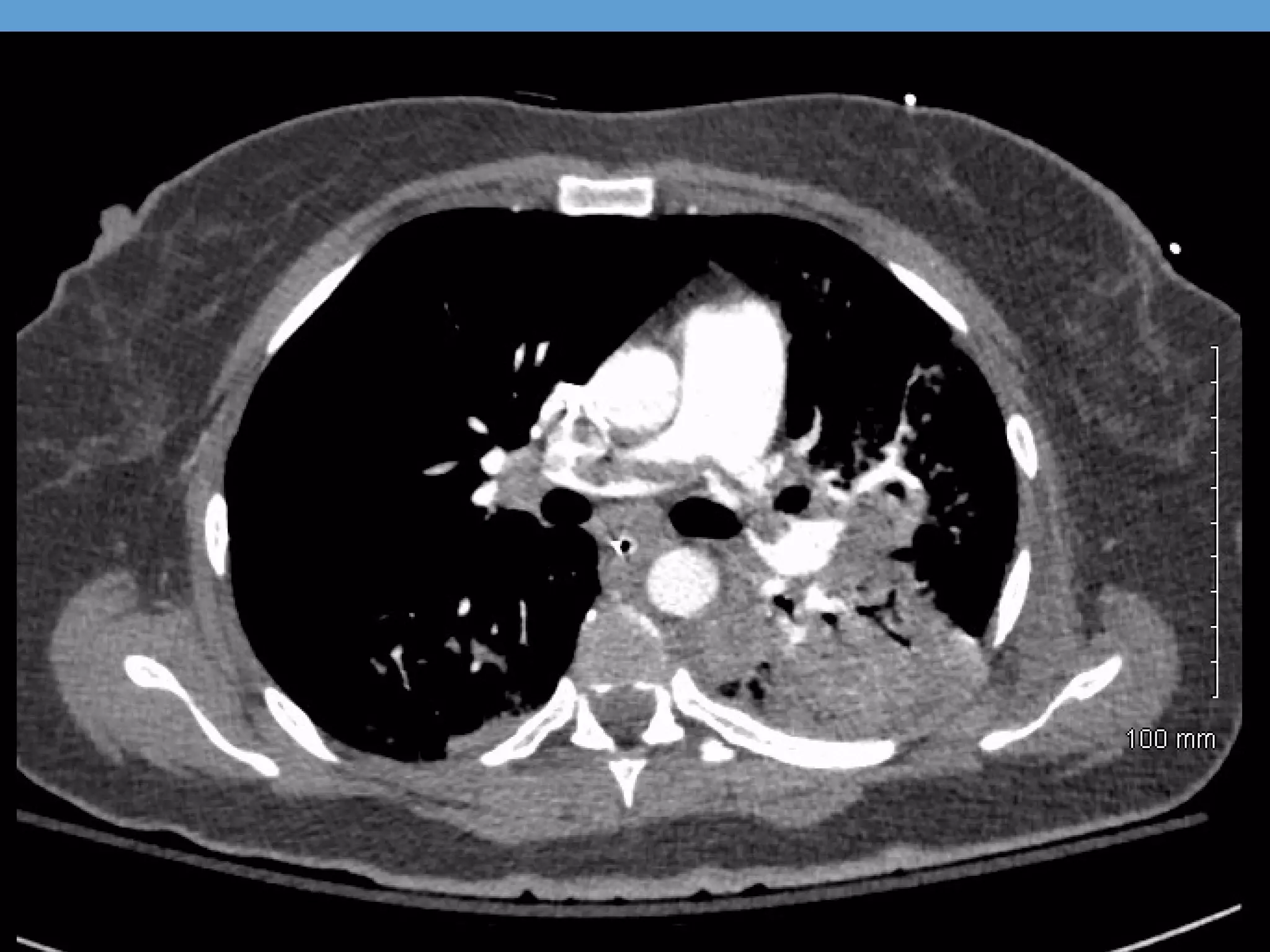
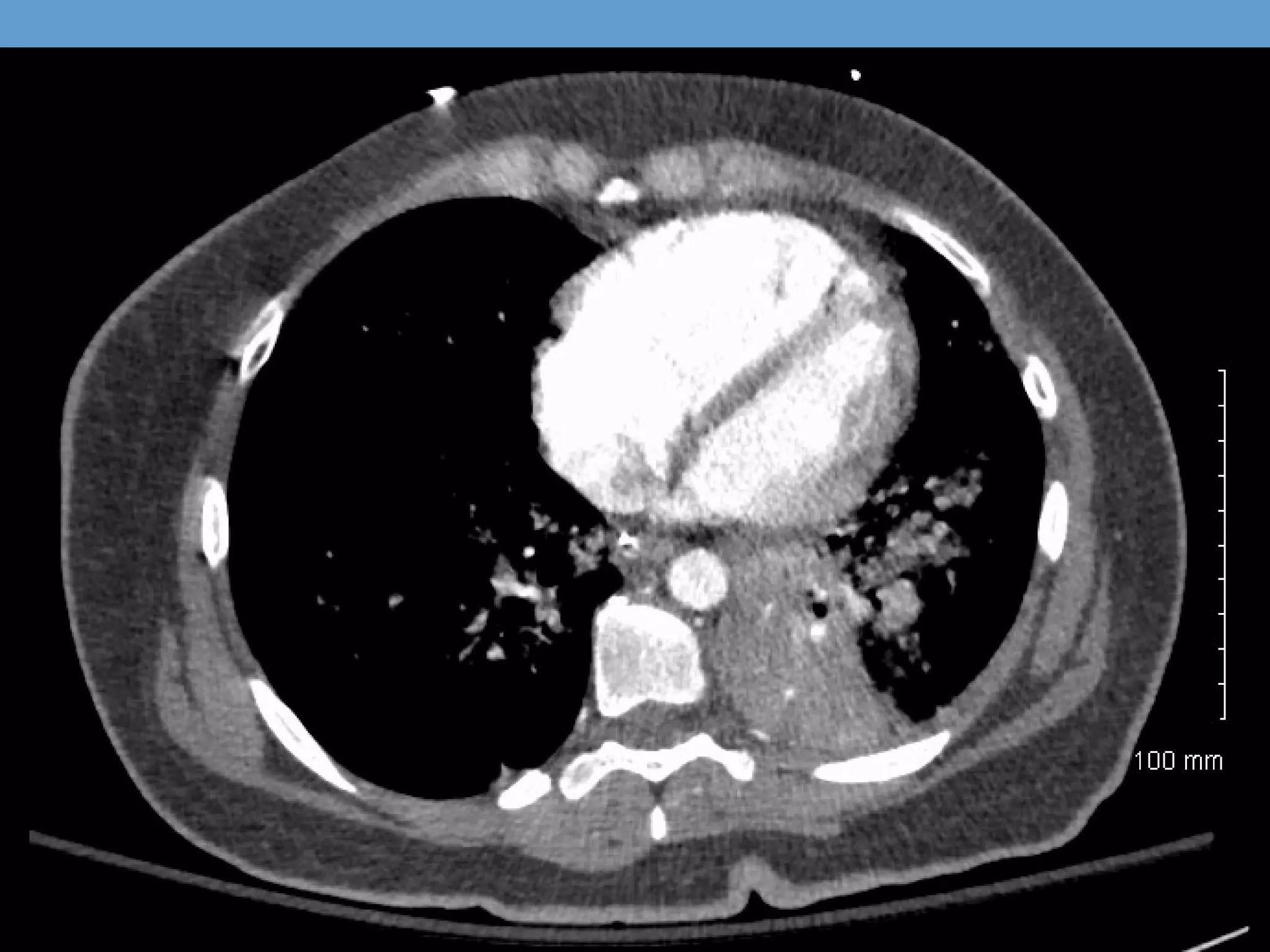
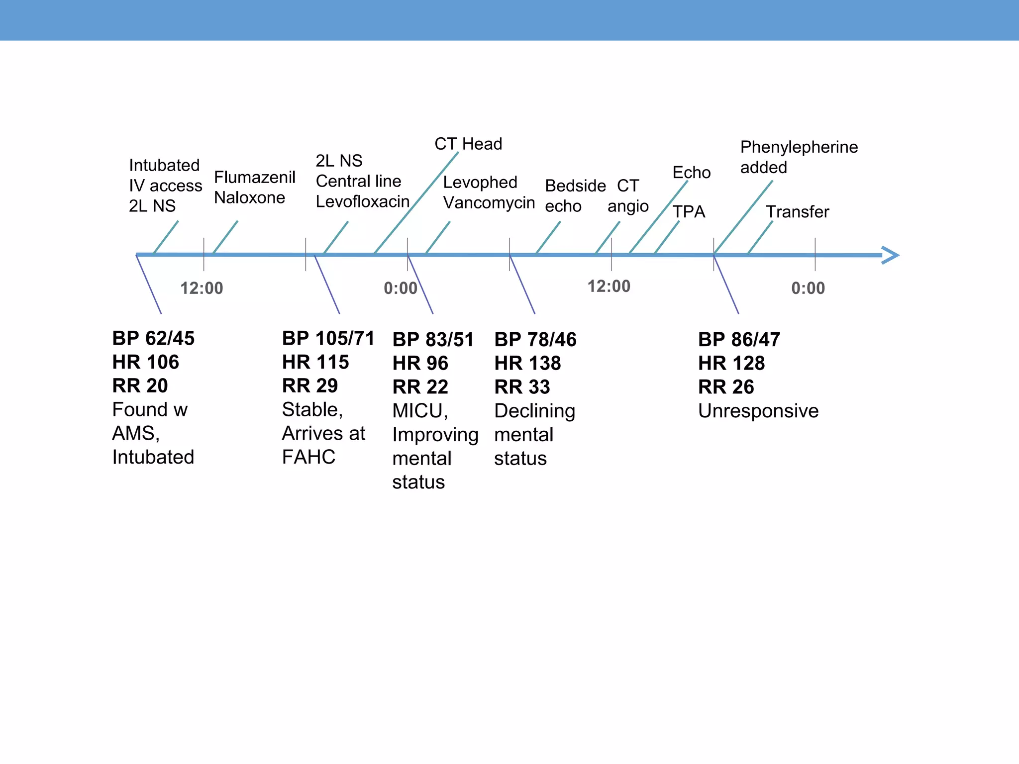
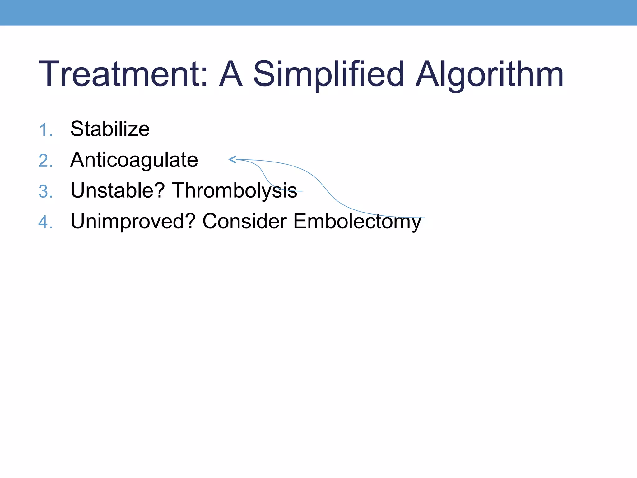
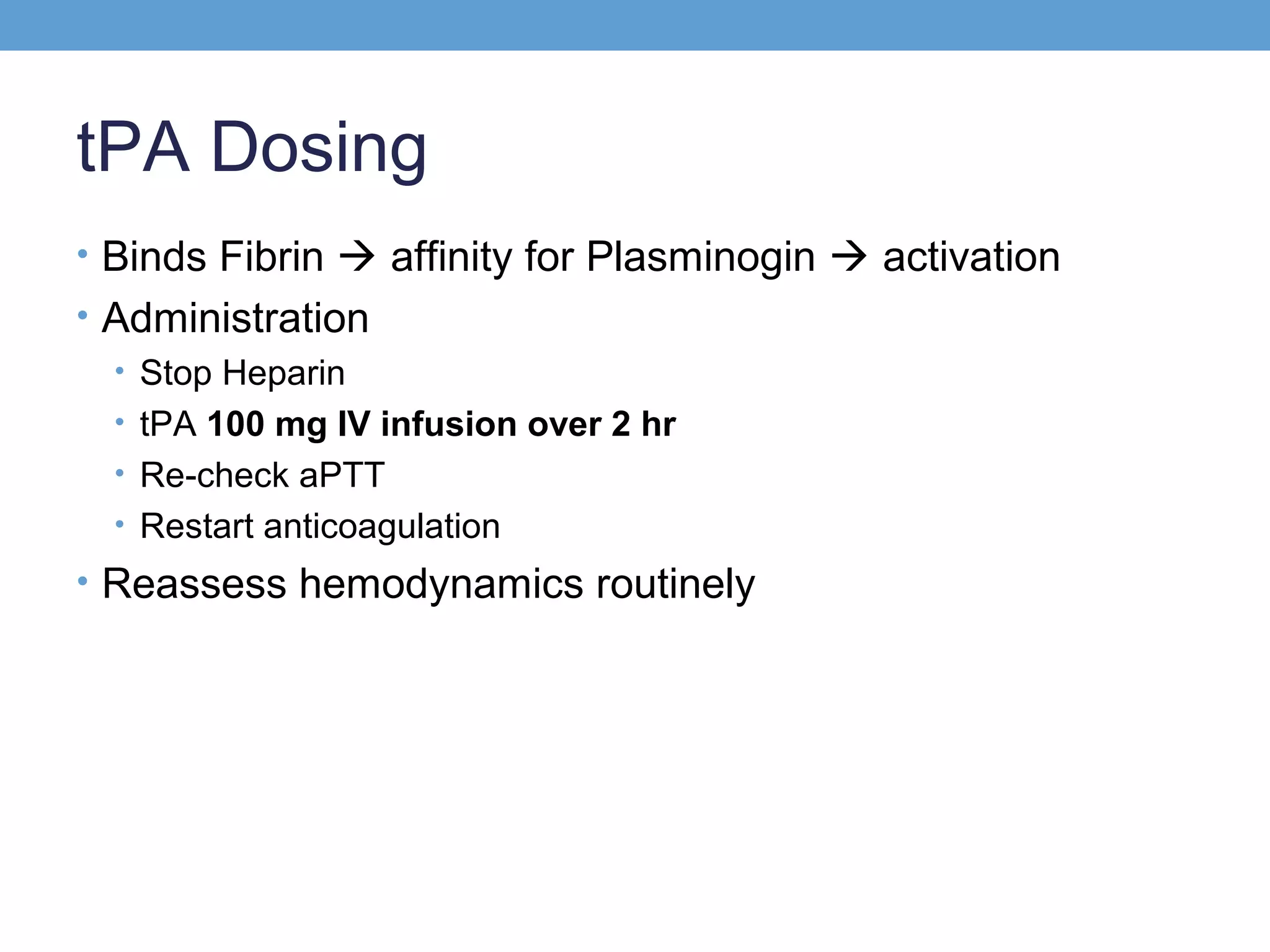
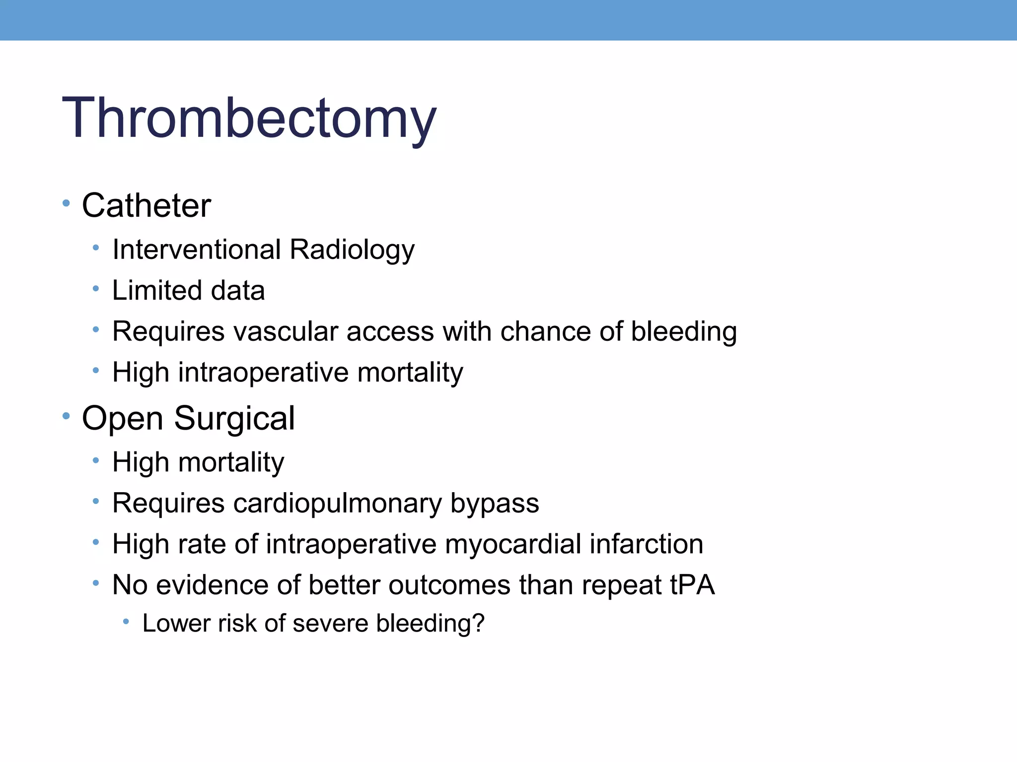
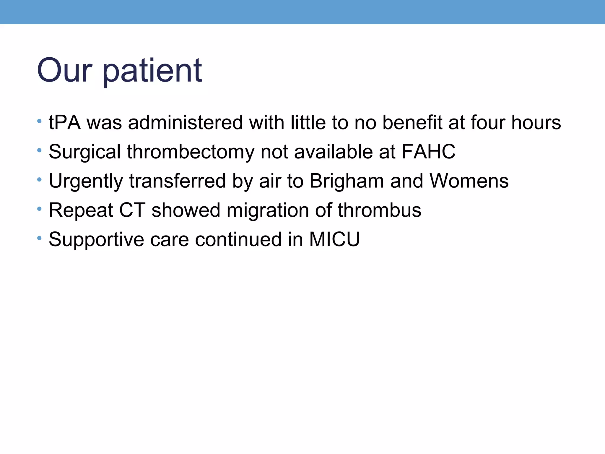
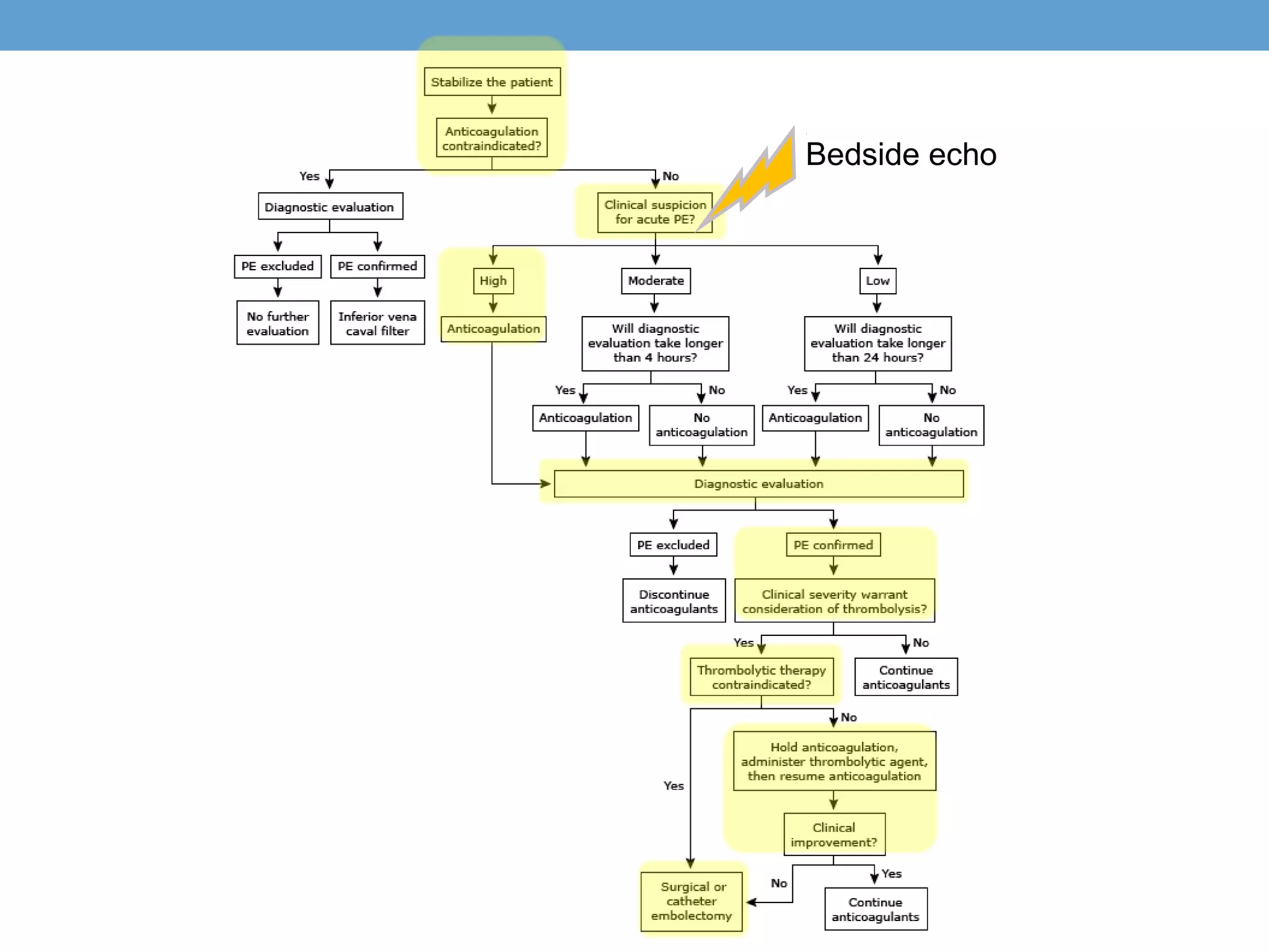
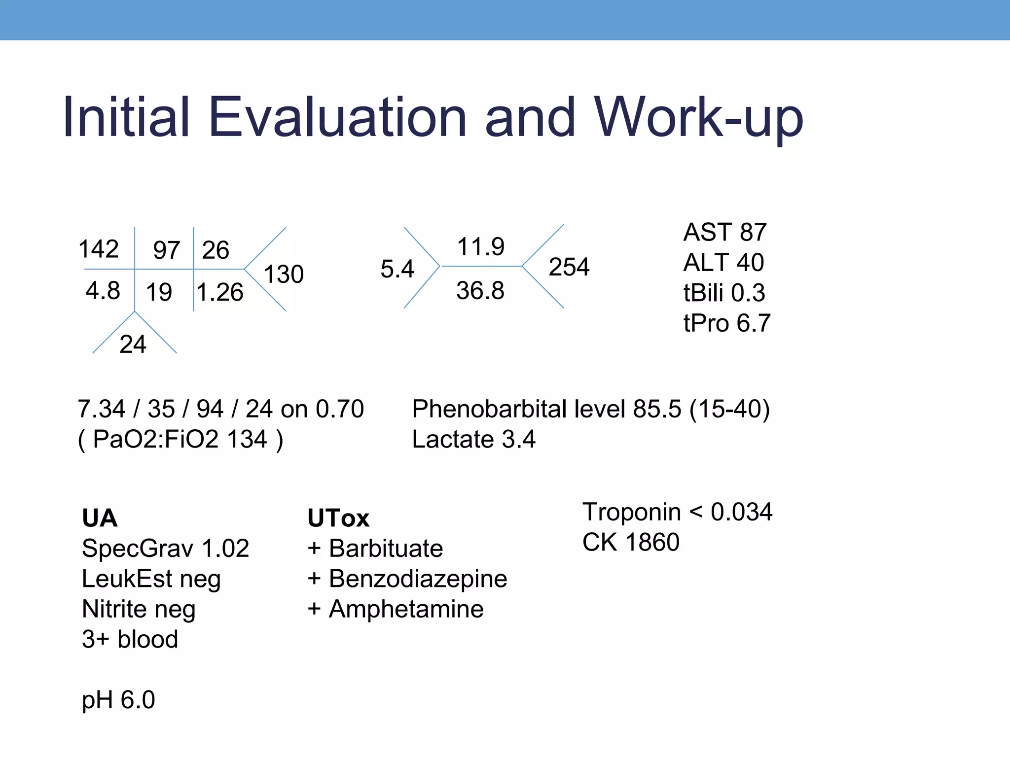
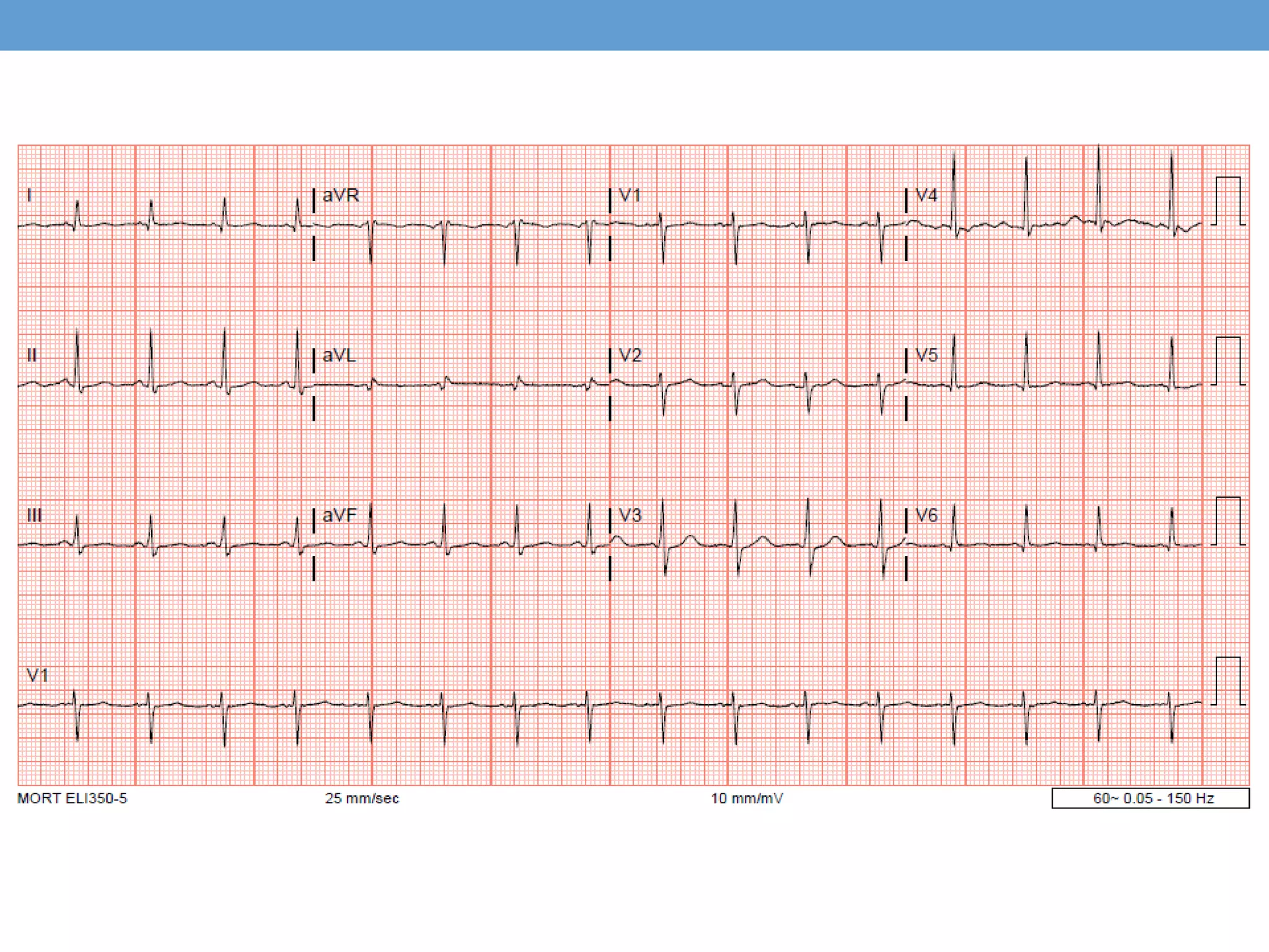
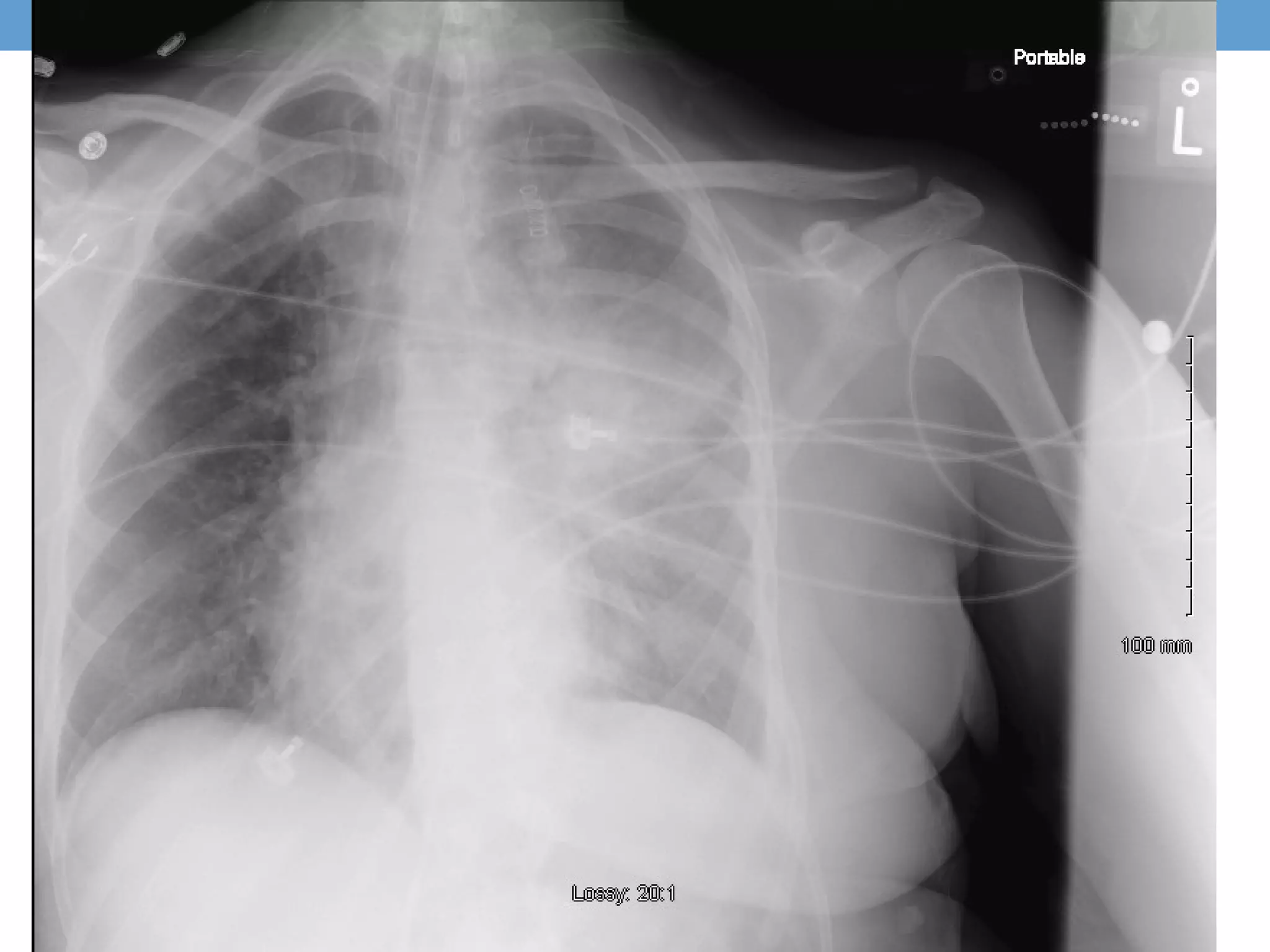
Call Girls Varanasi Just Call 9907093804 Top Class Call Girl Service Available
Pe massive wilfong
1. ALTERED MENTAL
STATUS IN A 36 Y/O F
Jonathan B Wilfong, MD
January 31, 2014
Fletcher Allen Health Care
2. Admission
36 y/o F is transported to FAHC after being found in bed
with altered mental status, responsive only to pain. Per
EMS report, two men, one being her husband, were found
deceased at the scene with drug paraphernalia scattered
close-by. There was no evidence of trauma.
Prior to arrival she was treated with Flumazenil 0.5 mg x2,
Naloxone 2 mg IV x4 at a local hospital. She briefly
regained consciousness following Flumazenil
administration but mental status declined rapidly again.
She arrives intubated, off sedation, and is admitted to the
MICU for management of hypoxemic respiratory failure.
3. History
PMH
•Substance abuse (benzos, barbituates, IV heroin)
•Depression w suicide attempt (overdose, laceration)
•Victim of domestic violence
•History of imprisonment
•Multiple psychiatric (MDD, BPD1, PTSD, ADHD)
•Recent aspiration pneumonia in the setting of recent drug
overdose
FHx
•Unknown
5. Social History
• Born in New York, NY
• Married, Living in Plattsburg
• Married, 3 teenage children in NYC
• Known hx of long-standing illicit and Rx drug use
6. Objective
In the field [AM, Day 1]
•62/45
•106
•20
•79% SpO2 on RA
Following intubation, I/O line x2, 2L NS [18:00, Day 1]
•105/71
•115
•29
•100% SpO2 on 0.70 FiO2
11. Management
• 33.7C , 83/51, 96, 22, GCS 8, ventilated [0:00, Day 2]
• Aspiration in the setting of poly-substance use was
suspected.
• Pulm: Levofloxacin, Vanc, tapering off ventilator
• Cor: IV fluid resuscitation, serial lactate, serial EKG
• RenalL: Foley catheter, monitor UOP
• Neuro: Head CT negative, Serial neuro exams
• GI: NPO
• ID: Sputum, Blood, Urine Cx. Abx as above
• PPx: PPI, SQ Heparin, SCD
12. Day of Hospitalization #2
• 7.38 / 30 / 60 / 18 on 0.55 (PaO2:FiO2 109) [05:00, Day 2]
• Improving mental status, responding to commands
but…
• Systolic pressure continued to drop despite 4L IVf
78/46, 138, 33, 98% on 0.60
• Levophederine gtt started
The patient was declining despite aggressive supportive
care.
13. Day of Hospitalization #2
• Bedside Echocardiogram was performed suggesting RV
ballooning with septal flattening
• A STAT CT PE protocol of chest was obtained
• A STAT echocardiogram was obtained
14.
15.
16. CT PE Protocol
CT CHEST W CONTRAST (PE) PROTOCOL 11/1/2013
1:01 PM
Findings: Opacification of the pulmonary vasculature is
good. There is a saddle embolus with clot extending
predominantly into the lower lobe pulmonary arteries, with
the most significant clot burden in the left lower lung.
Additional regions of emboli are seen in the right middle
lobe There is evidence of right heart strain with a right
ventricle to left ventricular ratio of greater than 1.
17. Echocardiogram
Impressions: Cor pulmonale. Summary: 1. Left ventricle:
The cavity size was below normal. … Systolic function was
hyperdynamic. The estimated ejection fraction was 6570%. Wall motion was normal; there were no regional wall
motion abnormalities. 2. Ventricular septum: The contour
showed diastolic flattening and systolic flattening. 3. Right
ventricle: The cavity size was severely dilated. Systolic
function was severely reduced. 4. Pulmonary arteries:
Pulmonary systolic pressure was moderately increased,
estimated to 50mm Hg.
18. Intubated
IV access Flumazenil
Naloxone
2L NS
2L NS
Central line
Levofloxacin
12:00
BP 62/45
HR 106
RR 20
Found w
AMS,
Intubated
CT Head
Levophed
Bedside CT
Vancomycin echo
angio
12:00
0:00
BP 105/71
HR 115
RR 29
Stable,
Arrives at
FAHC
BP 83/51
HR 96
RR 22
MICU,
Improving
mental
status
BP 78/46
HR 138
RR 33
Declining
mental
status
Echo
Phenylepherine
added
TPA
Transfer
0:00
BP 86/47
HR 128
RR 26
Unresponsive
21. Incidence
• “Obstruction of the pulmonary artery or one of its
branches by material (eg, thrombus, tumor, air, or fat) that
originated elsewhere”
• Massive = sustained hypotension
• 112.3 per 100,000
• Mortality estimated 30% if untreated
• Risk factors
• Immobilization
• Obesity, Heart Disease
• Malignancy, Autoimmune
• Central venous instrumentation, Surgery
• Smoking, Medications
22. Pathophysiology
• 50-80% from below popliteal vein
• Severe acute VQ mismatch
• Death is by R heart failure, acute cardiogenic shock
• Increase in mortality from PE and overall
• RV strain
• Mural thrombus
• DVT
• Associated w increased mortality
• BNP
• Troponin
• Hyponatremia
• Lactate
23. Diagnosis
• Clinical findings nonspecific
• DVT signs (47%)
• Diagnostic studies
• S1Q3T3 on ECG suggestive of cor pulmonale
• Lower extremity US for risk reduction – not diagnosis
• VQ scan 15-86% diagnostic accuracy
• D-Dimer high negative predictive value
• CT angiogram, 53-98% diagnostic accuracy
• Echocardiogram
• Increased RV size
• TR
• Mural thrombus
• McConnell sign (hypokinesis w apical sparing)
25. Treatment: A Simplified Algorithm
1. Stabilize
2. Anticoagulate
3. Unstable? Thrombolysis
4. Unimproved? Consider Embolectomy
26. Treatment of Acute Massive PE
• Correction of hypoxemia
• Hemodynamic Support
• IV fluids, 500-1000 mL
• Pressors: NorEpi/Epi/Dobutamine
• Empiric Anticoagulation within 24hr
• Weigh against risk of acute bleed (recurrent PE 25% > Bleed 3%)
• LMWH as tx in uncomplicated cases
• IV UFH if massive PE, risk of bleed, or considering lysis, thrombectomy
27. Dosing IV UFH
• Bolus 80 mg/kg
• Infusion 18 mg/hr
• Titrate to institution protocol goal aPTT
• If abnormal PTT response
• Follow anti-Xa
• When to consider thrombolysis?
28. Thrombolysis
• Indication:
• persisting SHOCK
• Isolated RV dysfunction..?
• Outcomes:
• Early hemodynamic improvement
• No proven mortality benefit
• (3.5 vs 6.1 percent, relative risk 0.7, 95% CI 0.37-1.31)
• Increased risk of major bleeding
• (9.0 vs 5.7 percent, relative risk 1.63, 95% CI 1.00-2.68)
• Risks for bleeding not well characterized
• Intracranial hemorrhage rate ~3%
29. Contraindications
• Surgery in last 10 days
• Bleeding diathesis
• Intracranial neoplasm
• Severe HTN (>200)
• Ischemic stroke within 2 months
• Past hemorrhagic stroke
• Intracranial trauma/surgery in last 6 months
• Thrombocytopenia (<100k)
31. Thrombectomy
• Catheter
• Interventional Radiology
• Limited data
• Requires vascular access with chance of bleeding
• High intraoperative mortality
• Open Surgical
• High mortality
• Requires cardiopulmonary bypass
• High rate of intraoperative myocardial infarction
• No evidence of better outcomes than repeat tPA
• Lower risk of severe bleeding?
32. Our patient
• tPA was administered with little to no benefit at four hours
• Surgical thrombectomy not available at FAHC
• Urgently transferred by air to Brigham and Womens
• Repeat CT showed migration of thrombus
• Supportive care continued in MICU
34. Take Away
• Think PE on the DDx for cardiogenic shock
• Bedside US can aid in diagnosis
• Urgent anticoagulation with IV UFH
• There are 8 strict contraindications to tPA
• No evidence of improved mortality w tPA
• Surgical thrombectomy as a last resort