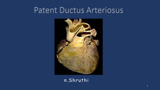
2. Introduction
1. The patent ductus arteriosus (PDA) is the most common cardiac condition in
neonates.
2. Galen was the first to describe a patent ductus arteriosus.
3. The PDA was the first congenital heart lesion that was surgically repaired by
Dr. Robert Gross in 1938.
4. In 1989, Krichenko et al. angiographically classified the PDA.
5. Closure of the PDA in a premature neonate with RDS was first detailed by Dr.
ML Powell in 1963.
2
3. VI Branchial arch arteries:
⢠The proximal part of the sixth right arch
persists as the proximal part of the right
pulmonary artery while the distal section
degenerates;
⢠The sixth left arch gives off the Lt.
pulmonary artery and the distal part
forms the ductus arteriosus;
3
6. Histology
1. Histologically, the walls of the DA are mainly muscular in contrast to the walls of the
adjacent aorta and pulmonary artery, which are fibro-elastic.
2. After birth, the medial smooth muscle fibers contract in response to the exposure to
oxygen-rich air. This leads to constriction of the lumen and functional closure between 24
and 48 h.
3. The second stage of closure involves proliferation of the medial and intimal connective
tissue and smooth muscle atrophy which leads to the formation of ligamentum arteriosum
over the next 3 weeks.
6
7. Term versus Preterm
1. The smooth muscle in the wall of the preterm ductus is less responsive to
high Po2 and therefore less likely to constrict soon after birth. But early
pharmacologic can close PDA and spontaneous closure can also occur.
2. In a term infant with PDA, the wall of the ductus is deficient in both the
mucoid endothelial layer and the muscular media. PDA persisting beyond
the 1st few weeks of life in a term infant rarely closes spontaneously or with
pharmacologic intervention.
7
8. Mechanisms Underlying Postnatal Closure of the DA
1. Initial DA constriction occurs as pulmonary vascular resistance (PVR) falls, systemic vascular resistance
increases, circulating prostaglandin E (PGE) levels decline, and respiration is initiated, triggering a sharp
increase in arterial oxygen tension.
2. Molecular Factors: Postnatal DA closure is facilitated by sharp reductions in dilating factors (nitric oxide,
prostaglandin E2, adenosine, atrial natriuretic peptide, carbon monoxide, and potassium channels) and
increases in intracellular calcium levels.
3. Structural Factors: Rudimentary or absent intimal cushions, less contractile smooth muscle cells, and lack of
vasa vasorum contribute to sustained ductal patency in preterm infants.
4. Hemodynamic Factors: Some investigators have observed an association between thrombocytopenia and
delayed ductal closure in very preterm infants.
5. Infection Factors: Congenital rubella syndrome and Zika virus may contribute to postnatal ductal patency
6. Prenatal teratogens: Tetrahydrocannabinol concentrations secondary to cannabis use among pregnant
women have been associated with a greater incidence of PDAs 8
9. Predisposing factors to PDA
1. Preterm infants
2. Perinatal hypoxia
3. Children born at extreme altitude
4. Familial; 5% among siblings
5. Congenital Rubella
6. Fetal alcohol syndrome
7. Maternal amphetamine and phenytoin use.
8. Genetic factors.
9
11. 1. 0.8 per 1000 live births.
2. 5-10% of CHDs.
3. More in female; 3:1 ratio.
4. The incidence of PDA is inversely related to the gestational age. It remains
open at 4 days of age in 10% of neonates born at 30â37 weeks, 80% of
those between 25 and 28 weeks gestation and 90% born <24 weeks
gestation.
11
12. Basic pathophysiology of Lt. to Rt. shunts
1. Clinical signs are not always apparent at birth
2. They manifest anytime during infancy or early childhood.
3. Mild forms may get worse during pregnancy.
4. increased pulmonary blood flow;
5. Increased 02 saturation in the right side of the heart
6. Physiologic effects:
1. Pulmonary hypertension
2. ventricular strain, dilation, and hypertrophy,
3. CCF
7. Eisenmenger complex 12
13. 1. High pressure gradient from aorta to PA; hence shunt occurs both in diastole and systole
producing continuous murmur
2. Small shunt: No PH and P2 is normal
3. large shunt:
1. Increased blood flow to the lungs.
2. Pulmonary Hypertension .
3. RVH and RAH
4. Volume overload to RA and RV
4. Decreased systemic blood flow and tissue oxygenation.
5. As PH progresses bidirectional shunt develops with differential cyanosis: upper limbs pink and
lower limbs blue
13
14. Pathophysiology
Effects on the Lungs
1. Increased lung water and pulmonary edema
2. Reduction in lung compliance
3. Increased ventilation and oxygenation
requirement, lung injury
4. Chronic Lung Disease
5. Pulmonary hemorrhage
Effect on GI Tract
1. Decreased mesenteric blood flow
2. GI injury
3. Translocation of bacteria
4. Necrotizing enterocolitis (NEC)
5. Local ischemia, spontaneous intestinal
perforation (SIP)
Effects on CNS
1. Alterations in cerebral blood flow,
2. Intraventricular hemorrhage (IVH)
Renal effects:
1. Decreased blood flow to kidney
2. Elevated BUN, creatinine
3. Decreased kidney function, poor urine output
14
15. Clinical Features
1. No symptoms in small ductus
2. Large ductus leads to:
1. Exertional dyspnoea â poor feeding; suck rest cycle; frontal sweating.
2. Poor weight gain
3. Recurrent respiratory infections
4. CHF
1. Tachycardia
2. Tachypnea
3. Presacral edema in infants; pedal edema in older children.
15
16. Signs
1. Bounding and collapsing pulse
1. Related to the high left ventricular stroke volume, which may cause systolic
hypertension.
2. The low diastolic pressure in the systemic circulation as blood runs off from the
aorta into the pulmonary circulation.
3. Wide pulse pressure
2. The apical impulse is down and out
3. A thrill in the suprasternal notch or in the left infraclavicular region.
4. S1 is normal. The second heart sound (S2) is obscured by the murmur.
5. Paradoxical splitting of S2 related to premature closure of the pulmonary valve
16
17. Murmur
1. Gibsonâs machinery murmur, continuous & loudest in the second left
intercostal space
2. In the small infant, it is uncommon to hear the diastolic component of
the murmur
3. Apical diastolic rumble, caused by high flow through mitral valve into the
left ventricle is audible in large PDA
17
18. Natural History And Complications
1. No spontaneous closure of PDA is possible in term infants
2. Recurrent pneumonia
3. CHF
4. Infective endocarditis
5. Aneurismal dilatation of PDA:
1. Rupture
2. Recurrent laryngeal nerve paralysis
3. Left bronchial obstruction
6. Atrial fibrillation that results from chronic and gradually progressive left atrial
enlargement
7. Eisenmenger complex
18
19. CXR
1. Radiographic studies in patients with a
large PDA show a prominent pulmonary
artery with increased pulmonary vascular
markings.
2. Cardiac size depends on the degree of
left-to-right shunting; it may be normal or
moderately to greatly enlarged.
3. The chambers involved are the left atrium
and left ventricle.
4. The aortic knob may be normal or
prominent.
19
20. ECG
1. With a small shunt the ECG is normal.
2. Left ventricular hypertrophy of the volume overload type, with deep Q
waves and increased R wave voltage in the left precordial leads, is noted
with increasing shunt size with left ventricular volume overload.
3. Right ventricular hypertrophy is seen with pulmonary hypertension.
20
21. Echocardiogram
1. On echocardiogram the cardiac chambers will be normal in size if the ductus is small.
2. With large shunts, left atrial and LV dimensions are increased.
3. The ductus can easily be visualized directly and its size estimated.
4. Color and pulsed Doppler examinations demonstrate systolic or diastolic (or both)
retrograde turbulent flow in the pulmonary artery, and aortic retrograde flow in
diastole in the presence of a large shunt.
5. Echocardiography can also be useful as a predictive measure of whether a PDA is
hemodynamically significant
21
22. Color Doppler evaluation in a
parasternal short axis view shows
flow (arrow) from the aorta into
the main pulmonary artery.
22
23. ECHO: Ductal size and shunt direction evaluation
a. Color flow of PDA.
b. Pulsatile pattern with left-to-
right low-velocity flow with wide
differential between systole and
diastole.
c. Restrictive pattern with higher
velocity in both systole and
diastole.
d. Bidirectional pattern with right-
to-Left ductal flow during systole.
23
24. Echocardiographic examination of left atrial and left ventricular enlargement and quantification of ductal
impact on cardiac performance.
a. Dilated left atrium (A) and ventricle (V).
b. M-mode measurement, demonstrating a dilated left
atrium indexed to the aortic diameter.
c. Elevated and pulsatile pulmonary venous flow,
demonstrating high pulmonary venous diastolic flow.
d. Shortened isovolumetric relaxation time.
e. Transmitral flow, demonstrating an early/atrial flow
ratio of 1.
f. Reversal of diastolic flow in the postductal
descending thoracic aorta. IVRT indicates
isovolumetric relaxation time;
LA:Ao, left atrial diameter to aortic root diameter ratio;
MPA, main pulmonary artery;
MV E/A, mitral valve early/atrial flow ratio; and
PV D, pulmonary vein diastolic velocity.
24
a c e
b d f
25. 2D Echocardiographic markers of hemodynamically significant PDA (Adapted from
Boradhouse KM, Price AN, Durighel G, et al.)
Measurement Hemodynamically significant.
PDA size >1.5 mm
LAâAorta ratio >1.5 mm
IVRT ( isovolumic relaxation time) <50 ms
LA size Increased âĽ2 SD
LV size Increased âĽ2 SD
LV output >1.5 Ă RV output
Mitral E wave >45 cm/sec
PV D-wave >30 cm/sec
Aortic diastolic flow Reversed
25
26. Cardiac catheterization
1. Cardiac catheterization will demonstrate either normal or increased pressure in the
right ventricle and pulmonary artery, depending on the size of the ductus.
2. The presence of oxygenated blood shunting into the pulmonary artery confirms the
left-to-right shunt.
3. The catheter may pass from the pulmonary artery through the ductus into the
descending aorta. It gives a specific appearance âHair pinâ appearance.
4. Injection of contrast medium into the ascending aorta shows opacification of the
pulmonary artery from the aorta and identifies the ductus.
26
28. PH leads to Eisenmenger syndrome which sets in as follows:
1. Cardiomegaly decreases
2. Pulmonary vascularity increases
3. More RVH and less LVH
4. Continuous murmur and diastolic rumble disappear
5. S2 becomes loud and single
6. Cyanosis in lower limbs
28
29. No Lesion Differentiating points
1 Coronary AV fistula Murmur at Rt. sternal border
2 Systemic AVF Bounding pulse but no murmur over precardium
3 Pulmonary AVF Murmur over back with cyanosis and clubbing
4 Venous hum Rt. infra and supra clavicular; disappears in supine
position
5 VSD with AR Murmur in lower left sternum
6 persistent truncus arteriosus Cyanosis; BVH; murmur in Rt intercostal spaces
7 Pulmonary artery stenosis Continuous murmur all over chest; RVH; in Rubella and
Williams syndrome
8 Total anomalous pul. venous
return
Venous hum over Rt. sternal border; cyanosis; RVH
29
30. PDA related morbidities in neonates
Epidemiological data that reveals an association between a PDA and multiple morbidities including:
1. Accentuated respiratory distress syndrome (RDS),
2. Pulmonary hemorrhage,
3. Bronchopulmonary dysplasia (BPD),
4. Necrotizing enterocolitis (NEC),
5. Renal insufficiency,
6. Intraventricular hemorrhage (IVH),
7. Cardiac dysfunction.
8. Retinopathy of prematurity (ROP)
30
31. PDA management in Preterm Neonate
1. There is no consensus on clinical or sonographic criteria that define the need for PDA closure.
2. Rationale to consider early treatment:
a. A ductal diameter > 1.5 mm during the first hours of life,
b. La-to-aortic root ratio > 1.4,
c. Lv enlargement,
d. Increased mean and diastolic pa flow velocities,
e. Reversed mitral e/a ratio
f. Retrograde diastolic flow in the
g. Descending aorta and low-antegrade
h. Retrograde diastolic flow in systemic arteries (eg, anterior cerebral artery and renal and mesenteric
arteries) indicate systemic hypoperfusion and ductal steal
31
32. PRETERM
Pharmacologic Ductus Closure - categories
1. Prophylactic, early: < 24 hours,
2. Symptomatic treatment: between 2 and 6 days after birth.
3. Third treatment category: asymptomatic infants,72 hours after birth
4. Fourth treatment category: late symptomatic treatment after watchful
waiting.
32
33. 1. Targeted prophylaxis in at-risk infants (6â24 h after birth):
a. Indomethacin 3 x 0.1 mg/kg per dose IV every 12 hrs (single dose prophylaxis may be
considered)
b. Do not start treatment within the first 6 h of life. It is recommended not to use ibuprofen (IV) in
the first 24 h of life (increased risks for renal failure, gastrointestinal hemorrhage, and possibly
PPHN)
c. Pros: Prevention of IVH (prophylaxis); risk reduction of pulmonary hemorrhage; association with
beneficial neurodevelopmental outcome in boys
d. Cons: Unnecessary treatment of many infants without an hsPDA
2. Early targeted treatment of infants with PDA (< 6 d after birth):
a. Indomethacina 1 x 0.2 mg/kg per dose IV, followed by 2 x 0.1 mg/kg per dose every 12 h
b. Ibuprofen 10 mg/kg per dose PO or IV, followed by 5 mg/kg at 24 and 48 h of treatment start
33
34. 3. Treatment in symptomatic infants with hsPDA (> 6 d after birth)
1. Ibuprofenc 10 mg/kg per dose PO or IV, followed by 5 mg/kg at 24 and 48 h of
treatment start
2. Higher doses might be considered.
3. Treatment only in infants with hsPDA
4. No evidence for beneficial longterm outcome if administered late (.6â14 d); might still
be associated with adverse outcome (eg, BPD) due to a longer duration of a significant
shunt
4. Rescue treatment:
a. Paracetamol 15 mg/kg per dose PO or IV every 6 h for 3â7 d
b. Might be attempted in selected infants after failed standard COX inhibitor treatment;
can also be applied earlier if contraindications for standard COX inhibitor use are
present
c. Might prevent the use of more invasive measures, such as ligation or catheter
intervention; no known renal or gastrointestinal side effects 34
35. Trials on drug based interventions
Indomethacin:
1. Thirty-nine randomized trials (23 as prophylaxis, 4 for early treatment of an asymptomatic PDA, and 12
for treatment of a symptomatic PDA) demonstrate consistent efficacy for achievement of ductal closure.
2. Randomized trials have not identified benefits with respect to most other outcomes (including
mortality, BPD, necrotizing enterocolitis, and neurodevelopment).
3. Indomethacin treatment is often followed by oliguria and increases in serum creatinine levels.
Ibuprofen:
1. 15 randomized controlled trials (6 as prophylaxis and 9 as treatment64). Collectively, these trials
demonstrated efficacy for PDA closure comparable to that of indomethacin
2. Oliguria and necrotizing enterocolitis are less likely in infants treated with ibuprofen.
Acetaminophen:
A recent systematic review of studies comparing oral paracetamol with intravenous ibuprofen (559 infants),
intravenous indomethacin (277 infants), and a placebo (80 infants) identified moderate-quality evidence
suggesting that paracetamol is as effective as ibuprofen, with less gastrointestinal bleeding and lower
serum creatinine levels.50 35
36. Catheter-Based Interventional Closure
1. Transcatheter PDA closure is routinely performed in the cardiac
catheterization laboratory.
2. Small PDAs are generally closed with intravascular coils.
3. Moderate to large PDAs may be closed with an umbrella-like device or with
a catheter-introduced sac into which several coils are released.
36
37. Gianturco coil occlusion of PDA
⢠Example of Gianturco coil occlusion of PDA.
⢠A, Views of a Gianturco coil in its stretched out
configuration (top) and in its natural coiled
configuration (bottom).
⢠Note the attached Dacron fibers, which promote
thrombosis, along its length.
⢠B through D, Lateral angiograms demonstrating
closure of a PDA with a single 0.038-in diameter
Gianturco coil.
37
38. Nit-Occlud PDA occlusion device
⢠Example of PDA closure with a Nit-Occlud
PDA occlusion device.
⢠A, Image of a Nit-Occlud coil with its
biconical configuration. Note the reversed
winding on the proximal end.
⢠B through D, Lateral angiograms
demonstrating closure of a PDA with a
single Nit-Occlud coil.
38
39. Amplatzer duct occluder device
⢠Example of PDA occlusion with
⢠an Amplatzer duct occluder device.
⢠A, Image of an Amplatzer duct occlude
device.
⢠B through D, Lateral angiograms
demonstrating closure of a PDA with an
Amplatzer duct occluder device.
39
40. 1. Indomethicin for preterm (not for term babies) : 0.2 mg IV infusion 12 hrly 3 doses; or
ibuprofen 10 mg/kg iv for 3 days;
2. SBE prophylaxis
3. CCF management
4. Non surgical closure: Amplatzer PDA device
5. Surgical: anytime when shunt becomes hemodynamically significant: thoracotomy or
thoracoscopy; clip ligation; or ligation and division.
40
41. Surgical closure
1. Surgical closure of a PDA can be accomplished by a standard left thoracotomy or using
thoracoscopic minimally invasive techniques.
2. Ligation and division through left posterolateral thoracotomy without cardiopulmonary bypass is
the standard procedure. The technique of video-assisted thoracoscopic surgery (VATS) clip ligation
has become the standard of care for surgical management of ductus with adequate length (to allow
safe ligation), which is performed through three small ports in the fourth intercostal space.
3. The case fatality rate with interventional or surgical treatment is considerably less than 1%; thus
closure of the ductus is indicated even in asymptomatic patients.
4. Pulmonary hypertension is not a contraindication to surgery at any age if it can be demonstrated
at cardiac catheterization that the shunt flow is still predominantly left to right and that severe
pulmonary vascular disease is not present.
41
43. Conservative Management in preterm PDA
1. In conservative management, health care providers avoid definitive (transcatheter or
surgical) closure, while awaiting the possibility of spontaneous closure.
2. In conservative management, several strategies to manage consequences of the ductus are
often used, including fluid restriction, diuretics, systemic afterload reduction, increases in
positive airway pressures, or maintenance of higher hematocrits.
3. Questions on the safety and effectiveness of conservative management versus alternative
treatments (definitive closure) remain unanswered.
4. The PDA TOLERATE trial provides evidence that conservative management may be more
beneficial compared to active treatment in select settings
43
44. References
1. CirculationVolume 114, Issue 17, 24 October 2006; Pages 1873-1882
https://doi.org/10.1161/CIRCULATIONAHA.105.592063 CONGENITAL HEART DISEASE FOR THE ADULT
CARDIOLOGIST Patent Ductus Arteriosus
2. J Am Heart Assoc. 2022;11:e025784. DOI 10.1161/JAHA.122.025784 Patent Ductus Arteriosus: A
Contemporary Perspective for the Pediatric and Adult Cardiac Care Provider Carl H. Backes et.al.
3. Frontiers in Pediatrics: Management of Patent Ductus Arteriosus in Premature Infants in 2020 Sarah
Parkerson1, Ranjit Philip2, Ajay Talati 3 and Shyam Sathanandam2*
4. PEDIATRICS Volume 146, number 5, November 2020:e20201209 Patent Ductus Arteriosus of the Preterm
Infant ; Shannon E.G. Hamrick, MD,a,b Hannes Sallmon, MD,c Allison T. Rose, MD, Diego Porras, MD, Elaine
L. Shelton, PhD, Jeff Reese, MD, Georg Hansmann, MD, PhD.
5. Nelson Text Book of Pediatrics 21 Edition
44