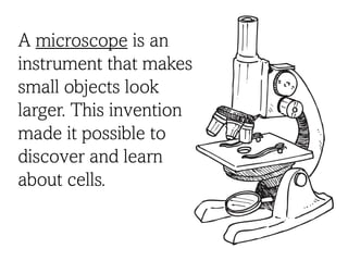
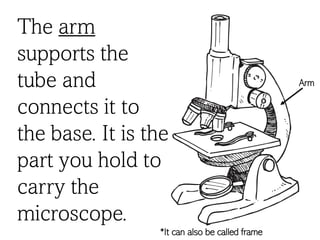
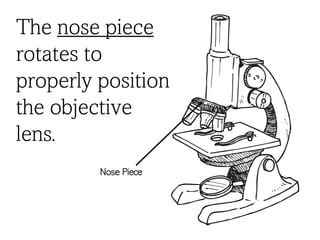
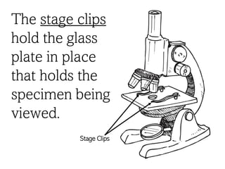
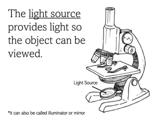
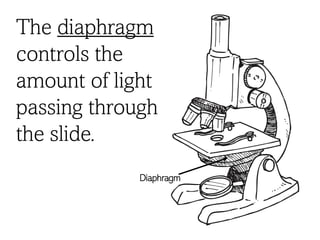
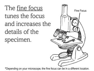
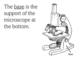
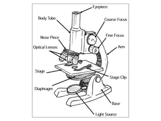
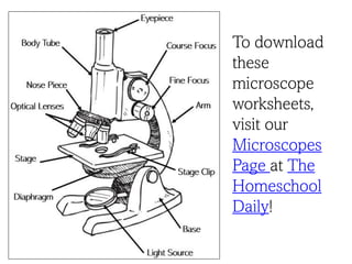
Parts of a
Microscope
Created by Marie @ thehomeschooldaily.com
A microscope is an
instrument that makes
small objects look
larger. This invention
made it possible to
discover and learn
about cells.
The eyepiece is
the lens you
look through to
see the
specimen. The
lens is usually a
10x or 15x lens.
Eyepiece
The body tube
connects the
eyepiece to the
objective lens.
Body Tube
The arm
supports the
tube and
connects it to
the base. It is the
part you hold to
carry the
microscope.
Arm
*It can also be called frame
The nose piece
rotates to
properly position
the objective
lens.
Nose Piece
The objective lens
magnifies the
image of the
specimen. A
compound
microscope has 3-
5 objective lens
that range in
power from 4x to
100x.
Objective lens
The stage holds
the specimen to
be viewed.
Stage
The stage clips
hold the glass
plate in place
that holds the
specimen being
viewed.
Stage Clips
The light source
provides light so
the object can be
viewed.
Light Source
*It can also be called illuminator or mirror
The diaphragm
controls the
amount of light
passing through
the slide.
Diaphragm
The course focus
brings the
specimen into
general view.
Course Focus
*Depending on your microscope, the course focus can be in a different location.
The fine focus
tunes the focus
and increases the
details of the
specimen.
Fine Focus
*Depending on your microscope, the fine focus can be in a different location.
The base is the
support of the
microscope at
the bottom.
Base
To download
these
microscope
worksheets,
visit our
Microscopes
Page at The
Homeschool
Daily!
Terms of Use: This document is provided to you for your own personal use. You agree that you will
not copy, reproduce, alter, modify, create derivative works, or publicly display contents as your
own. No Redistribution. You may not reproduce, repackage, or redistribute the contents of these
downloads, in whole or in part, for any reason. This includes “giving” someone your copy that you
are no longer using or hosting them on drop box or Facebook files. Please refer others to
www.thehomeschooldaily.com to download their own copy.
YOU MAY:
• Save the files on your computer and print off
copies for your family or classroom whenever
you would like.
• Link directly to my blog to share my files with
others.
• Post to your blog using pictures of your child
using my curriculum, as long as proper credit is
given to www.thehomeschooldaily.com
YOU MAY NOT:
• Host or store my files on your own or other
sites (this includes drop box, the cloud, and any
other site off your personal computer)
• Alter or Sell files to make a profit. All files are
for personal/classroom use only.
• All downloads are copyright protected. Not to
be distributed, transferred, or shared in any
form.
Hi! Thank you for your download. I’m so glad you were able to find
a school tool you can use. Please feel free to use this activity for
your own personal use or classroom. Hope it works out great!
Marie

Parts of a Compound Microscope

  • 1.
    Parts of a Microscope Createdby Marie @ thehomeschooldaily.com
  • 2.
    A microscope isan instrument that makes small objects look larger. This invention made it possible to discover and learn about cells.
  • 3.
    The eyepiece is thelens you look through to see the specimen. The lens is usually a 10x or 15x lens. Eyepiece
  • 4.
    The body tube connectsthe eyepiece to the objective lens. Body Tube
  • 5.
    The arm supports the tubeand connects it to the base. It is the part you hold to carry the microscope. Arm *It can also be called frame
  • 6.
    The nose piece rotatesto properly position the objective lens. Nose Piece
  • 7.
    The objective lens magnifiesthe image of the specimen. A compound microscope has 3- 5 objective lens that range in power from 4x to 100x. Objective lens
  • 8.
    The stage holds thespecimen to be viewed. Stage
  • 9.
    The stage clips holdthe glass plate in place that holds the specimen being viewed. Stage Clips
  • 10.
    The light source provideslight so the object can be viewed. Light Source *It can also be called illuminator or mirror
  • 11.
    The diaphragm controls the amountof light passing through the slide. Diaphragm
  • 12.
    The course focus bringsthe specimen into general view. Course Focus *Depending on your microscope, the course focus can be in a different location.
  • 13.
    The fine focus tunesthe focus and increases the details of the specimen. Fine Focus *Depending on your microscope, the fine focus can be in a different location.
  • 14.
    The base isthe support of the microscope at the bottom. Base
  • 16.
  • 17.
    Terms of Use:This document is provided to you for your own personal use. You agree that you will not copy, reproduce, alter, modify, create derivative works, or publicly display contents as your own. No Redistribution. You may not reproduce, repackage, or redistribute the contents of these downloads, in whole or in part, for any reason. This includes “giving” someone your copy that you are no longer using or hosting them on drop box or Facebook files. Please refer others to www.thehomeschooldaily.com to download their own copy. YOU MAY: • Save the files on your computer and print off copies for your family or classroom whenever you would like. • Link directly to my blog to share my files with others. • Post to your blog using pictures of your child using my curriculum, as long as proper credit is given to www.thehomeschooldaily.com YOU MAY NOT: • Host or store my files on your own or other sites (this includes drop box, the cloud, and any other site off your personal computer) • Alter or Sell files to make a profit. All files are for personal/classroom use only. • All downloads are copyright protected. Not to be distributed, transferred, or shared in any form. Hi! Thank you for your download. I’m so glad you were able to find a school tool you can use. Please feel free to use this activity for your own personal use or classroom. Hope it works out great! Marie