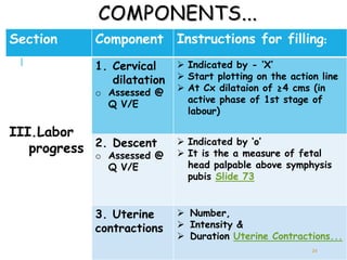
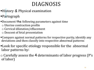
PARTOGRAPH &
ABNORMAL
LABOUR
PATTERNS
DEJENE A. (MD,
OBESTETRICIAN &
GYNECOLOGIST)
1
OUTLINE OF PRESNTATION
Introduction
Methods of labor progress documentation &
monitoring
Advantages of labour monitoring using a Partograph
History, components and of steps in documenting
findings on the partograph
Indicators of labor progress on the partograph
 Sample partograph
2
OUTLINE...
Definition of abnormal labor (dystocia)
Summary of normal labor
Friedman’s normal labor pattern curve
Incidence and etiologies(risk factors) of abnormal
labor
Classifications & diagnosis of abnormal labor
Management options of abnormal labor patterns
3
INTRODUCTION
 Each year >1/2 a million MD occur world wide
~ 99% of these deaths  developing countries
 Significant proportion of these deaths follow
prolonged labour (PL)
 PL occurs mainly due to CPD & it results in:
1. OL
2. Uterine rupture
3. Obstetrics fistula &
4. Less directly in:
a. PPH &
b. Neonatal infection
4
INTRODUCTION...
OL is one of the 5 major cause of MD in developing c.
5
RECORDING OF LABOR EVALUATIONS & IT’S
PROGRESS :
 Helps in early detection of abnormal labor &
prevention of prolonged labour:
► ↓Maternal & PN M & M.
 Two methods:
1. Chart documentation
Findings from the evaluation of the patient are periodically documented on her chart
2. Partographic Monitoring of labor
Documenting labor progress, maternal & fetal status on the partograph
6
RECORDING OF LABOR EVALUATIONS &
PROGRESS...
It is the graphic recording of the progress of
labour and the condition of the mother and
the fetus
It serves as an “early warning system” and
assists in early decision to:
Transfer,
Augmentation &
Termination of labour
7
ADVANTAGES OF PARTOGRAPH
I. Pictorial display of events of labor, thus
1. Clarifies recordings
2. Avoids lengthy written notes
3. Facilitates recognition of any omissions
4. Saves time → Companionship
II. Considerable educational value
All interrelated variables of labor can be seen on a single paper
III. Low cost, feasible
IV. Easy documentation of findings
V. Quick evaluation of findings
8
ADVANTAGES OF PARTOGRAPH...
Easy handing over of many laboring mothers
Can be easily understood by midlevel health
workers ( clear & easy indicators for referral)
Suitable for research purposes
Clear landmarks to assess when labor progress is
delayed( alert and action lines)
Prevention of prolonged labor
Avoids unnecessary use of augmentation
Improves out come of labor →↑Credibility of
formal health sector
9
ADVANTAGE OF PARTOGRAPH:
EVIDENCE/WHO TRIAL
WHO multicenter study, >35,000 women, 1990
Outcomes
Before
Implementation
After
Implementation
p
Labor > 18 hrs 6.4% 3.4% 0.002
Labor augmented 20.7% 9.1% 0.023
Postpartum
sepsis
0.70% 0.21% 0.028
Spontaneous
cephalic
83.9% 86.3% < 0.001
Emergency C/S 9.9% 8.7% 0.628
Forceps 3.4% 2.5% 0.005
Intrapartum still
births
0.50% 0.30% 0.024
10
HISTORY OF PARTOGRAPH
 In 1954 E. A. Freidman, following a study in a large
number of women in USA described a normal Cx dilation
 First to show plotting Cx dilation Vs time
 His work has been a foundation on which others built...
 He divided labour functionally into:
1. Early (latent phase)
Extends over 8-10hrs
Up to 3cm Cx dilation
2. Active phase
Characterized by acceleration from about 3-10cm at the end of which deceleration occurs
11
HISTORY OF PARTOGRAPH...
12
HISTORY OF PARTOGRAPH...
The WHO partograph (1987)
Safe motherhood conference in 1987  “A call to action”

The health workers involved in the care of mothers &
children take positive action to reduce M M & M.

“All pregnant women in labor are managed by:
o Appropriately trained personnel using practical & relevant technology”

 One of which is the partograph
13
14
THE WHO PARTOGRAPH...
 Principles:
1. The active phase commences at 3 cm cx dilation
2. The latent phase should not last >8 hrs
3. During active phase, the rate of Cx dilation
shouldn't be <1cm/hr
4. Vx examination
Infrequently as compatible with safe practice (Q 4 hrs is recommended)
5. Health personnel managing labor may have difficulty
in constructing alert & action line ►pre-drawn lines
15
THE WHO PARTOGRAPH...
 It has been modified to make it
simpler & easier to use(2001)
 The latent phase has been removed &
plotting begins in the active phase
when the cx is 4 cm dilated
16
MODIFIED WHO PARTOGRAPH
17
18
I II
USE OF THE PARTOGRAPH
Partograph is used for the assessment of:
Fetal well being
Maternal well being
Progress of labor
USING THE PARTOGRAPH
19
PARTS OF WHO PARTOGRAPH
 It has four Parts:
1. Patient information
2. Fetal condition
3. Progress of labour
4. Maternal condition
20
COMPONENTS OF PARTOGRAPH
Section Component Instructions for
filling:
I. Patient
informations
o Identification  Name
 Hospital number
 Date & time of
admission
o Reproductive
Performance
 Gravidity
 Parity
o Membrane
condition
 Time of rupture
of membranes
21
COMPONENTS...
Section Component Instructions for filling:
II.Fetal
1. FHR
o Count Q 30’
Indicated with a dot ()
2. Liquor
o Color of AF at
Q V/E
I- Intact membranes;
C- clear liquor;
M- meconium stained
B-Blood stained
3. Molding
o Degree of
molding at Q
V/E
0,
+1(apposed),
+2( reducible overlap),
+3 ( irreducible overlap)
22
LABOUR PROGRESS…
Alert line:
A line starts @ 4 cm of cervical dilatation to the point
of expected full dilatation @ the rate of 1 cm/hour.
Action line:
Parallel & 4 hrs to the right of the alert line.
Hours:
Refers to the time elapsed since onset of active phase
of labour (observed or extrapolated).
Time:
Record actual time.
23
COMPONENTS...
Section Component Instructions for filling:
III.Labor
progress
1. Cervical
dilatation
o Assessed @
Q V/E
 Indicated by - ‘X’
 Start plotting on the action line
 At Cx dilataion of ≥4 cms (in
active phase of 1st stage of
labour)
2. Descent
o Assessed @
Q V/E
 Indicated by ‘o’
 It is the a measure of fetal
head palpable above symphysis
pubis Slide 73
3. Uterine
contractions
 Number,
 Intensity &
 Duration Uterine Contractions...
24
UTERINE
CONTRACTIONS...
Contractions:
Chart Q 1/2 hrs;
Palpate the number of contractions in 10’ & their duration in seconds:
o < 20’’; mark with
o B/n 20 & 40’’; mark with
o > 40’’; mark with
25
COMPONENTS...
Section Component Instructions for filling:
IV.Mat.
Cond.
1. Vital signs  BP indicate by – “” & record
Q 4 hrs
 PR indicate by – “” & record Q
30’
 Tempratre in °C& record Q 2
hrs
2. Urine output  Protein, acetone & volume
 Record Q time urine is passed.
3. Medications  Oxytocin:
Amount per volume IV
fluids in dpm Q30’
 Drugs given: Any additional
drugs given. 26
STEPS IN FILLING THE
PARTOGRAPH
STEP DESCRIPTION
1 Cervical dilatation-
@ dilatation of ≥ 4 cms,
Start plotting on the action line
Mark with “X”
2 Time
On the same vertical line as the cervical dilatation
3 Descent- filled on the same vertical line as the cervical
dilatation
5th of fetal head palpable above the symphysis pubis
Mark with “O”
4 Uterine contractions-
 Filled by covering the number of vertical squres corresponding to
the number of contractions; 27
STEPS...
Step Description
5 Fetal condition
On the same vertical line as the cervical dilatation
6 Maternal condition
Filled on the same vertical line as the cervical
dilatation
7  Repeat evaluation:
 performed and filled four hours later or more
frequently as indicated
28
INDICATORS OF LABOR PROGRESS ON
THE PARTOGRAPH
Alert line- Cx dilatation of the least progressing 10%
of primi’s who had SVD
oA rate of 1cm/hr
oAny progress <1cm/hr ( crosses alert line ) - Slow
progress
Further evaluation as to the specific cause, OR
Referral
Action line – an arbitrary line four hours parallel and
to the right of the action line
oIf labor progress crosses the action line
significant delay and needs urgent evaluation & intervention
29
FOR WHOM TO USE A PARTHOGRAPH
1st make sure that:-
There are no complications of pregnancy that require
immediate action
The women is in labor
False labor is R/o & the partiunent in active phase of
1st stage of labour
It can be used for all labors:
In a hospital (including breech, multiple pregnancy,
previous C/S...)
In the peripheral health units
Inductions & augmentations – begin filling the
partograph when labor is established
Not in 2nd stage of labour
30
SAMPLE
PARTOGRAPH
FOR
NORMAL
LABOR
31
PARTOGRAPH
SHOWING
OL
32
PARTOGRAPH
SHOWING
INADEQUATE
UX
CONTRACTIONS
CORRECTED
WITH
OXYTOCIN
33
34
DYSTOCIA
 Synonyms:
o Difficult labor /Abnormal labor pattern
DYSTOCIA
Any labor in which the pattern of labor progress is
significantly different from accepted & recognized
patterns of labor progress in terms of:
Cervical changes,
Decent of fetal presenting part or
Profile of uterine contractions
35
NORMAL LABOR
Dynamic process
Uterine contractions that increase in :
 Regularity
 Intensity and
 Duration
Causing progressive dilatation & effacement of the cervix
Permit descent of the fetus through the birth canal
9/8/2022 36
NORMAL LABOR…
 Normal labor is characterized by:
1. Spontaneous onset
2. Rhythmic and regular uterine contraction
3. Vertex presentation
4. Vaginal delivery with out active interventions
5. Reasonable time
6. No maternal or fetal complications
FRIEDMAN’S NORMAL LABOR
PATTERN CURVE
Friedman describid normal labor pattern in
primigravids and multíparas in 1950’s
Using the 95th percentil valué as the upper limit of
normal, he described 4 abnormal patterns of labor
 He divided labor into three functional divisions:
1. The preparatory division,
2. Dilation division, and
3. Pelvic division
Sigmoid curve
9/8/2022 38
first stage of labor
second stage of labor
39
FRIEDMAN’S CURVE FOR NULLIPARA –
CERVICAL DILATATION AND DESCENT
9/8/2022 40
0
1
2
3
4
5
6
7
8
9
10
0 1 2 3 4 5 6 7 8 9 10 11 12 13 14 15 16 17 18 19 20
tiempo en trabajo de parto (hrs)
dilatación
cervical
(cm)
Acceleration
Phase of
maximum
slope
Deceleration
Latent phase Active phase 2nd Stage
ETIOLOGIES OF ABNORMAL LABOR
Related to one of the four p’s of labor determinants– 4
p’s
Abnormalities of :
1. Powers
2. Passage
3. Passanger
4. Psychologicology
9/8/2022 41
ETIOLOGIES OF ABNORMAL LABOR...
1) Abnormalities of the powers
o Primary power – uterine contraction
o Secondary power – maternal expulsive efforts…
2) Abnormalities of the passages
o Contraction of the bony pelvis –inlet, midpelvic , outlet
o Soft tissue dystocia – tumor previa, vaginal septa etc
3) Abnormalities of the passenger
4) Psychological factors
o Often due to stress of labor affecting autonomic
nervous system
42
ETIOLOGIES…
Abnormalities of the power–
Primary uterine inertia –
o Abnormal uterine contraction frequencies, duration & intensity
that is due to inherent myometrial dysfunction
o Mainly affects primigravid labors without other additional
factors
Secondary uterine inertia – causes
oProlonged labor
oMalpresentations/ malpositions
oEpidural analgesia
o Uterine myomata
oDehydration and electrolyte imbalances
oFetopelvic disproportion
oAbruptio placentae with couvaliare uterus
43
ABNORMALITIES OF THE PASSENGER
(FETUS) LEADING TO DYSTOCIA
 Macrosomia
 Multifetal gestation
 Congenital anomalies – e.g. hydrocephalus
 Malpresentations/ Malpositions
44
INCIDENCE
Nulliparas - 25% of all labors
Multiparas - 10% of all labors
40% of the indications for C/S, (EUA, 1994)
o 50% of primary C/S
o 21% of repeat C/S
45
CLASSIFICATIONS OF ABNORMAL
LABOR PATTERNS –
 Four major groups:
1) Prolongation disorders
2) Protraction disorders
3) Arrest Disorders
4) Precipitate labor
46
PROLONGATION DISORDERS
Only one prolongation disorder recognized in 1st stage
Disorders of the Latent Phase
o A latent phase lasting >14 hrs in a multigravida & 20 hrs in a
primigravida
oChallenge in diagnosis is often due to the problem in
diagnosing the exact time of onset of labor
 The 95th %iles for maximum length in latent labor:
o20 hours for nulliparous & 14 hours for multiparous
oThe upper limits for time spent in latent labor
 Mean duration of latent labor:
 Nullipara-6.4 hrs &
 Multi – 4.8 hrs
47
PROTRACTION DISORDERS
Two protraction disorders
1. Protracted cervical dilatation
o A cervical dilatation <1.2 cms in the multigravida and
1.5 cms in the primigravida during active labor
2. Protracted descent
o Descent of the fetal presentation less than 1 cms per
hour in the multigravida and 2 cms per hour in the
primigravida
48
ARREST DISORDERS
a) Prolonged deceleration Phase
 Deceleration phase > 3 hrs in nullipara
 > 1 hr in Multi
b) Secondary arrest of dilatation
 No progressive cervical dilatation for 2 hrs or more
c) Arrest of descent (Failure of descent)
 Descent fails to progress for > 1 hr
 Descent fails to occur in the deceleration phase
49
DISORDERS OF THE SECOND STAGE
The median duration of the second stage is 50 to 60 minutes
for nulliparas and 20 to 30 minutes for multiparas
Factors influencing the length of the second stage include
parity, maternal size, birth weight, OP position, fetal station
at complete dilation, and, potentially, conduction anesthesia
For nulliparous women, the diagnosis should be considered
when the second stage exceeds 3 hours if regional anesthesia
has been administered or 2 hours if no regional anesthesia is
used, and in multiparous women, the diagnosis can be made
when the second stage exceeds 2 hours with regional
anesthesia or 1 hour without
50
PRECIPITATE LABOR
 Precipitous labor refers to delivery of the infant in less
than 3 hours
This occurs in approximately 2% of all deliveries
Short labors can be associated with placental abruption,
uterine tachysystole, and recent maternal cocaine use—
all of which are major contributors to poor outcomes for
mothers and infants
Two precipitate labor disorders
a) Precipitate dilatation
 Primigravida > 5 cm/hr
 Multigravida > 10 cm/hr
b) Precipitate descent
 Primigravida > 5 cm/hr
 Multigravida > 10 cm/hr
51
DIAGNOSIS
History & Physical examination
Partograph
Document the following parameters against time
o Uterine contraction profile
o Cervical dilatation/effacement
o Descent of fetal presentation
Compare against normal patterns for respective parity, identify any
deviations and then classify into respective abnormal patterns
Look for specific etiology responsible for the abnormal
labor patterns by:
o Carefully assess the 4 determinants of labor progress (P’s
of labor)
52
EVALUATION FOR CAUSES OF ABNORMAL LABOR
PATTERNS – ASSESSMENT OF THE 4 P’S OF
LABOR
Assessment of powers of labor – three ways
1) Palpation of uterine contractions
2) External tocodynamometer
3) Intrauterine pressure catheter monitoring
oMaternal exhaustion, vital signs, blood glucose and
evidence of dehydration
Assessment of the passenger
oSize, number, presentation, position and anomalies of
the fetus by Leopold's palpations & ultrasonography
Assessment of the passages
oBony pelvis – clinical pelvimetry
oSoft tissue dystocia – vaginal exam
Assessment of maternal emotional status and pain
control 53
MANAGEMENT OF ABNORMAL LABOR –
DEPENDS ON SPECIFIC ETIOLOGY DIAGNOSED
 Power abnormalities
 Uterine inertia – Augmentation
 Secondary powers failure – Instrumental assistance
 Passenger abnormalities
 Often caesarean deliveries required
 Destructive deliveries in cases of fetal deaths
 Abnormalities of the passages
 Often Caesarean delivery
 Episiotomy for perineal level obstruction
54
9/8/2022 55
SUMMARY
56
CAUSES OF DYSTOCIA
Abnormalities of the
passage:
Pelvic Dystocia:
 Bony pelvis - contracted
pelvis
 Soft tissues
abnormalities
Fetal Dystocia:
 Excessive size
 Malposition
 Congenital anomalies
 Multiple gestation
57
Uterine Dystocia:
Hypotonia
Hypertonia
Lack of voluntary expulsive effort in 2nd stage
Abnormalities of the
passager:
Abnormalities of the power:
58
59

Partograph Labour abnormalities1_2.ppt

  • 1.
    PARTOGRAPH & ABNORMAL LABOUR PATTERNS DEJENE A.(MD, OBESTETRICIAN & GYNECOLOGIST) 1
  • 2.
    OUTLINE OF PRESNTATION Introduction Methodsof labor progress documentation & monitoring Advantages of labour monitoring using a Partograph History, components and of steps in documenting findings on the partograph Indicators of labor progress on the partograph  Sample partograph 2
  • 3.
    OUTLINE... Definition of abnormallabor (dystocia) Summary of normal labor Friedman’s normal labor pattern curve Incidence and etiologies(risk factors) of abnormal labor Classifications & diagnosis of abnormal labor Management options of abnormal labor patterns 3
  • 4.
    INTRODUCTION  Each year>1/2 a million MD occur world wide ~ 99% of these deaths  developing countries  Significant proportion of these deaths follow prolonged labour (PL)  PL occurs mainly due to CPD & it results in: 1. OL 2. Uterine rupture 3. Obstetrics fistula & 4. Less directly in: a. PPH & b. Neonatal infection 4
  • 5.
    INTRODUCTION... OL is oneof the 5 major cause of MD in developing c. 5
  • 6.
    RECORDING OF LABOREVALUATIONS & IT’S PROGRESS :  Helps in early detection of abnormal labor & prevention of prolonged labour: ► ↓Maternal & PN M & M.  Two methods: 1. Chart documentation Findings from the evaluation of the patient are periodically documented on her chart 2. Partographic Monitoring of labor Documenting labor progress, maternal & fetal status on the partograph 6
  • 7.
    RECORDING OF LABOREVALUATIONS & PROGRESS... It is the graphic recording of the progress of labour and the condition of the mother and the fetus It serves as an “early warning system” and assists in early decision to: Transfer, Augmentation & Termination of labour 7
  • 8.
    ADVANTAGES OF PARTOGRAPH I.Pictorial display of events of labor, thus 1. Clarifies recordings 2. Avoids lengthy written notes 3. Facilitates recognition of any omissions 4. Saves time → Companionship II. Considerable educational value All interrelated variables of labor can be seen on a single paper III. Low cost, feasible IV. Easy documentation of findings V. Quick evaluation of findings 8
  • 9.
    ADVANTAGES OF PARTOGRAPH... Easyhanding over of many laboring mothers Can be easily understood by midlevel health workers ( clear & easy indicators for referral) Suitable for research purposes Clear landmarks to assess when labor progress is delayed( alert and action lines) Prevention of prolonged labor Avoids unnecessary use of augmentation Improves out come of labor →↑Credibility of formal health sector 9
  • 10.
    ADVANTAGE OF PARTOGRAPH: EVIDENCE/WHOTRIAL WHO multicenter study, >35,000 women, 1990 Outcomes Before Implementation After Implementation p Labor > 18 hrs 6.4% 3.4% 0.002 Labor augmented 20.7% 9.1% 0.023 Postpartum sepsis 0.70% 0.21% 0.028 Spontaneous cephalic 83.9% 86.3% < 0.001 Emergency C/S 9.9% 8.7% 0.628 Forceps 3.4% 2.5% 0.005 Intrapartum still births 0.50% 0.30% 0.024 10
  • 11.
    HISTORY OF PARTOGRAPH In 1954 E. A. Freidman, following a study in a large number of women in USA described a normal Cx dilation  First to show plotting Cx dilation Vs time  His work has been a foundation on which others built...  He divided labour functionally into: 1. Early (latent phase) Extends over 8-10hrs Up to 3cm Cx dilation 2. Active phase Characterized by acceleration from about 3-10cm at the end of which deceleration occurs 11
  • 12.
  • 13.
    HISTORY OF PARTOGRAPH... TheWHO partograph (1987) Safe motherhood conference in 1987  “A call to action”  The health workers involved in the care of mothers & children take positive action to reduce M M & M.  “All pregnant women in labor are managed by: o Appropriately trained personnel using practical & relevant technology”   One of which is the partograph 13
  • 14.
  • 15.
    THE WHO PARTOGRAPH... Principles: 1. The active phase commences at 3 cm cx dilation 2. The latent phase should not last >8 hrs 3. During active phase, the rate of Cx dilation shouldn't be <1cm/hr 4. Vx examination Infrequently as compatible with safe practice (Q 4 hrs is recommended) 5. Health personnel managing labor may have difficulty in constructing alert & action line ►pre-drawn lines 15
  • 16.
    THE WHO PARTOGRAPH... It has been modified to make it simpler & easier to use(2001)  The latent phase has been removed & plotting begins in the active phase when the cx is 4 cm dilated 16
  • 17.
  • 18.
  • 19.
    USE OF THEPARTOGRAPH Partograph is used for the assessment of: Fetal well being Maternal well being Progress of labor USING THE PARTOGRAPH 19
  • 20.
    PARTS OF WHOPARTOGRAPH  It has four Parts: 1. Patient information 2. Fetal condition 3. Progress of labour 4. Maternal condition 20
  • 21.
    COMPONENTS OF PARTOGRAPH SectionComponent Instructions for filling: I. Patient informations o Identification  Name  Hospital number  Date & time of admission o Reproductive Performance  Gravidity  Parity o Membrane condition  Time of rupture of membranes 21
  • 22.
    COMPONENTS... Section Component Instructionsfor filling: II.Fetal 1. FHR o Count Q 30’ Indicated with a dot () 2. Liquor o Color of AF at Q V/E I- Intact membranes; C- clear liquor; M- meconium stained B-Blood stained 3. Molding o Degree of molding at Q V/E 0, +1(apposed), +2( reducible overlap), +3 ( irreducible overlap) 22
  • 23.
    LABOUR PROGRESS… Alert line: Aline starts @ 4 cm of cervical dilatation to the point of expected full dilatation @ the rate of 1 cm/hour. Action line: Parallel & 4 hrs to the right of the alert line. Hours: Refers to the time elapsed since onset of active phase of labour (observed or extrapolated). Time: Record actual time. 23
  • 24.
    COMPONENTS... Section Component Instructionsfor filling: III.Labor progress 1. Cervical dilatation o Assessed @ Q V/E  Indicated by - ‘X’  Start plotting on the action line  At Cx dilataion of ≥4 cms (in active phase of 1st stage of labour) 2. Descent o Assessed @ Q V/E  Indicated by ‘o’  It is the a measure of fetal head palpable above symphysis pubis Slide 73 3. Uterine contractions  Number,  Intensity &  Duration Uterine Contractions... 24
  • 25.
    UTERINE CONTRACTIONS... Contractions: Chart Q 1/2hrs; Palpate the number of contractions in 10’ & their duration in seconds: o < 20’’; mark with o B/n 20 & 40’’; mark with o > 40’’; mark with 25
  • 26.
    COMPONENTS... Section Component Instructionsfor filling: IV.Mat. Cond. 1. Vital signs  BP indicate by – “” & record Q 4 hrs  PR indicate by – “” & record Q 30’  Tempratre in °C& record Q 2 hrs 2. Urine output  Protein, acetone & volume  Record Q time urine is passed. 3. Medications  Oxytocin: Amount per volume IV fluids in dpm Q30’  Drugs given: Any additional drugs given. 26
  • 27.
    STEPS IN FILLINGTHE PARTOGRAPH STEP DESCRIPTION 1 Cervical dilatation- @ dilatation of ≥ 4 cms, Start plotting on the action line Mark with “X” 2 Time On the same vertical line as the cervical dilatation 3 Descent- filled on the same vertical line as the cervical dilatation 5th of fetal head palpable above the symphysis pubis Mark with “O” 4 Uterine contractions-  Filled by covering the number of vertical squres corresponding to the number of contractions; 27
  • 28.
    STEPS... Step Description 5 Fetalcondition On the same vertical line as the cervical dilatation 6 Maternal condition Filled on the same vertical line as the cervical dilatation 7  Repeat evaluation:  performed and filled four hours later or more frequently as indicated 28
  • 29.
    INDICATORS OF LABORPROGRESS ON THE PARTOGRAPH Alert line- Cx dilatation of the least progressing 10% of primi’s who had SVD oA rate of 1cm/hr oAny progress <1cm/hr ( crosses alert line ) - Slow progress Further evaluation as to the specific cause, OR Referral Action line – an arbitrary line four hours parallel and to the right of the action line oIf labor progress crosses the action line significant delay and needs urgent evaluation & intervention 29
  • 30.
    FOR WHOM TOUSE A PARTHOGRAPH 1st make sure that:- There are no complications of pregnancy that require immediate action The women is in labor False labor is R/o & the partiunent in active phase of 1st stage of labour It can be used for all labors: In a hospital (including breech, multiple pregnancy, previous C/S...) In the peripheral health units Inductions & augmentations – begin filling the partograph when labor is established Not in 2nd stage of labour 30
  • 31.
  • 32.
  • 33.
  • 34.
    34 DYSTOCIA  Synonyms: o Difficultlabor /Abnormal labor pattern
  • 35.
    DYSTOCIA Any labor inwhich the pattern of labor progress is significantly different from accepted & recognized patterns of labor progress in terms of: Cervical changes, Decent of fetal presenting part or Profile of uterine contractions 35
  • 36.
    NORMAL LABOR Dynamic process Uterinecontractions that increase in :  Regularity  Intensity and  Duration Causing progressive dilatation & effacement of the cervix Permit descent of the fetus through the birth canal 9/8/2022 36
  • 37.
    NORMAL LABOR…  Normallabor is characterized by: 1. Spontaneous onset 2. Rhythmic and regular uterine contraction 3. Vertex presentation 4. Vaginal delivery with out active interventions 5. Reasonable time 6. No maternal or fetal complications
  • 38.
    FRIEDMAN’S NORMAL LABOR PATTERNCURVE Friedman describid normal labor pattern in primigravids and multíparas in 1950’s Using the 95th percentil valué as the upper limit of normal, he described 4 abnormal patterns of labor  He divided labor into three functional divisions: 1. The preparatory division, 2. Dilation division, and 3. Pelvic division Sigmoid curve 9/8/2022 38 first stage of labor second stage of labor
  • 39.
  • 40.
    FRIEDMAN’S CURVE FORNULLIPARA – CERVICAL DILATATION AND DESCENT 9/8/2022 40 0 1 2 3 4 5 6 7 8 9 10 0 1 2 3 4 5 6 7 8 9 10 11 12 13 14 15 16 17 18 19 20 tiempo en trabajo de parto (hrs) dilatación cervical (cm) Acceleration Phase of maximum slope Deceleration Latent phase Active phase 2nd Stage
  • 41.
    ETIOLOGIES OF ABNORMALLABOR Related to one of the four p’s of labor determinants– 4 p’s Abnormalities of : 1. Powers 2. Passage 3. Passanger 4. Psychologicology 9/8/2022 41
  • 42.
    ETIOLOGIES OF ABNORMALLABOR... 1) Abnormalities of the powers o Primary power – uterine contraction o Secondary power – maternal expulsive efforts… 2) Abnormalities of the passages o Contraction of the bony pelvis –inlet, midpelvic , outlet o Soft tissue dystocia – tumor previa, vaginal septa etc 3) Abnormalities of the passenger 4) Psychological factors o Often due to stress of labor affecting autonomic nervous system 42
  • 43.
    ETIOLOGIES… Abnormalities of thepower– Primary uterine inertia – o Abnormal uterine contraction frequencies, duration & intensity that is due to inherent myometrial dysfunction o Mainly affects primigravid labors without other additional factors Secondary uterine inertia – causes oProlonged labor oMalpresentations/ malpositions oEpidural analgesia o Uterine myomata oDehydration and electrolyte imbalances oFetopelvic disproportion oAbruptio placentae with couvaliare uterus 43
  • 44.
    ABNORMALITIES OF THEPASSENGER (FETUS) LEADING TO DYSTOCIA  Macrosomia  Multifetal gestation  Congenital anomalies – e.g. hydrocephalus  Malpresentations/ Malpositions 44
  • 45.
    INCIDENCE Nulliparas - 25%of all labors Multiparas - 10% of all labors 40% of the indications for C/S, (EUA, 1994) o 50% of primary C/S o 21% of repeat C/S 45
  • 46.
    CLASSIFICATIONS OF ABNORMAL LABORPATTERNS –  Four major groups: 1) Prolongation disorders 2) Protraction disorders 3) Arrest Disorders 4) Precipitate labor 46
  • 47.
    PROLONGATION DISORDERS Only oneprolongation disorder recognized in 1st stage Disorders of the Latent Phase o A latent phase lasting >14 hrs in a multigravida & 20 hrs in a primigravida oChallenge in diagnosis is often due to the problem in diagnosing the exact time of onset of labor  The 95th %iles for maximum length in latent labor: o20 hours for nulliparous & 14 hours for multiparous oThe upper limits for time spent in latent labor  Mean duration of latent labor:  Nullipara-6.4 hrs &  Multi – 4.8 hrs 47
  • 48.
    PROTRACTION DISORDERS Two protractiondisorders 1. Protracted cervical dilatation o A cervical dilatation <1.2 cms in the multigravida and 1.5 cms in the primigravida during active labor 2. Protracted descent o Descent of the fetal presentation less than 1 cms per hour in the multigravida and 2 cms per hour in the primigravida 48
  • 49.
    ARREST DISORDERS a) Prolongeddeceleration Phase  Deceleration phase > 3 hrs in nullipara  > 1 hr in Multi b) Secondary arrest of dilatation  No progressive cervical dilatation for 2 hrs or more c) Arrest of descent (Failure of descent)  Descent fails to progress for > 1 hr  Descent fails to occur in the deceleration phase 49
  • 50.
    DISORDERS OF THESECOND STAGE The median duration of the second stage is 50 to 60 minutes for nulliparas and 20 to 30 minutes for multiparas Factors influencing the length of the second stage include parity, maternal size, birth weight, OP position, fetal station at complete dilation, and, potentially, conduction anesthesia For nulliparous women, the diagnosis should be considered when the second stage exceeds 3 hours if regional anesthesia has been administered or 2 hours if no regional anesthesia is used, and in multiparous women, the diagnosis can be made when the second stage exceeds 2 hours with regional anesthesia or 1 hour without 50
  • 51.
    PRECIPITATE LABOR  Precipitouslabor refers to delivery of the infant in less than 3 hours This occurs in approximately 2% of all deliveries Short labors can be associated with placental abruption, uterine tachysystole, and recent maternal cocaine use— all of which are major contributors to poor outcomes for mothers and infants Two precipitate labor disorders a) Precipitate dilatation  Primigravida > 5 cm/hr  Multigravida > 10 cm/hr b) Precipitate descent  Primigravida > 5 cm/hr  Multigravida > 10 cm/hr 51
  • 52.
    DIAGNOSIS History & Physicalexamination Partograph Document the following parameters against time o Uterine contraction profile o Cervical dilatation/effacement o Descent of fetal presentation Compare against normal patterns for respective parity, identify any deviations and then classify into respective abnormal patterns Look for specific etiology responsible for the abnormal labor patterns by: o Carefully assess the 4 determinants of labor progress (P’s of labor) 52
  • 53.
    EVALUATION FOR CAUSESOF ABNORMAL LABOR PATTERNS – ASSESSMENT OF THE 4 P’S OF LABOR Assessment of powers of labor – three ways 1) Palpation of uterine contractions 2) External tocodynamometer 3) Intrauterine pressure catheter monitoring oMaternal exhaustion, vital signs, blood glucose and evidence of dehydration Assessment of the passenger oSize, number, presentation, position and anomalies of the fetus by Leopold's palpations & ultrasonography Assessment of the passages oBony pelvis – clinical pelvimetry oSoft tissue dystocia – vaginal exam Assessment of maternal emotional status and pain control 53
  • 54.
    MANAGEMENT OF ABNORMALLABOR – DEPENDS ON SPECIFIC ETIOLOGY DIAGNOSED  Power abnormalities  Uterine inertia – Augmentation  Secondary powers failure – Instrumental assistance  Passenger abnormalities  Often caesarean deliveries required  Destructive deliveries in cases of fetal deaths  Abnormalities of the passages  Often Caesarean delivery  Episiotomy for perineal level obstruction 54
  • 55.
  • 56.
  • 57.
    CAUSES OF DYSTOCIA Abnormalitiesof the passage: Pelvic Dystocia:  Bony pelvis - contracted pelvis  Soft tissues abnormalities Fetal Dystocia:  Excessive size  Malposition  Congenital anomalies  Multiple gestation 57 Uterine Dystocia: Hypotonia Hypertonia Lack of voluntary expulsive effort in 2nd stage Abnormalities of the passager: Abnormalities of the power:
  • 58.
  • 59.

Editor's Notes