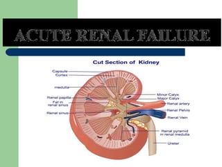
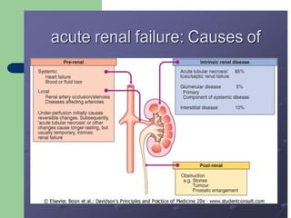
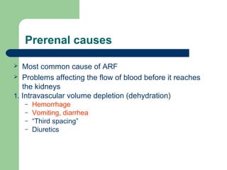
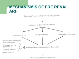
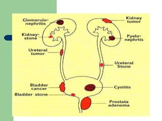
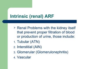
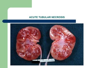
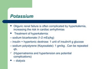
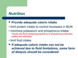
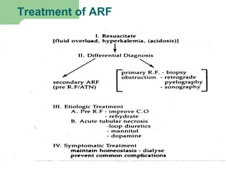
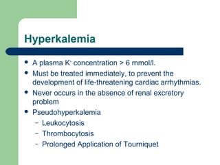
East Africa University
Faculty of Medicine
semester:11
DEFINITIONS
 Acute renal failure is a sudden reduction in
kidney function that results in nitrogenous
wastes accumulating in the blood.
 ARF is an abrupt decline in glomerular and
tubular function, resulting in the failure of the
kidneys to excrete nitrogenous waste products
and to maintain fluid and electrolyte
homeostasis.
 Acute renal failure (ARF) refers to a sudden
and usually reversible loss of renal function,
which develops over a period of days or weeks
and is usually accompanied by a reduction in
urine volume.
ACUTE RENAL FAILURE
CLASSIFICATION BY URINE
VOLUME
OLIGURIC: <400 CC/ 24 Hrs.
NON-OLIGURIC: >500 CC/24 Hrs.
ANURIC : <50 CC/24 Hrs. (some
references <100 cc/24 hrs.)
ETIOLOGY
 There are many possible causes and it is frequently
multifactorial. The clinical picture is often dominated by
the underlying condition (e.g. septic shock, trauma). If
the cause cannot be rapidly corrected and renal
function restored, temporary renal replacement therapy
may be required.
The causes can be:
I. PRE-RENAL 55-60%
II. POST RENAL <5%
III. RENAL 35-40%
Prerenal causes
 Most common cause of ARF
 Problems affecting the flow of blood before it reaches
the kidneys
1. Intravascular volume depletion (dehydration)
– Hemorrhage
– Vomiting, diarrhea
– “Third spacing”
– Diuretics
2. Disruption of blood flow to the kidneys .
 Major surgery with blood loss, severe injury or burns, or
infection in the bloodstream.
 Blockage or narrowing of a blood vessel carrying blood to
the kidneys.
 Heart failure or heart attacks causing low blood flow.
 Liver failure causing changes in hormones that affect blood
flow and pressure to the kidney.
MECHANISMIS OF PRE RENAL
ARF
Post renal
 Problems affecting the movement of urine out
of the kidneys.
a) Kidney stone: usually only on one side.
b) Cancer of the urinary tract organs or structures
near the urinary tract that may obstruct the
outflow of urine.
c) Medications.
d) Bladder stone.
e) Benign prostate hyperplasia (the most
common cause in men).
f) Blood clot.
g)Bladder cancer.
Must be bilateral to result in ARF
– Unless : single kidney or prior chronic renal failure
 Suspect obstruction in anuria
 Etiology may be age dependent
– Young = congenital abnormality
– Older male = prostatic enlargement
 ARF most often associated with lesions in:
– Bladder, prostate or urethra
Intrinsic (renal) ARF
 Renal Problems with the kidney itself
that prevent proper filtration of blood
or production of urine, those include:
1. Tubular (ATN)
2. Interstitial (AIN)
3. Glomerular (Glomerulonephritis)
4. Vascular
Renal damage
 Vascular disease
– Vasculitis (SLE, polyarteritis etc.)
– Scleroderma
– Thromboembolic disease
– Malignant hypertension
 Glomerular disease
– Acute glomerulonephritis
Post infectious GN
Crescentic GN
– ANCA positive diseases
Goodpasture’s dis.
– Anti- glomerular basement antibody
ACUTE INTERSTITIAL NEPHRITIS
DRUG INDUCED
 Penicillins
 Sulfonamides
 Cephalosporin
 Rifampin ( 2nd time)
 Quinolones
 NSAID (fenoprofen)
 Allopurinol
 Phenytoin
 Thiazides
 Furosemide
 Cimetidine
 Fever
 Rash
 Eosinophilia
 Pyuria
 Eosinophiluria
 WBC Casts
Acute Interstitial Nephritis
Clinical picture
 Acute tubular necrosis
– Ischemic injury
– Toxic injury
 Endogenous toxins
– Hemoglobinuria
– Myoblobinuria (rhabdomyolysis)
– Endotoxemia
 Acute tubular necrosis
– Exogenous toxins
 Aminoglycosides
 Radiographic contrast
 Heavy metal compounds
 Ethylene glycol
 Methanol
 Carbon tetrachloride
 Cisplatin
ACUTE TUBULAR NECROSIS
Contrast-Induced ARF
Prevalence
Less than 1% in patients with normal renal
function
 Increases significantly with renal insufficiency
Risk Factors of contrast-induced ARF
 Renal insufficiency
 Diabetes mellitus
 Multiple myeloma
 High osmolar (ionic) contrast media
 Contrast medium volume
Clinical Characteristics
 Onset - 24 to 48 hrs. after exposure
 Duration - 5 to 7 days
 Non-oliguric (majority)
 Dialysis - rarely needed
 Urinary sediment - variable
 Low fractional excretion of Na
Prophylactic Strategies
 Use I.V. contrast only when necessary
 Hydration
 Minimize contrast volume
 Low-osmolar (nonionic) contrast media
 N-acetylcysteine, fenoldopam
RISK FACTORS
 Advanced age
 Blockages in the blood vessels in your arms or
legs
 Diabetes
 High blood pressure
 Heart failure
 Kidney diseases
 Liver disease
Common clinical features of ARF
 Azotemia “Is a medical condition characterized by abnormally high levels of
nitrogen-containing compounds (such as urea, creatinine, various body waste compounds,
and other nitrogen-rich compounds) in the blood.”
 Hypervolemia
 Electrolytes abnormalities:
 Increase K+ & phosphate
 Na+ & calcium
 Metabolic acidosis
 Hypertension
 Oliguria - anuria
 Weight loss.
 Nocturnal urination.
 pale urine.
 Less frequent urination, or in smaller amounts than usual, with
dark coloured urine
 Bone damage.
 Non-union in broken bones.
 Muscle cramps (caused by low levels of calcium which can cause
hypocalcaemia).:
 Abnormal heart rhythms.
 Muscle paralysis.
PHASES OF ARF
 Initiating phase
 Oliguric phase
 Diuretic phase
 Recovery phase
DIAGNOSTIC APPROACH TO ARF
 HISTORY
 PHYSICAL EXAMINATION
 ASSMENT OF URINE VOLUME
 URINE ANALYSIS
 BLOOD CHEMISTRY
 BLOOD AND URINE INDICES
 RADIOLOGIC STUDIES
 Biopsy
Urine Volume
 Anuria (< 100 ml/24h)
 – Acute bilateral arterial or venous occlusion
 – Bilateral cortical necrosis
 – Acute necrotizing glomerulonephritis
 – Obstruction (complete)
 – ATN (very rare)
 Oliguria (100-500 ml/24h)
 – Pre-renal azotemia
 – ATN
 Non-Oliguria (> 500 ml/24h)
 – ATN
 – Obstruction (partial)
Distinguishing tubulo-interstitial disease
from Pre-Renal ARF
 Most difficult classification of ARF
  Urine sediment helpful
(i) Bland
 Pre-renal azotemia
 Urinary outlet obstruction
(ii) RBC casts or dysmorphic RBCs
 Acute glomerulonephritis
 Small vessel vasculitis
(iii) WBC Cells and WBC Casts
 Acute interstitial nephritis
 Acute pyelonephritis
(iv) RTE cells, RTE cell casts, pigmented granular (“muddy brown”)
casts
 Acute tubular necrosis
 Urine chemistries is helpful
1) ATN = tubular necrosis; tubular function is
impaired, thus high urine Na
 (Reabsorption of water and sodium impaired in tubulo-
interstitial disease and ATN)
2) Pre-renal = intact tubular function, thus urine Na is
low
 (Reabsorption of water and sodium intact in pre-renal
failure).
Red Blood Cell Cast
 Two examples of red
blood cell casts, typical
of glomerular
bleeding.
Red Blood Cells
 Monomorphic
(nondysmorphic)RBC
suggest non-glomerular
source of bleeding – i.e.,
bleeding from the
calyces, pelvis, ureter(s),
bladder, prostate or
urethra.
 Dysmorphic red blood
cells suggest glomerular
injury.
Dysmorphic
Prevention of ARF
 Recognize patients at risk (postoperative
states, cardiac surgery, septic shock)
 Prevent progression from prerenal to renal
 Preserve renal perfusion
– isovolemia, cardiac output, normal blood pressure
 Avoid nephrotoxins (aminoglycosides,
NSAIDS, amphotericin)
Treatment of ARF
 Based on type/etiology of AKI (acute kidney injury) i.e.,
pre-renal, post-renal, or intrinsic renal initially
 – Pre-renal – volume, improve renal perfusion
 – Post-renal – relieve obstruction
 – Intrinsic – glomerular, tubular, interstitial, vascular
 Depends on type
 Follow clinically; attention to volume status, avoid
additional insults; treat complications of ARF
GENERAL PROTOCOL FOR
MANAGEMENT
 Treat the underlying disease
 Strictly monitor intake and output (weight,
urine output, insensible losses, IVF)
 Monitor serum electrolytes
 Adjust medication dosages according to GFR
 Avoid highly nephrotoxic drugs
 Attempt to convert oliguric to non-oliguric renal
failure (furosemide x 3)
Fluid therapy
 If patient is fluid overloaded
 • fluid restriction (insensible losses)
 • attempt furosemide 1-2 mg/kg
 • Renal replacement therapy
 If patient is dehydrated:
 • restore intravascular volume first
 • then treat as euvolemic (below)
 If patient is euvolemic:
 • restrict to insensible losses (30-35 ml/100kcal/24 hours) +
other losses (urine, chest tubes, etc)
Sodium
 most patients have dilutional hyponatremia
which should be treated with fluid restriction
 severe hyponatremia (Na< 125 mEq/L) or
hypernatremia (Na> 150 mEq/L): dialysis or
hemofiltration
Potassium
 Oliguric renal failure is often complicated by hyperkalemia,
increasing the risk in cardiac arrhythmias
 Treatment of hyperkalemia:
– sodium bicarbonate (1-2 mEq/kg)
– insulin + hypertonic dextrose: 1 unit of insulin/4 g glucose
– sodium polystyrene (Kayexalate): 1 gm/kg . Can be repeated
qh.
 (Hypernatremia and hypertension are potential
complications)
 – dialysis
Nutrition
 Provide adequate caloric intake
• limit protein intake to control increases in BUN
• minimize potassium and phosphorus intake
“Foods and fluid containing potassium or phosphorous (banana,
coffee) are restricted.”
• limit fluid intake
 If adequate caloric intake can not be
achieved due to fluid limitations, some form
of dialysis should be considered
Treatment of ARF
Hyperkalemia
 A plasma K+
concentration > 6 mmol/l.
 Must be treated immediately, to prevent the
development of life-threatening cardiac arrhythmias.
 Never occurs in the absence of renal excretory
problem
 Pseudohyperkalemia
– Leukocytosis
– Thrombocytosis
– Prolonged Application of Tourniquet
Hyperkalemia
 Significance of urine output
 Role of increased catabolism or tissue
breakdown
 Factors affecting shift of Potassium out of cells
 Etiololgy of the renal failure
Treatment of Hyperkalemia
 Urgency
 Role of the EKG in making the decision
 Clinical setting in which it occurs
– Acute renal failure
– Chronic renal failure
Table 5-3. Treatment of hyperkalemia
Medication Mechanism of action Dosage Peak effect
Calcium Antagonism of 10-30 ml of 10% solution IV -5 min
gluconate membrane over 2 min
Insulin and Increased K+
entry Insulin, 10 U IV bolus 30-60 min
Glucose into the cells followed by 0.5 mU/kg of
body weight per minute in
50 ml of 20% glucose
Sodium Increased K+
entry 44-50 mEq IV over 5 min; 30-60 min
bicarbonate into the cells can be repeated within 30
min
Albuterol Increased K+
entry
into the cells 20 mg in the nebulized form 30-60 min
Kayexalate Removal of the 20 g of resin with 100 ml of 2-4 hr
excess K+
20% sorbitol; can be
repeated every 4-6 hr
Hemodialysis Removal of the Dialysis bath K+
concentration 30-60 min
excess K+
variable
Indications for Dialysis
 A – acidosis
 E – electrolyte disturb., usually hyperkalemia
 I – intoxications (lithium, ethylene glycol, etc)
 O – overload (volume overload)
 U – uremia (symptoms, signs)
COMPLICATIONS
 ARF can affect the entire body
 Infection
 Hyperkalaemia, Hyperphosphataemia,
hyponatraemia
 water overload
 Pericarditis
 Pulmonary oedema.
 Reduced level of consciousness.
 Immune deficiency.
ARF -- Summary
 Dx AKI by falling GFR [rising BUN and
creatinine]
 Classify into pre-renal, renal, or postrenal by
history, PE, BUN:creat ratio, urine analysis
 Sometimes also need urine chemistries [urine
Na, FxExNa and/or FxExurea] to distinguish
pre-renal from ATN.
ARF – Summary (cont…)
 Sometimes need renal ultrasound to verify
obstruction [post-renal ARF]
 Rarely need other studies, esp to dx type of
intrinsic ARF [e.g., kidney biopsy: GN vs
interstitial nephritis vs ATN].
ARF – Summary (cont…)
 Distinguishing the types of intrinsic ARF
usually depends on history, PE, urine analysis
 – Vascular – large or small vessels
 – Tubular = ATN
 – Interstitial = AIN
 – Glomerular = acute GN
Acute Renal failure

Acute Renal failure

  • 1.
    East Africa University Facultyof Medicine semester:11
  • 3.
    DEFINITIONS  Acute renalfailure is a sudden reduction in kidney function that results in nitrogenous wastes accumulating in the blood.  ARF is an abrupt decline in glomerular and tubular function, resulting in the failure of the kidneys to excrete nitrogenous waste products and to maintain fluid and electrolyte homeostasis.
  • 4.
     Acute renalfailure (ARF) refers to a sudden and usually reversible loss of renal function, which develops over a period of days or weeks and is usually accompanied by a reduction in urine volume.
  • 5.
    ACUTE RENAL FAILURE CLASSIFICATIONBY URINE VOLUME OLIGURIC: <400 CC/ 24 Hrs. NON-OLIGURIC: >500 CC/24 Hrs. ANURIC : <50 CC/24 Hrs. (some references <100 cc/24 hrs.)
  • 6.
    ETIOLOGY  There aremany possible causes and it is frequently multifactorial. The clinical picture is often dominated by the underlying condition (e.g. septic shock, trauma). If the cause cannot be rapidly corrected and renal function restored, temporary renal replacement therapy may be required. The causes can be: I. PRE-RENAL 55-60% II. POST RENAL <5% III. RENAL 35-40%
  • 8.
    Prerenal causes  Mostcommon cause of ARF  Problems affecting the flow of blood before it reaches the kidneys 1. Intravascular volume depletion (dehydration) – Hemorrhage – Vomiting, diarrhea – “Third spacing” – Diuretics
  • 9.
    2. Disruption ofblood flow to the kidneys .  Major surgery with blood loss, severe injury or burns, or infection in the bloodstream.  Blockage or narrowing of a blood vessel carrying blood to the kidneys.  Heart failure or heart attacks causing low blood flow.  Liver failure causing changes in hormones that affect blood flow and pressure to the kidney.
  • 10.
  • 11.
    Post renal  Problemsaffecting the movement of urine out of the kidneys. a) Kidney stone: usually only on one side. b) Cancer of the urinary tract organs or structures near the urinary tract that may obstruct the outflow of urine. c) Medications.
  • 12.
    d) Bladder stone. e)Benign prostate hyperplasia (the most common cause in men). f) Blood clot. g)Bladder cancer. Must be bilateral to result in ARF – Unless : single kidney or prior chronic renal failure
  • 13.
     Suspect obstructionin anuria  Etiology may be age dependent – Young = congenital abnormality – Older male = prostatic enlargement  ARF most often associated with lesions in: – Bladder, prostate or urethra
  • 15.
    Intrinsic (renal) ARF Renal Problems with the kidney itself that prevent proper filtration of blood or production of urine, those include: 1. Tubular (ATN) 2. Interstitial (AIN) 3. Glomerular (Glomerulonephritis) 4. Vascular
  • 16.
  • 17.
     Vascular disease –Vasculitis (SLE, polyarteritis etc.) – Scleroderma – Thromboembolic disease – Malignant hypertension
  • 18.
     Glomerular disease –Acute glomerulonephritis Post infectious GN Crescentic GN – ANCA positive diseases Goodpasture’s dis. – Anti- glomerular basement antibody
  • 19.
    ACUTE INTERSTITIAL NEPHRITIS DRUGINDUCED  Penicillins  Sulfonamides  Cephalosporin  Rifampin ( 2nd time)  Quinolones  NSAID (fenoprofen)  Allopurinol  Phenytoin  Thiazides  Furosemide  Cimetidine
  • 20.
     Fever  Rash Eosinophilia  Pyuria  Eosinophiluria  WBC Casts Acute Interstitial Nephritis Clinical picture
  • 21.
     Acute tubularnecrosis – Ischemic injury – Toxic injury  Endogenous toxins – Hemoglobinuria – Myoblobinuria (rhabdomyolysis) – Endotoxemia
  • 22.
     Acute tubularnecrosis – Exogenous toxins  Aminoglycosides  Radiographic contrast  Heavy metal compounds  Ethylene glycol  Methanol  Carbon tetrachloride  Cisplatin
  • 23.
  • 24.
    Contrast-Induced ARF Prevalence Less than1% in patients with normal renal function  Increases significantly with renal insufficiency
  • 25.
    Risk Factors ofcontrast-induced ARF  Renal insufficiency  Diabetes mellitus  Multiple myeloma  High osmolar (ionic) contrast media  Contrast medium volume
  • 26.
    Clinical Characteristics  Onset- 24 to 48 hrs. after exposure  Duration - 5 to 7 days  Non-oliguric (majority)  Dialysis - rarely needed  Urinary sediment - variable  Low fractional excretion of Na
  • 27.
    Prophylactic Strategies  UseI.V. contrast only when necessary  Hydration  Minimize contrast volume  Low-osmolar (nonionic) contrast media  N-acetylcysteine, fenoldopam
  • 28.
    RISK FACTORS  Advancedage  Blockages in the blood vessels in your arms or legs  Diabetes  High blood pressure  Heart failure  Kidney diseases  Liver disease
  • 29.
    Common clinical featuresof ARF  Azotemia “Is a medical condition characterized by abnormally high levels of nitrogen-containing compounds (such as urea, creatinine, various body waste compounds, and other nitrogen-rich compounds) in the blood.”  Hypervolemia  Electrolytes abnormalities:  Increase K+ & phosphate  Na+ & calcium  Metabolic acidosis  Hypertension  Oliguria - anuria
  • 30.
     Weight loss. Nocturnal urination.  pale urine.  Less frequent urination, or in smaller amounts than usual, with dark coloured urine  Bone damage.  Non-union in broken bones.  Muscle cramps (caused by low levels of calcium which can cause hypocalcaemia).:  Abnormal heart rhythms.  Muscle paralysis.
  • 31.
    PHASES OF ARF Initiating phase  Oliguric phase  Diuretic phase  Recovery phase
  • 35.
    DIAGNOSTIC APPROACH TOARF  HISTORY  PHYSICAL EXAMINATION  ASSMENT OF URINE VOLUME  URINE ANALYSIS  BLOOD CHEMISTRY  BLOOD AND URINE INDICES  RADIOLOGIC STUDIES  Biopsy
  • 37.
    Urine Volume  Anuria(< 100 ml/24h)  – Acute bilateral arterial or venous occlusion  – Bilateral cortical necrosis  – Acute necrotizing glomerulonephritis  – Obstruction (complete)  – ATN (very rare)  Oliguria (100-500 ml/24h)  – Pre-renal azotemia  – ATN  Non-Oliguria (> 500 ml/24h)  – ATN  – Obstruction (partial)
  • 38.
    Distinguishing tubulo-interstitial disease fromPre-Renal ARF  Most difficult classification of ARF   Urine sediment helpful (i) Bland  Pre-renal azotemia  Urinary outlet obstruction (ii) RBC casts or dysmorphic RBCs  Acute glomerulonephritis  Small vessel vasculitis (iii) WBC Cells and WBC Casts  Acute interstitial nephritis  Acute pyelonephritis (iv) RTE cells, RTE cell casts, pigmented granular (“muddy brown”) casts  Acute tubular necrosis
  • 39.
     Urine chemistriesis helpful 1) ATN = tubular necrosis; tubular function is impaired, thus high urine Na  (Reabsorption of water and sodium impaired in tubulo- interstitial disease and ATN) 2) Pre-renal = intact tubular function, thus urine Na is low  (Reabsorption of water and sodium intact in pre-renal failure).
  • 40.
    Red Blood CellCast  Two examples of red blood cell casts, typical of glomerular bleeding.
  • 41.
    Red Blood Cells Monomorphic (nondysmorphic)RBC suggest non-glomerular source of bleeding – i.e., bleeding from the calyces, pelvis, ureter(s), bladder, prostate or urethra.  Dysmorphic red blood cells suggest glomerular injury. Dysmorphic
  • 42.
    Prevention of ARF Recognize patients at risk (postoperative states, cardiac surgery, septic shock)  Prevent progression from prerenal to renal  Preserve renal perfusion – isovolemia, cardiac output, normal blood pressure  Avoid nephrotoxins (aminoglycosides, NSAIDS, amphotericin)
  • 43.
    Treatment of ARF Based on type/etiology of AKI (acute kidney injury) i.e., pre-renal, post-renal, or intrinsic renal initially  – Pre-renal – volume, improve renal perfusion  – Post-renal – relieve obstruction  – Intrinsic – glomerular, tubular, interstitial, vascular  Depends on type  Follow clinically; attention to volume status, avoid additional insults; treat complications of ARF
  • 44.
    GENERAL PROTOCOL FOR MANAGEMENT Treat the underlying disease  Strictly monitor intake and output (weight, urine output, insensible losses, IVF)  Monitor serum electrolytes  Adjust medication dosages according to GFR  Avoid highly nephrotoxic drugs  Attempt to convert oliguric to non-oliguric renal failure (furosemide x 3)
  • 45.
    Fluid therapy  Ifpatient is fluid overloaded  • fluid restriction (insensible losses)  • attempt furosemide 1-2 mg/kg  • Renal replacement therapy  If patient is dehydrated:  • restore intravascular volume first  • then treat as euvolemic (below)  If patient is euvolemic:  • restrict to insensible losses (30-35 ml/100kcal/24 hours) + other losses (urine, chest tubes, etc)
  • 46.
    Sodium  most patientshave dilutional hyponatremia which should be treated with fluid restriction  severe hyponatremia (Na< 125 mEq/L) or hypernatremia (Na> 150 mEq/L): dialysis or hemofiltration
  • 47.
    Potassium  Oliguric renalfailure is often complicated by hyperkalemia, increasing the risk in cardiac arrhythmias  Treatment of hyperkalemia: – sodium bicarbonate (1-2 mEq/kg) – insulin + hypertonic dextrose: 1 unit of insulin/4 g glucose – sodium polystyrene (Kayexalate): 1 gm/kg . Can be repeated qh.  (Hypernatremia and hypertension are potential complications)  – dialysis
  • 48.
    Nutrition  Provide adequatecaloric intake • limit protein intake to control increases in BUN • minimize potassium and phosphorus intake “Foods and fluid containing potassium or phosphorous (banana, coffee) are restricted.” • limit fluid intake  If adequate caloric intake can not be achieved due to fluid limitations, some form of dialysis should be considered
  • 49.
  • 50.
    Hyperkalemia  A plasmaK+ concentration > 6 mmol/l.  Must be treated immediately, to prevent the development of life-threatening cardiac arrhythmias.  Never occurs in the absence of renal excretory problem  Pseudohyperkalemia – Leukocytosis – Thrombocytosis – Prolonged Application of Tourniquet
  • 51.
    Hyperkalemia  Significance ofurine output  Role of increased catabolism or tissue breakdown  Factors affecting shift of Potassium out of cells  Etiololgy of the renal failure
  • 52.
    Treatment of Hyperkalemia Urgency  Role of the EKG in making the decision  Clinical setting in which it occurs – Acute renal failure – Chronic renal failure
  • 54.
    Table 5-3. Treatmentof hyperkalemia Medication Mechanism of action Dosage Peak effect Calcium Antagonism of 10-30 ml of 10% solution IV -5 min gluconate membrane over 2 min Insulin and Increased K+ entry Insulin, 10 U IV bolus 30-60 min Glucose into the cells followed by 0.5 mU/kg of body weight per minute in 50 ml of 20% glucose Sodium Increased K+ entry 44-50 mEq IV over 5 min; 30-60 min bicarbonate into the cells can be repeated within 30 min Albuterol Increased K+ entry into the cells 20 mg in the nebulized form 30-60 min Kayexalate Removal of the 20 g of resin with 100 ml of 2-4 hr excess K+ 20% sorbitol; can be repeated every 4-6 hr Hemodialysis Removal of the Dialysis bath K+ concentration 30-60 min excess K+ variable
  • 55.
    Indications for Dialysis A – acidosis  E – electrolyte disturb., usually hyperkalemia  I – intoxications (lithium, ethylene glycol, etc)  O – overload (volume overload)  U – uremia (symptoms, signs)
  • 56.
    COMPLICATIONS  ARF canaffect the entire body  Infection  Hyperkalaemia, Hyperphosphataemia, hyponatraemia  water overload  Pericarditis  Pulmonary oedema.  Reduced level of consciousness.  Immune deficiency.
  • 57.
    ARF -- Summary Dx AKI by falling GFR [rising BUN and creatinine]  Classify into pre-renal, renal, or postrenal by history, PE, BUN:creat ratio, urine analysis  Sometimes also need urine chemistries [urine Na, FxExNa and/or FxExurea] to distinguish pre-renal from ATN.
  • 58.
    ARF – Summary(cont…)  Sometimes need renal ultrasound to verify obstruction [post-renal ARF]  Rarely need other studies, esp to dx type of intrinsic ARF [e.g., kidney biopsy: GN vs interstitial nephritis vs ATN].
  • 59.
    ARF – Summary(cont…)  Distinguishing the types of intrinsic ARF usually depends on history, PE, urine analysis  – Vascular – large or small vessels  – Tubular = ATN  – Interstitial = AIN  – Glomerular = acute GN