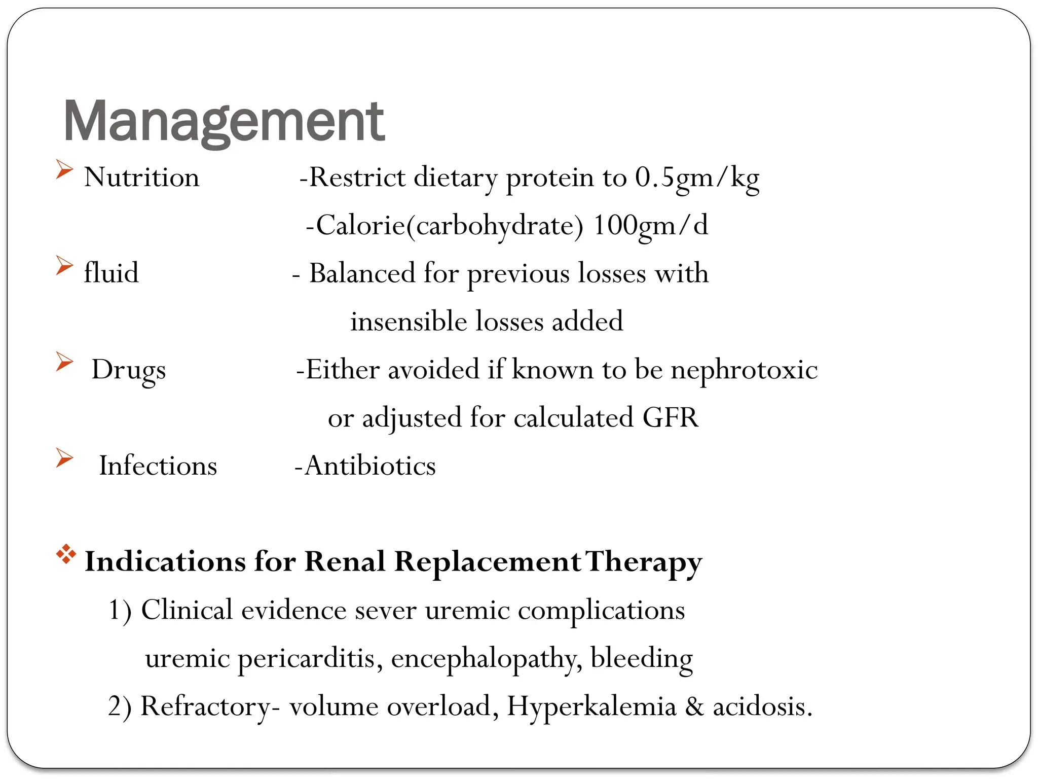
Renal Disorders
Overview
Manifestation of renal disorders
Acute Kidney Injury (AKI)
Chronic Kidney Disease (CKD)
Glomerular disorders
Nephritic Syndrome
Nephrotic Syndrome
Urinary tract infections
Acute Kidney Injury (AKI)
Acute Kidney Injury
AKI ,previously known as acute renal failure,
Is defined as a sudden impairment of kidney function
which results in the retention of urea and other
nitrogenous waste products normally cleared by the
kidneys.
AKI is not a single disease, it also reflects all spectrum of
kidney
damage including non clinical one, unlike acute renal
failure.
Acute: Develops over hours to days.
AKI
ARF ------>AKI ……Why?
Renal ----->Kidney……….easily understood by
the general pop.
Failure----->Injury………..failure indicates only
part of the spectrum
of damage to the kidney
that occurs clinically.
Cont.
It is recommended (AKIN) that acute kidney injury be defined
as any of the following :
1. Increase in Scr by >0.3 mg/dl within 48 hours, or
2. Increase in serum Cr by >1.5-fold above baseline which is known or
presumed to have occurred within 7 days or
3. Urine volume <0.5 ml/kg/h for 6 hours.
Epidemiology
 AKI complicates 5-7% of hospital admission and up to 30% of admission to ICU
particularly in the setting of diarrheal illnesses ,infectious diseases like malaria and
leptospirosis, and natural disasters such as earthquakes.
 AKI can be prevented if predisposing conditions are promptly recognized and treated.
 AKI is reversible even from complete lose function if early recognized
and managed
AKI is associated with a marked increased risk of death in hospitalized
individuals, particularly in those admitted to the ICU where in-hospital
mortality rates may exceed 50%.
AKI increases the risk for the development or worsening of chronic
kidney disease.
AKI may be community acquired or hospital-acquired.
Common causes of community-acquired AKI include volume
depletion, adverse effects of medications and obstruction of the
urinary tract.
The most common clinical settings for hospital-acquiredAKI are
sepsis, major surgical procedures, critical illness involving heart or
liver failure, intravenous iodinated contrast administration and
nephrotoxic medication administration.
Etiology of AKI
I. Pre-renal
II. Renal (intrinsic)
III. Post renal
01/24/2025
Amanuel.A
12
Pre-renal
55% of AKI
Causes includ
1. True Hypovolumia:-Massive bleeding
-Diarrhoea
-Vigorous diuresis
2.Relative hypovolumia- sepsis, anaphylaxis, Cirrhosis, CHF ...
3. Impaired renal auto regulation- NSAIDS
-ACE-I
Pathophysiology
 Tubular function is normal
 Decreased renal perfusion leads to decreased GFR
 With lesser degree of renal hypoperfusion glomerular filtration
pressure and GFR is maintained renal autoregulatory system.
 Mild hypovolumia ….> vasodilatation of afferent arterioles
by prostaglandins(prostacyclin,PgE2)
……>and vasoconstriction of efferent art.
Angiotensin II.
 Usually the renal autoregulation fails once the systolic BP falls below
80 mmHg.
Intravascular volume depletion
Gastrointestinal lose
Diarrhea ,vomiting ,prolonged NG tube drainage
Renal loss(eg.diuretics therapy)
Dermal loss(eg.burn)
Altered intrarenal
hemodynamics
Decreased effective arterial
blood volume
Third space sequstration
Etiologies of
PrerenalAKI
Diagnosis of pre-renal AKI
 History of volume loss or, signs for volume depletion or an edematous state
(CHF, cirrhosis).
 Lab tests show evidence of preserved renal tubular function :
high osmolality, urinary
Na(<20mEq/l),low fractional excretion of Na(<1%) and urea(<35%),
high
BUN/creat ratio(>20) and bland urinary sediment.
FeNa= UNa/PNa∕ UCr/PCr×100
 Definite diagnosis often made in retrospect when reversal occurs( in
urine output and in creatinine) with restoration of effective ECF volume
↓
II. Intrinsic causes
40% of AKI
1. Glomerular-AGN
2. Tubules and Interstitium-ischemia
-sepsis
-nephrotoxins
3.Vascular-vasculitis
-malignant HTN
Intrinsic causes
Acute tubular necrosis is the most common form of
intrinsicARF (85 %)
Tubular injury
 Nephrotoxic (35%)
 Ischemic (50%)
 Multifactorial
Profound ischemic injury may result in bilateral cortical
necrosis.
ATN
 ATN :- the renal parenchymal injury that follows renal ischemia or exposure to
nephrotoxins which particularly injure the tubular endothelium.
 ATN may occur with severe hemodynamic insult or with exposure to endo or
exogenous toxins but usually a combination of ischemia and toxic injury occurs.
 Oliguric and nonoliguric forms on the basis of the urinary output.
 Clinically it may have 3 phases, The
initial phase …..>daily increases in creatinine and ↓ in urinary volume,
The maintenance phase ….>
the GFR is stable and low and The recovery phase in which urine
output , creatinine falls and tubular function is restored.
Initiation
Maintenanc
e
Recovery
Clinical course
Causes of ATN(acute tubular necrosis)
Ischemia-sever hypoperfussion or uncorrected, prolonged pre-
renal causes.
Toxic injury
Exogenous:radiocontrast agents,drugs like
aminoglycosides, mphtericineB
acetaminophine, chemotherapy
Endogenous:hemoglobin,myoglobin,urate
I.Postrenal AKI
Contributes to 5% of AKI
AKI requires obstruction to urine flow
-between the external urethral meatus and bladder neck,
-bilateral ureteric obstruction, or unilateral ureteric
obstruction in a patient with one functioning kidney or with
significant preexisting chronic kidney disease
Etiologies of Post renal AKI
Intrinsic- Stone ,Papillary necrosis, Blood clot
Extrinsic- Retroperitoneal fibrosis
-Aortic aneurysm
- Retroperitoneal or pelvic malignancy
Lower tract obstruction-Urethral stricture
-BPH
- Prostate CA
-TCC of bladder
-Bladder stones
- Blood clot
- Neurogenic bladder
Clinical evaluation of a patient
The clinical evaluation of AKI should aim answering the following
questions
1.Is the kidney injury acute, acute on chronic , or chronic?
2) Is it due to pre-renal, renal or post renal cause?
3) Is there evidence of true hypovolemia (pre renal cause)?
3) Has there been major vascular accident?
5) Is there urinary tract obstruction?
History
 History of vomiting or diarrhea ,thirst, orthostatic light headedness, history
of cardiac illness
 History of systemic infection
 Systemic condition like DM,HTN,IHD
 Urinary symptom
 Recent procedure
 Drug history including herbal medication
 Travel history of malaria endemic area
 Previous diagnosis of kidney problem.
 Sign symptom of BOO
Physical examination
 G/A : Periorbital edema( facal puffiness)
V/S : tachycardia and hypotension or
hypertention
 Chest: rales-PE
 CVS : jvp may increase
 Abdomen: abdominal mass, ascites, distended bladder,
stigma of liver disease
 Integumentary: skin rash
 Peripheral oedema
Investigations
1.Urinalysis
-acellular(bland),hyaline cast in prerenal
- Pigmented "muddy brown" granular casts and casts containing tubule
epithelial cells in ATN
2.Serum Cr and BUN
3.Renal failure indices: FENa(fractional excretion of sodium) <1 in case of
pre-renal and >1 in case of ATN.
4.Imaging to r/o obstruction-USG is easiest and the most important (size of
kidney, to exclude obstruction...), KUB,
IVP...CT scan
5.Renal biopsy- uncertain causes of AKI,parenchymal
disease like RPGN is suspected...
Pre-Renal vs. Renal Failure
Prerenal Renal
BUN/Cr >20 <20
FENa <1% >2%
UNa <20 mEq/L >40 mEq/L
Specific Gravity >1.020 <1.010
Uosm >500 mOsm/L <350 mOsm/L
Urinary sedment hyaline cast granular
casts,epi.casts
Renal Lecture Required Picture #3
01/24/2025
Amanuel.A
30
Urine-to-
plasma urea
ratio
>8
< 3
RENAL FAILURE INDICE
Investigation
 Serum electrolyt-Na,K,Ca
 CBC with differential
 Novel biomarkers
Complications of AKI
1) CVS -Fluid overload
-HTN -Pericardial effusion
-MI -Arrhythmia
2) Metabolic & electrolyte disturbance
Hyperkalemia - Hyper/Hypomangesimia
-Hyponatremia -Hyperuricemia
-Hypocalcaemia -Metabolic acidosis
-Hyperphosphatemia
3. Hematologic - Anemia
-Bleeding tendency
Complications
4) GI : Nausea, vomiting, GI ulcer
malnutrition.
5) Neurologic: Change in mental stat, astrixis,
Sz, impaired cognition
6) Infection : Pneumonia, UTI, sepsis
Management
I. Supportive mx including complications
II.Specific mx
III.Prevention mx
I. Supportive Management
Complications Rx
 Volume overload -Salt restriction to 1-2gm/d; water to 1l/d
-Diuretics, dopamine,
-Ultrafiltration,dialysis
 Hyponatremia -Restriction of water intake
 Hyperkalemia -Mild (<5.5) –Restriction of dietary K
- Eliminate K sparing diuretics
- Moderate (5.5-6.5) –K binding exchange resin
-NaHCO3 therapy
- Sever (>6.5) -Calcium gluconate
-Glucose + insulin
-Dialysis
Management
 Metabolic acidosis -Restriction of dietary protein
- NaHCO3 too maintain HCO3> 15
-dialysis
Hyperphosphatemia -Restriction of dietary phosphate
-Phosphate binding agents
Hypocalcaemia -CaCO3 or Ca gluconate if sxic
Management
 Nutrition -Restrict dietary protein to 0.5gm/kg
-Calorie(carbohydrate) 100gm/d
 fluid - Balanced for previous losses with
insensible losses added
 Drugs -Either avoided if known to be nephrotoxic
or adjusted for calculated GFR
 Infections -Antibiotics
 Indications for Renal ReplacementTherapy
1) Clinical evidence sever uremic complications
uremic pericarditis, encephalopathy, bleeding
2) Refractory- volume overload, Hyperkalemia & acidosis.
Management
Forms of Renal ReplacementTherapy
Hemodialysis:-can be done continously or
intermittently.
The most common form of RRT forAKI
Remove solutes through diffusive and convective clearance.
Performed 3-4hrs/d,3-4 times/week
Has complications, mainly hypotension
Peritoneal Dialysis
Renal Replacement Therapy
 Goals of RRT
Acid-base, electrolyte and volume homeostasis
Prevention of uremia and its complications
Maintenance of optimal cardiopulmonary performance and
normal hemodynamics
Maximum nutritional support
I. Specific Management
 Pre-renal:- correct hypovolumia
 Post renal:- relieve obstruction
 Intrinsic;- d/c nephrotoxic drugs,mgt.of sepsis etc.
III. Prevention of AKI
Many cases ofARF can be prevented if attention to volume status
is given or nephrotoxic agents are avoided.
Outcome & prognosis
Mortality -50%; usual cause of death is underlying illnesses
Recovery -40%
Progression to CRF 5%
No complete recovery in 5%
Poor pxic factors
1)Oliguria at presentation 3) Old age
2)Rise in serum Cr. >3mg/day 4) Multiple organ failure
Chronic Kidney Disease
Definition of CKD:
The presence of kidney damage or decreased kidney
function for three or more months, irrespective of the cause
.
The persistence of the damage or decreased function for at
least three months is necessary to distinguish CKD from acute
kidney disease.
Kidney damage:_ pathologic abnormalities, whether established
via renal biopsy or imaging studies, or inferred from markers such as
urinary sediment abnormalities or increased rates of urinary
albumin excretion.
Definition
Decreased kidney function refers to a decreased GFR,
which is usually estimated (eGFR) using serum creatinine and
one of several available equations.
 The Cockgroft-Gault formula simple to use:
CrCl(ml/min)=(140-age)x body wt. in Kg
----------------------------------
serum Cr in mg/dl x 72
Multiply by 0.85 in females
MDRD equation………..
Classification
The term CRF applies to the process of continuing significant irreversible
reduction in nephron number & typically corresponds to CKD stages 3–5.
The term ESRD represents a stage of CKD where the accumulation of
toxins, fluid, and electrolytes normally excreted by the kidneys results in the
uremic syndrome.
Etiology
1. Prerenal causes
-Sever long standing renal artery stenosis -
Bilateral renal artery embolism
2. Renal causes
-Chronic glomerulonephritis:
primary or secondary forms (30%)
Etiology
-Chronic tubulointerstitial disease: - vesicouretral reflux, and
pyelonephritis
• Vascular disease: - hypertensive nephrosclerosis
• Diabetic nephropathy
• Connective tissue diseases : SLE , scleroderma
• Hereditary disease: -polycystic kidney disease
3. Post renal cause:- Obstructive
nephropathy, urolithiasis, BPH
Etiology
 Diabetes : the most common cause of ESRD in theWesternWorld
 Hypertension : a common cause of ESRD among African Americans
 Structural anomalies of the kidneys and urinary tract: important
causes of ESRD among children
 Chronic tubulointerstitial nephritis
Leading Categories of Etiologies of CKD
WorldWide(90% causes of CKD)
 Diabetic glomerular disease
 Glomerulonephritis
 Hypertensive nephropathy
Primary glomerulopathy with hypertension
Vascular and ischemic renal disease
 Autosomal dominant polycystic kidney disease
Other cystic and tubulointerstitial nephropathy
Pathophysiology of CKD
Involves two broad sets of mechanisms:-
1) Initiating mechanisms specific to the underlying etiology like
genetic abnormalities, immune complex deposition and
inflammation in a certain types of GN or direct toxin exposure
to tubules or interstitium.
2) A set of progressive mechanisms, involving hyper filtration and
hypertrophy of the remaining viable nephrons.
Clinical manifestations
Manifestations dependent upon severity of renal impairment
and underlying disease.
Most patients with early(Stage 1&2) CKD are
asymptomatic, but there may be symptoms from
the underlying renal diseases, like edema in NS ,HTN.
If GFR progresses to 3 and 4, symptoms related to
decreased GFR and complications become prominent.
In late CKD uremic manifestations like easy fatigability,
anorexia, vomiting, deficits in cognitive function, etc will be
present.
Complications
Disorders of fluid and electrolytes
Disorders of mineral metabolism and renal osteodystrophy
Cardiovascular disorders: Hypertension, myocardial ischemia, Heart
Failure,Anemia
Dyslipidemia,
Endocrine abnormalities: Hyperphosphatemia, Hypocalceamia,↑PTH
Growth impairment
Decreased clearance of renal excreted substances from the body
History
o Hypertension
oDiabetes mellitus
o Systemic infectious or inflammatory diseases
o Metabolic diseases
o Exposure to drugs and toxins
o Family history of renal and urologic
disease.
• Uremic syndrome:-questions about appetite, diet, nausea,
and vomiting, hiccoughing, shortness of breath, edema, weight change,
muscle cramps, bone pain, mental acuity,
P/E
 Blood presure
 Funduscopy
 Precordial examination
 Examination of the abdomen for bruits and palpable renal masses
• Extremity examination for edema
• Neurologic examination for the presence of
asterixis, muscle weakness, and neuropathy
• In addition, the evaluation of prostate size in men
and potential pelvic masses in women should be undertaken by appropriate
physical examination.
CKD :Screening
 Targeted evaluation of those at increased risk : those with diabetes,
hypertension, CVD, family history of diabetes/hypertension or CKD,
etc
 Urinary exam for protein/albumin, creatinine assessment ± additional
tests
 Once diagnosis is established identify and treat cause & potentially
reversible factors and classify Stage of CKD.
 Calculating GFR
Investigation
Normal to high
normal
High Very high
AER (mg/day) <30 30-300 >300
PER (mg/day) <150 150-500 >500
AER=Albumin
PER=Protien(total)
Protein dipstick Negative to trace Trace to 1+
>1+
Investigations
Detection of complications
Serum electrolyte,
Hct with RBC indicies
CKD: Management
Establish the cause of CKD and treat (if possible)
Identify and manage unexpected (acute) deterioration in
kidney function.
Slow progression.
Manage complications of CKD.
Prepare for Renal ReplacementTherapy.
Reversible Causes of CKD
Identify and treat causes of reversible renal
dysfunction.
I) Decreased renal perfusion: hypovolemia,
hypotension(e.g. Myocardial dysfunction),
sepsis, drugs that ↓GFR
II) Nephrotoxic Drugs
III)Urinary Tract Obstruction
IV)Others: Disease relapse,
hypertension, hypocalcaemia
Slowing Progression
Focus on modifiable risk factors
I) Dietary interventions: Protein restriction, low
phosphate diet, manipulation of lipids including fish oil
supplements, salt restriction
II) Pharmacologic interventions: BP control &
proteinuria reduction- ACE-I/ARB ± other
antihypertensive. ? Lipid lowering agents
III) Other interventions : Cessation of Smoking, restricting
alcohol consumption
Treatment of Complications
A) Hypertension: Present in ≥80% of those with advanced
CKD.
BP target:<130/80 if proteinuria < 1gm/24 hrs,<125/75 if
proteinuria>1gm/24 hrs
ACE-I, Salt restriction/diuretic,ARB, Non-dihydropyridine
Ca channel blockers, alpha blockers, etc
Treatment of Complications
B ) Anemia: Normocytic-normochromic & primarily due to
EPO deficiency,stablises at~Hct 25% if untreated.
-associated with increased morbidity & mortality. Is an
important & independent risk factor for the development
and progression of LVH and CHF in CKD as well as for
increased CV mortality
-evaluation to begin when Hgb < 12gm/dl in females & <
13.5 gm/dl in males.
-Diagnosis of anemia of CKD made after excluding non-
renal causes in a person with ↓GFR
Treatment of Complications
B) Anemia: evaluation to include RBC indices, reticulocyte count,
Ferritin, %transferrin sat., stool occult blood
-Treatment target: Hemoglobin 11- 12gm/Dl.
- Iron : oral or IV
Treatment of Complications
C) Hyperphosphatemia:Phosphate retention from early
CKD but normal balance maintained until
GFR~30ml/min
(but at the cost of developing secondary
hyperparathyroidism & renal osteodystrophy).
- Hyperphosphatemia is associated with vascular
calcifications and increased mortality.
-Rx includes dietary phosphate restriction
Treatment of Complications
D) Renal Osteodystrophy:The principal types of renal
bone disease include osteitis fibrosa,osteomalacia and
adynamic bone disease.
Osteitis fibrosa results from secondary
hyperparathyroidism- PTH when GFR<
40-70ml/minute.
Treatment includes dietary PO4 restriction,PO4 binders,
activeVitamin D sterols (Calcitriol,ά calcidol, paricalcitol,
doxercalciferol, etc)
Treatment of Complications
E) Cardiovascular Disease (CVD):CKD is an independent &
important risk factor for CVD.
 The risk of death from CVD(CAD,CHF) is higher than the risk
of eventually requiring RRT.
 CV risk factors :HTN usually with LVH, DM, dyslipidemia,
smoking, old age are more prevalent in CKD populations.
Treatment of Complications
F) Other complications:Volume overload, metabolic acidosis,
hyperkalemia are issues to be addressed.
 Malnutrition: common in advanced CKD due to ↓food intake,
↓intestinal absorption/digestion and metabolic
acidosis. Serially monitor weight & serum albumin.
Involve a dietician if possible. Avoid over restriction
of proteins.
Renal Replacement Therapy
 Early identification of those with CKD and referral to a
specialist(nephrologist/?internist) may contribute to
improvement in outcome.
 Choice of Renal ReplacementTherapy: Counsel patient
about Hemodialysis, Peritoneal Dialysis and renal
transplantation.
 Transplantation is the treatment of choice for most patients
with ESRD.
 Prepare for the chosen RRT modality.
THANK YOU

5. Renal disease.pptx internal medicines

  • 1.
  • 2.
    Overview Manifestation of renaldisorders Acute Kidney Injury (AKI) Chronic Kidney Disease (CKD) Glomerular disorders Nephritic Syndrome Nephrotic Syndrome Urinary tract infections
  • 3.
  • 4.
    Acute Kidney Injury AKI,previously known as acute renal failure, Is defined as a sudden impairment of kidney function which results in the retention of urea and other nitrogenous waste products normally cleared by the kidneys. AKI is not a single disease, it also reflects all spectrum of kidney damage including non clinical one, unlike acute renal failure. Acute: Develops over hours to days.
  • 5.
    AKI ARF ------>AKI ……Why? Renal----->Kidney……….easily understood by the general pop. Failure----->Injury………..failure indicates only part of the spectrum of damage to the kidney that occurs clinically.
  • 6.
    Cont. It is recommended(AKIN) that acute kidney injury be defined as any of the following : 1. Increase in Scr by >0.3 mg/dl within 48 hours, or 2. Increase in serum Cr by >1.5-fold above baseline which is known or presumed to have occurred within 7 days or 3. Urine volume <0.5 ml/kg/h for 6 hours.
  • 7.
    Epidemiology  AKI complicates5-7% of hospital admission and up to 30% of admission to ICU particularly in the setting of diarrheal illnesses ,infectious diseases like malaria and leptospirosis, and natural disasters such as earthquakes.  AKI can be prevented if predisposing conditions are promptly recognized and treated.  AKI is reversible even from complete lose function if early recognized and managed
  • 8.
    AKI is associatedwith a marked increased risk of death in hospitalized individuals, particularly in those admitted to the ICU where in-hospital mortality rates may exceed 50%. AKI increases the risk for the development or worsening of chronic kidney disease.
  • 9.
    AKI may becommunity acquired or hospital-acquired. Common causes of community-acquired AKI include volume depletion, adverse effects of medications and obstruction of the urinary tract.
  • 10.
    The most commonclinical settings for hospital-acquiredAKI are sepsis, major surgical procedures, critical illness involving heart or liver failure, intravenous iodinated contrast administration and nephrotoxic medication administration.
  • 11.
    Etiology of AKI I.Pre-renal II. Renal (intrinsic) III. Post renal
  • 12.
  • 13.
    Pre-renal 55% of AKI Causesinclud 1. True Hypovolumia:-Massive bleeding -Diarrhoea -Vigorous diuresis 2.Relative hypovolumia- sepsis, anaphylaxis, Cirrhosis, CHF ... 3. Impaired renal auto regulation- NSAIDS -ACE-I
  • 14.
    Pathophysiology  Tubular functionis normal  Decreased renal perfusion leads to decreased GFR  With lesser degree of renal hypoperfusion glomerular filtration pressure and GFR is maintained renal autoregulatory system.  Mild hypovolumia ….> vasodilatation of afferent arterioles by prostaglandins(prostacyclin,PgE2) ……>and vasoconstriction of efferent art. Angiotensin II.  Usually the renal autoregulation fails once the systolic BP falls below 80 mmHg.
  • 15.
    Intravascular volume depletion Gastrointestinallose Diarrhea ,vomiting ,prolonged NG tube drainage Renal loss(eg.diuretics therapy) Dermal loss(eg.burn) Altered intrarenal hemodynamics Decreased effective arterial blood volume Third space sequstration Etiologies of PrerenalAKI
  • 16.
    Diagnosis of pre-renalAKI  History of volume loss or, signs for volume depletion or an edematous state (CHF, cirrhosis).  Lab tests show evidence of preserved renal tubular function : high osmolality, urinary Na(<20mEq/l),low fractional excretion of Na(<1%) and urea(<35%), high BUN/creat ratio(>20) and bland urinary sediment. FeNa= UNa/PNa∕ UCr/PCr×100  Definite diagnosis often made in retrospect when reversal occurs( in urine output and in creatinine) with restoration of effective ECF volume ↓
  • 17.
    II. Intrinsic causes 40%of AKI 1. Glomerular-AGN 2. Tubules and Interstitium-ischemia -sepsis -nephrotoxins 3.Vascular-vasculitis -malignant HTN
  • 18.
    Intrinsic causes Acute tubularnecrosis is the most common form of intrinsicARF (85 %) Tubular injury  Nephrotoxic (35%)  Ischemic (50%)  Multifactorial Profound ischemic injury may result in bilateral cortical necrosis.
  • 19.
    ATN  ATN :-the renal parenchymal injury that follows renal ischemia or exposure to nephrotoxins which particularly injure the tubular endothelium.  ATN may occur with severe hemodynamic insult or with exposure to endo or exogenous toxins but usually a combination of ischemia and toxic injury occurs.  Oliguric and nonoliguric forms on the basis of the urinary output.  Clinically it may have 3 phases, The initial phase …..>daily increases in creatinine and ↓ in urinary volume, The maintenance phase ….> the GFR is stable and low and The recovery phase in which urine output , creatinine falls and tubular function is restored.
  • 20.
  • 21.
    Causes of ATN(acutetubular necrosis) Ischemia-sever hypoperfussion or uncorrected, prolonged pre- renal causes. Toxic injury Exogenous:radiocontrast agents,drugs like aminoglycosides, mphtericineB acetaminophine, chemotherapy Endogenous:hemoglobin,myoglobin,urate
  • 22.
    I.Postrenal AKI Contributes to5% of AKI AKI requires obstruction to urine flow -between the external urethral meatus and bladder neck, -bilateral ureteric obstruction, or unilateral ureteric obstruction in a patient with one functioning kidney or with significant preexisting chronic kidney disease
  • 23.
    Etiologies of Postrenal AKI Intrinsic- Stone ,Papillary necrosis, Blood clot Extrinsic- Retroperitoneal fibrosis -Aortic aneurysm - Retroperitoneal or pelvic malignancy Lower tract obstruction-Urethral stricture -BPH - Prostate CA -TCC of bladder -Bladder stones - Blood clot - Neurogenic bladder
  • 25.
    Clinical evaluation ofa patient The clinical evaluation of AKI should aim answering the following questions 1.Is the kidney injury acute, acute on chronic , or chronic? 2) Is it due to pre-renal, renal or post renal cause? 3) Is there evidence of true hypovolemia (pre renal cause)? 3) Has there been major vascular accident? 5) Is there urinary tract obstruction?
  • 26.
    History  History ofvomiting or diarrhea ,thirst, orthostatic light headedness, history of cardiac illness  History of systemic infection  Systemic condition like DM,HTN,IHD  Urinary symptom  Recent procedure  Drug history including herbal medication  Travel history of malaria endemic area  Previous diagnosis of kidney problem.  Sign symptom of BOO
  • 27.
    Physical examination  G/A: Periorbital edema( facal puffiness) V/S : tachycardia and hypotension or hypertention  Chest: rales-PE  CVS : jvp may increase  Abdomen: abdominal mass, ascites, distended bladder, stigma of liver disease  Integumentary: skin rash  Peripheral oedema
  • 28.
    Investigations 1.Urinalysis -acellular(bland),hyaline cast inprerenal - Pigmented "muddy brown" granular casts and casts containing tubule epithelial cells in ATN 2.Serum Cr and BUN 3.Renal failure indices: FENa(fractional excretion of sodium) <1 in case of pre-renal and >1 in case of ATN. 4.Imaging to r/o obstruction-USG is easiest and the most important (size of kidney, to exclude obstruction...), KUB, IVP...CT scan 5.Renal biopsy- uncertain causes of AKI,parenchymal disease like RPGN is suspected...
  • 29.
    Pre-Renal vs. RenalFailure Prerenal Renal BUN/Cr >20 <20 FENa <1% >2% UNa <20 mEq/L >40 mEq/L Specific Gravity >1.020 <1.010 Uosm >500 mOsm/L <350 mOsm/L Urinary sedment hyaline cast granular casts,epi.casts Renal Lecture Required Picture #3
  • 30.
  • 31.
    Investigation  Serum electrolyt-Na,K,Ca CBC with differential  Novel biomarkers
  • 32.
    Complications of AKI 1)CVS -Fluid overload -HTN -Pericardial effusion -MI -Arrhythmia 2) Metabolic & electrolyte disturbance Hyperkalemia - Hyper/Hypomangesimia -Hyponatremia -Hyperuricemia -Hypocalcaemia -Metabolic acidosis -Hyperphosphatemia 3. Hematologic - Anemia -Bleeding tendency
  • 33.
    Complications 4) GI :Nausea, vomiting, GI ulcer malnutrition. 5) Neurologic: Change in mental stat, astrixis, Sz, impaired cognition 6) Infection : Pneumonia, UTI, sepsis
  • 34.
    Management I. Supportive mxincluding complications II.Specific mx III.Prevention mx
  • 35.
    I. Supportive Management ComplicationsRx  Volume overload -Salt restriction to 1-2gm/d; water to 1l/d -Diuretics, dopamine, -Ultrafiltration,dialysis  Hyponatremia -Restriction of water intake  Hyperkalemia -Mild (<5.5) –Restriction of dietary K - Eliminate K sparing diuretics - Moderate (5.5-6.5) –K binding exchange resin -NaHCO3 therapy - Sever (>6.5) -Calcium gluconate -Glucose + insulin -Dialysis
  • 36.
    Management  Metabolic acidosis-Restriction of dietary protein - NaHCO3 too maintain HCO3> 15 -dialysis Hyperphosphatemia -Restriction of dietary phosphate -Phosphate binding agents Hypocalcaemia -CaCO3 or Ca gluconate if sxic
  • 37.
    Management  Nutrition -Restrictdietary protein to 0.5gm/kg -Calorie(carbohydrate) 100gm/d  fluid - Balanced for previous losses with insensible losses added  Drugs -Either avoided if known to be nephrotoxic or adjusted for calculated GFR  Infections -Antibiotics  Indications for Renal ReplacementTherapy 1) Clinical evidence sever uremic complications uremic pericarditis, encephalopathy, bleeding 2) Refractory- volume overload, Hyperkalemia & acidosis.
  • 38.
    Management Forms of RenalReplacementTherapy Hemodialysis:-can be done continously or intermittently. The most common form of RRT forAKI Remove solutes through diffusive and convective clearance. Performed 3-4hrs/d,3-4 times/week Has complications, mainly hypotension Peritoneal Dialysis
  • 39.
    Renal Replacement Therapy Goals of RRT Acid-base, electrolyte and volume homeostasis Prevention of uremia and its complications Maintenance of optimal cardiopulmonary performance and normal hemodynamics Maximum nutritional support
  • 40.
    I. Specific Management Pre-renal:- correct hypovolumia  Post renal:- relieve obstruction  Intrinsic;- d/c nephrotoxic drugs,mgt.of sepsis etc.
  • 41.
    III. Prevention ofAKI Many cases ofARF can be prevented if attention to volume status is given or nephrotoxic agents are avoided. Outcome & prognosis Mortality -50%; usual cause of death is underlying illnesses Recovery -40% Progression to CRF 5% No complete recovery in 5% Poor pxic factors 1)Oliguria at presentation 3) Old age 2)Rise in serum Cr. >3mg/day 4) Multiple organ failure
  • 42.
  • 43.
    Definition of CKD: Thepresence of kidney damage or decreased kidney function for three or more months, irrespective of the cause . The persistence of the damage or decreased function for at least three months is necessary to distinguish CKD from acute kidney disease.
  • 44.
    Kidney damage:_ pathologicabnormalities, whether established via renal biopsy or imaging studies, or inferred from markers such as urinary sediment abnormalities or increased rates of urinary albumin excretion.
  • 45.
    Definition Decreased kidney functionrefers to a decreased GFR, which is usually estimated (eGFR) using serum creatinine and one of several available equations.  The Cockgroft-Gault formula simple to use: CrCl(ml/min)=(140-age)x body wt. in Kg ---------------------------------- serum Cr in mg/dl x 72 Multiply by 0.85 in females MDRD equation………..
  • 46.
    Classification The term CRFapplies to the process of continuing significant irreversible reduction in nephron number & typically corresponds to CKD stages 3–5. The term ESRD represents a stage of CKD where the accumulation of toxins, fluid, and electrolytes normally excreted by the kidneys results in the uremic syndrome.
  • 47.
    Etiology 1. Prerenal causes -Severlong standing renal artery stenosis - Bilateral renal artery embolism 2. Renal causes -Chronic glomerulonephritis: primary or secondary forms (30%)
  • 48.
    Etiology -Chronic tubulointerstitial disease:- vesicouretral reflux, and pyelonephritis • Vascular disease: - hypertensive nephrosclerosis • Diabetic nephropathy • Connective tissue diseases : SLE , scleroderma • Hereditary disease: -polycystic kidney disease 3. Post renal cause:- Obstructive nephropathy, urolithiasis, BPH
  • 49.
    Etiology  Diabetes :the most common cause of ESRD in theWesternWorld  Hypertension : a common cause of ESRD among African Americans  Structural anomalies of the kidneys and urinary tract: important causes of ESRD among children  Chronic tubulointerstitial nephritis
  • 50.
    Leading Categories ofEtiologies of CKD WorldWide(90% causes of CKD)  Diabetic glomerular disease  Glomerulonephritis  Hypertensive nephropathy Primary glomerulopathy with hypertension Vascular and ischemic renal disease  Autosomal dominant polycystic kidney disease Other cystic and tubulointerstitial nephropathy
  • 51.
    Pathophysiology of CKD Involvestwo broad sets of mechanisms:- 1) Initiating mechanisms specific to the underlying etiology like genetic abnormalities, immune complex deposition and inflammation in a certain types of GN or direct toxin exposure to tubules or interstitium. 2) A set of progressive mechanisms, involving hyper filtration and hypertrophy of the remaining viable nephrons.
  • 52.
    Clinical manifestations Manifestations dependentupon severity of renal impairment and underlying disease. Most patients with early(Stage 1&2) CKD are asymptomatic, but there may be symptoms from the underlying renal diseases, like edema in NS ,HTN.
  • 53.
    If GFR progressesto 3 and 4, symptoms related to decreased GFR and complications become prominent. In late CKD uremic manifestations like easy fatigability, anorexia, vomiting, deficits in cognitive function, etc will be present.
  • 54.
    Complications Disorders of fluidand electrolytes Disorders of mineral metabolism and renal osteodystrophy Cardiovascular disorders: Hypertension, myocardial ischemia, Heart Failure,Anemia Dyslipidemia, Endocrine abnormalities: Hyperphosphatemia, Hypocalceamia,↑PTH Growth impairment Decreased clearance of renal excreted substances from the body
  • 55.
    History o Hypertension oDiabetes mellitus oSystemic infectious or inflammatory diseases o Metabolic diseases o Exposure to drugs and toxins o Family history of renal and urologic disease. • Uremic syndrome:-questions about appetite, diet, nausea, and vomiting, hiccoughing, shortness of breath, edema, weight change, muscle cramps, bone pain, mental acuity,
  • 56.
    P/E  Blood presure Funduscopy  Precordial examination  Examination of the abdomen for bruits and palpable renal masses • Extremity examination for edema • Neurologic examination for the presence of asterixis, muscle weakness, and neuropathy • In addition, the evaluation of prostate size in men and potential pelvic masses in women should be undertaken by appropriate physical examination.
  • 57.
    CKD :Screening  Targetedevaluation of those at increased risk : those with diabetes, hypertension, CVD, family history of diabetes/hypertension or CKD, etc  Urinary exam for protein/albumin, creatinine assessment ± additional tests  Once diagnosis is established identify and treat cause & potentially reversible factors and classify Stage of CKD.  Calculating GFR
  • 58.
    Investigation Normal to high normal HighVery high AER (mg/day) <30 30-300 >300 PER (mg/day) <150 150-500 >500 AER=Albumin PER=Protien(total) Protein dipstick Negative to trace Trace to 1+ >1+
  • 59.
    Investigations Detection of complications Serumelectrolyte, Hct with RBC indicies
  • 60.
    CKD: Management Establish thecause of CKD and treat (if possible) Identify and manage unexpected (acute) deterioration in kidney function. Slow progression. Manage complications of CKD. Prepare for Renal ReplacementTherapy.
  • 61.
    Reversible Causes ofCKD Identify and treat causes of reversible renal dysfunction. I) Decreased renal perfusion: hypovolemia, hypotension(e.g. Myocardial dysfunction), sepsis, drugs that ↓GFR II) Nephrotoxic Drugs III)Urinary Tract Obstruction IV)Others: Disease relapse, hypertension, hypocalcaemia
  • 62.
    Slowing Progression Focus onmodifiable risk factors I) Dietary interventions: Protein restriction, low phosphate diet, manipulation of lipids including fish oil supplements, salt restriction II) Pharmacologic interventions: BP control & proteinuria reduction- ACE-I/ARB ± other antihypertensive. ? Lipid lowering agents III) Other interventions : Cessation of Smoking, restricting alcohol consumption
  • 63.
    Treatment of Complications A)Hypertension: Present in ≥80% of those with advanced CKD. BP target:<130/80 if proteinuria < 1gm/24 hrs,<125/75 if proteinuria>1gm/24 hrs ACE-I, Salt restriction/diuretic,ARB, Non-dihydropyridine Ca channel blockers, alpha blockers, etc
  • 64.
    Treatment of Complications B) Anemia: Normocytic-normochromic & primarily due to EPO deficiency,stablises at~Hct 25% if untreated. -associated with increased morbidity & mortality. Is an important & independent risk factor for the development and progression of LVH and CHF in CKD as well as for increased CV mortality -evaluation to begin when Hgb < 12gm/dl in females & < 13.5 gm/dl in males. -Diagnosis of anemia of CKD made after excluding non- renal causes in a person with ↓GFR
  • 65.
    Treatment of Complications B)Anemia: evaluation to include RBC indices, reticulocyte count, Ferritin, %transferrin sat., stool occult blood -Treatment target: Hemoglobin 11- 12gm/Dl. - Iron : oral or IV
  • 66.
    Treatment of Complications C)Hyperphosphatemia:Phosphate retention from early CKD but normal balance maintained until GFR~30ml/min (but at the cost of developing secondary hyperparathyroidism & renal osteodystrophy). - Hyperphosphatemia is associated with vascular calcifications and increased mortality. -Rx includes dietary phosphate restriction
  • 67.
    Treatment of Complications D)Renal Osteodystrophy:The principal types of renal bone disease include osteitis fibrosa,osteomalacia and adynamic bone disease. Osteitis fibrosa results from secondary hyperparathyroidism- PTH when GFR< 40-70ml/minute. Treatment includes dietary PO4 restriction,PO4 binders, activeVitamin D sterols (Calcitriol,ά calcidol, paricalcitol, doxercalciferol, etc)
  • 68.
    Treatment of Complications E)Cardiovascular Disease (CVD):CKD is an independent & important risk factor for CVD.  The risk of death from CVD(CAD,CHF) is higher than the risk of eventually requiring RRT.  CV risk factors :HTN usually with LVH, DM, dyslipidemia, smoking, old age are more prevalent in CKD populations.
  • 69.
    Treatment of Complications F)Other complications:Volume overload, metabolic acidosis, hyperkalemia are issues to be addressed.  Malnutrition: common in advanced CKD due to ↓food intake, ↓intestinal absorption/digestion and metabolic acidosis. Serially monitor weight & serum albumin. Involve a dietician if possible. Avoid over restriction of proteins.
  • 70.
    Renal Replacement Therapy Early identification of those with CKD and referral to a specialist(nephrologist/?internist) may contribute to improvement in outcome.  Choice of Renal ReplacementTherapy: Counsel patient about Hemodialysis, Peritoneal Dialysis and renal transplantation.  Transplantation is the treatment of choice for most patients with ESRD.  Prepare for the chosen RRT modality.
  • 71.

Editor's Notes

  • #18 Single agent(insult) is usually insufficient to cause ATN
  • #20 The initiation phase refers to that period during which patients are subject to factors known to cause ATN—e.g., hypotension, ischemia, sepsis, and nephrotoxins—but have not yet developed overt parenchymal injury. It is during this period that ARF is potentially preventable. With the development of overt tubular injury, there is an abrupt fall in GFR and the clinical syndrome of ATN becomes manifest. During the maintenance phase, which typically lasts 1 to 2 wk but is highly variable and may be as short as a few days or as long as 4 to 6 wk, GFR remains markedly depressed. Urine volume during this phase is variable—although many patients are oliguric, patients may be non-oliguric with urine volumes greater than 400 to 500 ml/d. During the recovery phase, cellular regeneration and repair restore tubular integrity and GFR increases to normal or near normal values.
  • #24 Postrenal acute renal failure is caused by the anatomic obstruction to the flow of urine. When urinary tract obstruction is unilateral and the contralateral kidney is normal, the plasma creatinine concentration is usually normal or only minimally elevated and clinical ARF does not develop. Renal failure becomes manifest when the obstruction is bilateral (or below the level of the bladder) or is unilateral with dysfunction or absence of the contralateral kidney.