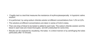
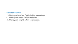
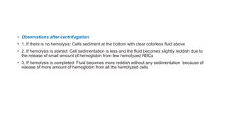
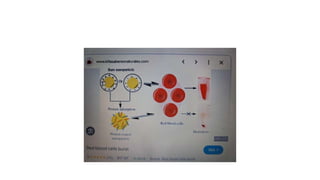
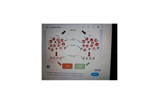
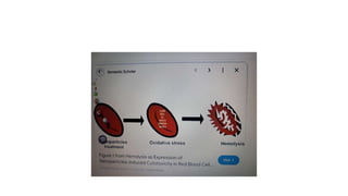
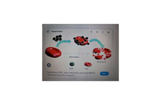
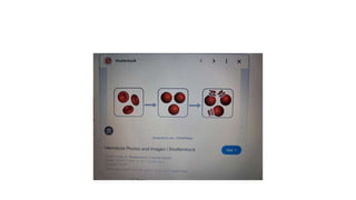
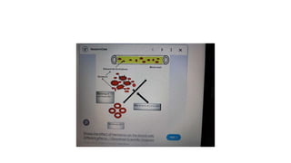
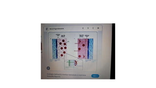
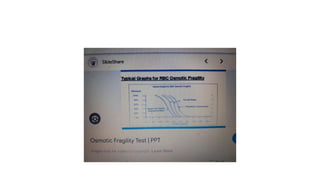
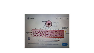
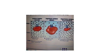
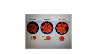
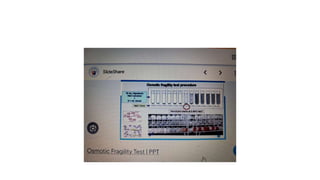
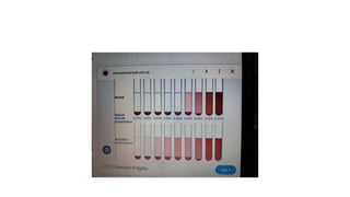
FRAGILITY TEST
• Fragility test is a test that measures the resistance of erythrocytesespecially in hypotonic saline
solution.
• It is performed by using sodium chloride solution at different concentrations from 1.2% to 0.2%.
• The solutions at different concentrations are taken in series of Cohn’s tubes.
• Then one drop of blood to be tested is added to each tube. The sodium chloride solution and the
blood in each tube are mixed thoroughly and left undisturbed for some time.
• Results can be analyzed by visualising the tubes in a direct manner or by centrifuging the tubes
particularly after 15 minutes.
• Direct observations
• 1. If there is no hemolysis: Fluid in the tube appears turbid
• 2. If hemolysis is started: Turbidity is reduced
• 3. If hemolysis is completed: Fluid becomes clear.
• Observations after centrifugation
• 1. If there is no hemolysis: Cells sediment at the bottom with clear colorless fluid above
• 2. If hemolysis is started: Cell sedimentation is less and the fluid becomes slightly reddish due to
the release of small amount of hemoglobin from few hemolyzed RBCs
• 3. If hemolysis is completed: Fluid becomes more reddish without any sedimentation because of
release of more amount of hemoglobin from all the hemolyzed cells
• Index for Fragility
• After 20 minutes:
•
• No hemolysis
• = up to 0.6%
• Onset of hemolysis
• = around 0.45%
• Completion of hemolysis = around 0.35%
•
• 1)At 0.45%, the destruction of only the older cells happens because, their membrane is fragile.
• 2)So, these cells cannot withstand specifically this hypotonicity. But, younger cells are not affected.
At 0.35%, even the destruction of younger cells takes place.
• CONDITIONS WHEN HEMOLYSIS OCCURS
• 1. Hemolytic jaundice
• 2. Antigenantibody reactions
• 3. Poisoning by chemicals or toxins
• 4. While using artificial kidney for hemodialysis or heartlung machine during cardiac surgery (rare
occasions)
• HEMOLYSINS
• Hemolysins or hemolytic agents are the substances, which lead to the destruction of RBCs. The
hemolysins are of two types:
• A. Chemical substances
• B. Substances of bacterial origin or substances found in body
• A. CHEMICAL SUBSTANCES
• 1. Alcohol
• 2. Benzene
• 3. Chloroform
• 4. Ether
• 5. Acids
• 6. Alkalis
• 7. Bile salts
• 8. Saponin
• 9. Chemical poisons like:
• i. Arsenial preparations
• ii. Carbolic acid
• iii. Nitrobenzene
• iv. Resin.
• B. SUBSTANCES OF BACTERIAL ORIGIN OR SUBSTANCES FOUND IN BODY
• 1. Toxic substances or toxins from bacteria:
• i. Streptococcus
• ii. Staphylococcus
• iii. Tetanus bacillus, etc.
• 2. Venom of poisonous snakes such as cobra
• 3. Hemolysins from normal tissues.
• REFERENCES:-
• Pagana KD, Pagana TJ, Pagana TN. Mosby's Diagnostic & Laboratory Test Reference. 14th ed. St. Louis,
MO: Elsevier; 2019. 371.
• Parpart AK, Lorenz PB, Parpart ER, Gregg JR, Chase AM. The osmotic resistance (fragilità) of human red
cells. J Clin Invest. 1947. 26:636-40.
• Yamamoto A, Saito N, Yamauchi Y, Takeda M, Ueki S, Itoga M, et al. Flow Cytometric Analysis of Red
Blood Cell Osmotic Fragility. J Lab Autom. 2014 Apr 21. [QxMD MEDLINE Link].
• Walski T, Chludzinska L, Komorowska M, Witkiewicz W. Individual osmotic fragility distribution: a new
parameter for determination of the osmotic properties of human red blood cells. Biomed Res Int. 2014.
2014:162102. [QxMD MEDLINE Link]. [Full Text].
• Gallagher PG. The Red Blood Cell Membrane and Its Disorders: Hereditary Spherocytosis, Elliptocytosis,
and Related Diseases. Prchal JT, Kaushansky K, Lichtman MA, Kipps TJ, Seligsohn U, eds. Williams
Hematology 2010. 8th ed. New York: McGraw-Hill; September 5, 2012. Chapter 45. [Full Text].
• Kattamis C, Efremov G, Pootrakul S. Effectiveness of one tube osmotic fragility screening in detecting beta-
thalassaemia trait. J Med Genet. 1981 Aug. 18(4):266-70. [QxMD MEDLINE Link]

Fragility test_causes_diagnosis_alleviation.pptx

  • 1.
  • 2.
    • Fragility testis a test that measures the resistance of erythrocytesespecially in hypotonic saline solution. • It is performed by using sodium chloride solution at different concentrations from 1.2% to 0.2%. • The solutions at different concentrations are taken in series of Cohn’s tubes. • Then one drop of blood to be tested is added to each tube. The sodium chloride solution and the blood in each tube are mixed thoroughly and left undisturbed for some time. • Results can be analyzed by visualising the tubes in a direct manner or by centrifuging the tubes particularly after 15 minutes.
  • 3.
    • Direct observations •1. If there is no hemolysis: Fluid in the tube appears turbid • 2. If hemolysis is started: Turbidity is reduced • 3. If hemolysis is completed: Fluid becomes clear.
  • 4.
    • Observations aftercentrifugation • 1. If there is no hemolysis: Cells sediment at the bottom with clear colorless fluid above • 2. If hemolysis is started: Cell sedimentation is less and the fluid becomes slightly reddish due to the release of small amount of hemoglobin from few hemolyzed RBCs • 3. If hemolysis is completed: Fluid becomes more reddish without any sedimentation because of release of more amount of hemoglobin from all the hemolyzed cells
  • 5.
    • Index forFragility • After 20 minutes: • • No hemolysis • = up to 0.6% • Onset of hemolysis • = around 0.45% • Completion of hemolysis = around 0.35% • • 1)At 0.45%, the destruction of only the older cells happens because, their membrane is fragile. • 2)So, these cells cannot withstand specifically this hypotonicity. But, younger cells are not affected. At 0.35%, even the destruction of younger cells takes place.
  • 6.
    • CONDITIONS WHENHEMOLYSIS OCCURS • 1. Hemolytic jaundice • 2. Antigenantibody reactions • 3. Poisoning by chemicals or toxins • 4. While using artificial kidney for hemodialysis or heartlung machine during cardiac surgery (rare occasions)
  • 7.
    • HEMOLYSINS • Hemolysinsor hemolytic agents are the substances, which lead to the destruction of RBCs. The hemolysins are of two types: • A. Chemical substances • B. Substances of bacterial origin or substances found in body
  • 8.
    • A. CHEMICALSUBSTANCES • 1. Alcohol • 2. Benzene • 3. Chloroform • 4. Ether • 5. Acids • 6. Alkalis • 7. Bile salts • 8. Saponin • 9. Chemical poisons like: • i. Arsenial preparations • ii. Carbolic acid • iii. Nitrobenzene • iv. Resin.
  • 9.
    • B. SUBSTANCESOF BACTERIAL ORIGIN OR SUBSTANCES FOUND IN BODY • 1. Toxic substances or toxins from bacteria: • i. Streptococcus • ii. Staphylococcus • iii. Tetanus bacillus, etc. • 2. Venom of poisonous snakes such as cobra • 3. Hemolysins from normal tissues.
  • 26.
    • REFERENCES:- • PaganaKD, Pagana TJ, Pagana TN. Mosby's Diagnostic & Laboratory Test Reference. 14th ed. St. Louis, MO: Elsevier; 2019. 371. • Parpart AK, Lorenz PB, Parpart ER, Gregg JR, Chase AM. The osmotic resistance (fragilità) of human red cells. J Clin Invest. 1947. 26:636-40. • Yamamoto A, Saito N, Yamauchi Y, Takeda M, Ueki S, Itoga M, et al. Flow Cytometric Analysis of Red Blood Cell Osmotic Fragility. J Lab Autom. 2014 Apr 21. [QxMD MEDLINE Link]. • Walski T, Chludzinska L, Komorowska M, Witkiewicz W. Individual osmotic fragility distribution: a new parameter for determination of the osmotic properties of human red blood cells. Biomed Res Int. 2014. 2014:162102. [QxMD MEDLINE Link]. [Full Text]. • Gallagher PG. The Red Blood Cell Membrane and Its Disorders: Hereditary Spherocytosis, Elliptocytosis, and Related Diseases. Prchal JT, Kaushansky K, Lichtman MA, Kipps TJ, Seligsohn U, eds. Williams Hematology 2010. 8th ed. New York: McGraw-Hill; September 5, 2012. Chapter 45. [Full Text]. • Kattamis C, Efremov G, Pootrakul S. Effectiveness of one tube osmotic fragility screening in detecting beta- thalassaemia trait. J Med Genet. 1981 Aug. 18(4):266-70. [QxMD MEDLINE Link]