Download to read offline


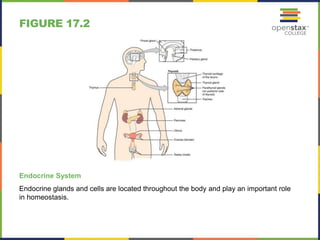
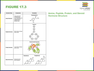


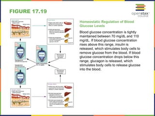


The document discusses the endocrine system's role in regulating various bodily functions, including growth, metabolism, and homeostasis through hormones. It details the functions of different endocrine glands such as the hypothalamus, pituitary gland, thyroid, and pancreas, explaining mechanisms like hormone binding and feedback loops. Additionally, it highlights the importance of maintaining blood glucose and calcium levels within specific ranges through the actions of insulin, glucagon, and parathyroid hormone.