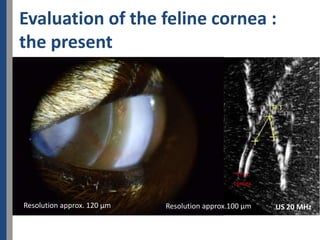
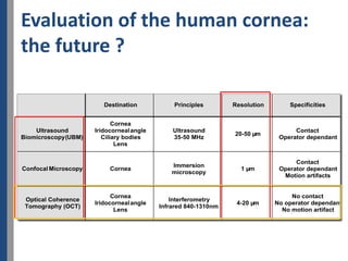
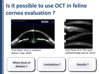
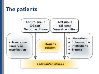
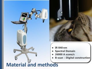
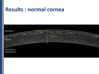
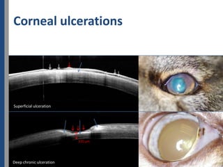
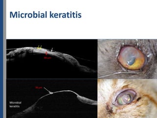
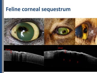
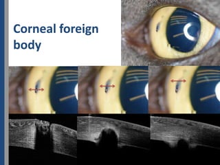
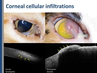
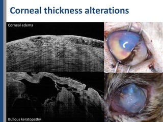
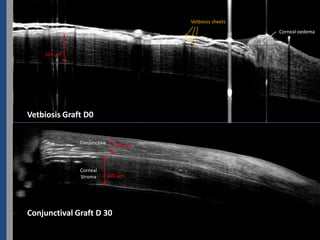
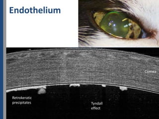

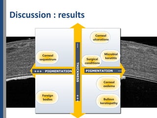
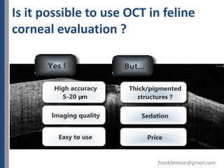

This document discusses the use of optical coherence tomography (OCT) to evaluate normal and pathological features of the feline cornea. It presents OCT images of the normal cornea layers including epithelium, stroma, and endothelium/Descemet's membrane. Pathological features imaged include corneal ulcerations, microbial keratitis, corneal sequestrum, and corneal foreign bodies. Corneal thickness alterations from edema, bullous keratopathy, and conjunctival grafts are also shown. The discussion evaluates the material and methods used and analyzes the results, concluding that OCT is possible and useful for feline corneal evaluation.


