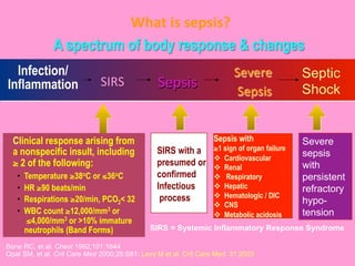
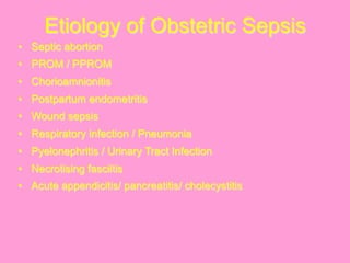
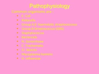
Obstetrics
Sepsis	


  Dr.	
  Kawita	
  Bapat
 
What	
  is	
  sepsis?	
  
Bone RC, et al. Chest 1992;101:1644
Opal SM, et al. Crit Care Med 2000;28:S81; Levy M et al. Crit Care Med 31:2003
A spectrum of body response & changes
Sepsis	
  SIRS	
  
Infection/
Inflammation
Severe	
  	
  
Sepsis	
  
Septic
Shock


  Clinical response arising from
a nonspecific insult, including
≥ 2 of the following:
•  Temperature ≥38oC or ≤36oC
•  HR ≥90 beats/min
•  Respirations ≥20/min, PCO2< 32
•  WBC count ≥12,000/mm3 or
≤4,000/mm3 or >10% immature
neutrophils (Band Forms) SIRS = Systemic Inflammatory Response Syndrome


SIRS with a
presumed or
confirmed
Infectious
process
Sepsis with
≥1 sign of organ failure
v  Cardiovascular
v  Renal
v  Respiratory
v  Hepatic
v  Hematologic / DIC
v  CNS
v  Metabolic acidosis
Severe
sepsis
with
persistent
refractory
hypo-
tension
Etiology of Obstetric Sepsis
•  Septic abortion
•  PROM / PPROM
•  Chorioamnionitis
•  Postpartum endometritis
•  Wound sepsis
•  Respiratory infection / Pneumonia
•  Pyelonephritis / Urinary Tract Infection
•  Necrotising fasciitis
•  Acute appendicitis/ pancreatitis/ cholecystitis
Pathophysiology
	
  Common	
  organisms	
  are	
  :	
  
§  E.	
  Coli	
  
§  Klebsiella	
  
§  Group	
  A	
  β	
  	
  haemoly@c	
  streptococcous	
  
§  Group	
  B	
  Streptococcus	
  (GBS).	
  
§  Staphylococcus	
  
§  Bacteroids	
  
§  N.	
  Gonorrhoea	
  
§  C.	
  Trachoma@s	
  
§  Cl.	
  Welchii	
  
§  Mycoplasma	
  hominis	
  
§  H.	
  influenzae	
  
Pathophysiology
§  TNF - α
§  IL – 1 β and I L – 6
§  Arachidonic acid metabolites
(Leukotrienes, PG, Tx)
§  complement system
§  coagulation cascade
§  Fibrinolytic system
§  Prekallikrein
§  Bradykinin
§  Histamine
§  β- endorphine / Encephalin
§  Mycocardial depressant factors.
Inflammatory
mediators are :
	
  
Mechanism
These	
  inflammatory	
  mediators	
  	
  cause	
  :	
  
§  Endothelial	
  dysfunc@on	
  
§  Increased	
  vascular	
  permeability	
  
§  Myocardial	
  suppression	
  
§  Ac@va@on	
   of	
   coagula@on	
   cascade	
   leading	
   to	
  
DIC.	


Evolu@on	
  of	
  Sepsis	
  
	
  In	
  Early	
  Stage	
  of	
  sepsis	
  
Released	
  Vasoac@ve	
  
Mediators	
  	
  cause	
  
§  Vasodila@on	
  
§  Platelet	
  aggrega@on	
  
§  Capillary	
  plugging	
  
§  Endothelial	
  damage	
  
	
  Resul@ng	
  in	
  	
  
§  Cellular	
  hypoxia	
  
§  lac@c	
  acidosis	
  
§  Worsening	
  of	
  @ssue	
  
perfusion	
  
In Late Stage of sepsis
Poor tissue perfusion causes :
v  Decreased vascular resistance
v  Decreased Cardiac output
v  Vasoconstriction
v  Further decrease in tissue
perfusion
v  End organ damage.
Many cytokines cause global
myocardial dysfunction , which
results in septic shock.
Investigations
§  CBC, ESR, BIood grouping and Rh typing
§  Serum β HCG
§  Urine – Routine, Microscopic culture and
sensitivity.
§  Blood culture
§  Blood Gas Analysis
§  Blood Glucose, electrolytes, BUN,
creatinine
§  Coagulation Profile
§  Liver function test
Special	
  Inves@ga@ons	
  
§  Endocervical and high vaginal swab culture must
be taken prior to internal examination.
§  The material is sent for
§  Gram staining
§  Culture both in aerobic and anaerobic medium
§  Sensitivity test
§  USG
§  Supine and upright x-ray of abdomen – Air, FB
§  CT, MRI – special cases to see for myometrial
necrosis.
Management
§ Hospitalisation and isolation if possible.
§ Overall assessment of case and Patient is put
in accordance to clinical grading
§ Aim of treatment:
v  Control of sepsis
v  Removal of source of infection
v  Supportive therapy
v  Assessment of response to treatment
Management
GENERAL	
  MANAGEMENT	
  :	
  
1.  Control	
  by	
  administering	
  	
  Broad-­‐spectrum	
  an@micirobial	
  therapy.	
  
Mul@ple	
  drugs	
  are	
  preferable	
  as	
  it	
  is	
  	
  commonly	
  a	
  mixed	
  infec@on.	
  
2.  Prophylac@c	
  TIG	
  along	
  with	
  Tetanus	
  Toxoid.	
  
3.  Maintenance	
  of	
  haemodynamic	
  status	


  Early	
  Goal	
  directed	
  therapy	
  (EGDT)	
  
v  Rapid	
  crystalloid	
  infusion.	
  MAP	
  should	
  be	
  maintained	
  at	
  65	
  mm	
  
Hg	
  and	
  urine	
  output	
  should	
  be	
  30	
  ml/hr	
  
v  Blood	
  transfusion.	
  
v  Inser@on	
  of	
  CVC,	
  PAC.	
  CVP	
  is	
  ideally	
  maintained	
  at	
  8-­‐12	
  mm	
  Hg	
  
v  Administra@on	
  of	
  ionotropes	
  like	
  Dopamine,	
  Norepinephrine	
  or	
  
Dobutamine	
  
General Management…contd
4.  Tissue oxygenation
v  Supplemental oxygenation maintaining oxygen
saturation > 70%
v  B.T if haematocrit < 30%
v  Mechanical ventilation in ARDS
5.  Treatment of acute renal failure, DIC etc as indicated.
6.  Use of anti – inflammatory agents like Hydrocortisone or
Dexamethasone.
7.  Human recombinant activated Protein C (HRAPC)
8.  Supportive measures:
v  GI haemorrhage prophylaxis by H2 receptor blockers
v  Maintainance of nutrition: Enteral nutrition is preferable
but total parenteral nutrition may be required.
v  DVT prophylaxis by Prophylactic heparin
v  Skin care
Specific	
  Management	
  -­‐	
  Sep@c	
  Abor@on	
  	
  
SPECIFIC MANAGEMENT :
Grade – I :
§  Evacuation should be performed after 24 hrs of antibiotic therapy unless
haemorrhage is profuse.
§  Gentle approach avoiding vigorous curettage.
Grade – II :
1.  Evacuation of uterus.
2.  Posterior colpotomy.
Grade – III :
Patients with septic shock are to be managed by both critical care physician and
gynaecolgists.
After the patient is stabilised after active resuscitation and critical care, surgery is
to be undertaken.
Specific	
  Management	
  -­‐	
  Sep@c	
  Abor@on	
  	
  
Indica@ons	
  for	
  laparotomy	
  are	
  :	
  
1.  	
  Injury	
  to	
  the	
  uterus	
  /	
  gut	
  
2.  	
  Presence	
  of	
  FB	
  
3.  	
  Unresponsive	
  peritoni@s	
  sugges@ve	
  of	
  	
  collec@on	
  of	
  pus	
  in	
  abdominal	
  cavity.	
  
4.  	
  Uterus	
  too	
  big	
  to	
  be	
  safely	
  evacuated	
  per	
  vagina.	


ON	
  	
  LAPAROTOMY	
  :	


Ø  Explora@ve	
  laparotomy	
  by	
  senior	
  	
  gynaec	
  and	
  skilled	
  anaesthesist.	


  When	
  gut	
  injury	
  is	
  suspected	
  general	
  surgeon	
  should	
  be	
  consulted.	
  
Ø  Removal	
  of	
  uterus	
  	
  irrespec@ve	
  of	
  parity	
  if	
  it	
  is	
  gangrenous.	
  
Ø  If	
  perfora@on	
  is	
  small	
  and	
  uterus	
  is	
  healthy	
  debridement	
  and	
  repair.	
  
Uterus	


  can	
  be	
  evacuated	
  through	
  the	
  perfora@on	
  site.	
  
Ø  Adnexa	
  to	
  be	
  removed	
  or	
  preserved	
  according	
  to	
  pathology.	
  It	
  should	
  be	
  
removed	
  in	
  Cl.	
  Welchii	
  infec@on.	
  
Ø  Thorough	
  inspec@on	
  of	
  gut	
  and	
  omentum.	
  Exteriorisa@on	
  of	
  gut	
  is	
  ideal	
  in	
  	
  
shock.	
  
Ø  If	
  nothing	
  is	
  found	
  simple	
  drainage	
  of	
  pus	
  is	
  effec@ve.	
  
Specific Management : Role of
Antibiotics
General	
  consensus	
  
•  Women	
  <	
  37	
  weeks	
  will	
  benefit	
  from	
  
an@bio@cs	
  
–  Oracle	
  study	
  2001 	
  	
  
–  Mercer	
  et	
  al	
  1997 	
  	
  
–  Lewis	
  et	
  al	
  1996	


Controversial	
  issues	
  
•  Which	
  an@bio@cs?	
  
•  Dura@on	
  of	
  course	
  
PROM	
  /	
  PPROM	
  
An@bio@cs	
  in	
  PROM	
  /	
  PPROM	
  
•  Erythromycin in PROM had beneficial effects on
the occurrence of major neonatal disease,
•  Administration of co-amoxiclav was associated
with a significant increase in the occurrence of
neonatal necrotizing enterocolitis.
•  No difference in rates of Preterm birth, Low Birth
Weight, neonatal death, chronic lung disease,
abnormal cerebral USG, or composite neonatal
outcome between any antibiotic & placebo.
•  Neither beta-lactam nor macrolide antibiotic in
women with spontaneous preterm labour
prolonged pregnancy or improved neonatal
health.
Preven@ng	
  Preterm	
  Birth	
  
•  Erythromycin – most studied and well accepted
•  Co-amoxyclav and Ampicillin have drawbacks
•  2nd/3rd generation cephalosporins may be used
•  New evidence emerging with Clindamycin
•  Oral 300mg BD X 5 days or intravaginal 2%
crème X 3 days
–  Low risk women detected to have abnormal genital flora
early in 2nd trimester (12-22 wk)
–  60% reduction in preterm labour and NICU admission.
–  Reduces late miscarriage rate.
Specific Management
Antibiotics in Chorioamnionitis
•  Gilstrap	
  Bawdon	
  &	
  Burris	
  measured	
  Levels	
  of	
  5	
  
an@bio@cs	
  in	
  maternal	
  blood,	
  cord	
  blood	
  &	
  
placental	
  membranes	
  
•  Ampicillin	
  provided	
  highest	
  ra@o	
  of	
  placental	
  to	
  
maternal	
  blood:	
  3.97	
  
•  Hence	
  Ampicillin	
  -­‐	
  Gentamycin	
  coverage	
  is	
  
appropriate	
  although	
  addi@onal	
  anaerobic	
  
coverage	
  may	
  be	
  needed	


  Gilstrap	
  Bawdon	
  &	
  Burris	
  1988	
  
Factors Influencing Outcome
•  Immune response of the host
•  Virulence of the micro organism
•  Burden of infection
•  Presence of super antigen and other
virulence factors
•  Resistance to opsonization and
phagocytosis
•  Antibiotic resistance
Scope	
  of	
  Surgery	
  
•  Caesarean section for obstetric indications
Chorioamnionitis and PROM are not
indications of C-section as such.
•  Drainage of abscess
•  Wound debridement and secondary suture
•  Dilatation & Evacuation
•  Exploration of uterus
•  Hysterectomy
Activated Protein C
•  Anticoagulant / Anti-inflammatory agent
•  No study on pregnant mothers.
•  24 µg / kg / min for 4 days.
•  M/A : Stimulates fibrinolysis.
•  Decreases thrombin formation by
–  Inhibiting platelet activation
–  Inhibiting neutrophil recruitment
–  Inhibiting mast cell degranulation
Liaw PC et al. Blood 104:2003.
•  Trials like ADDRESS trial or PROWESS trial have not shown any
benefit. Also, there is increased chance of haemorrhage. (Bernard GR
et al. NEJM 344:2001.)
•  Contraindications are Recent trauma or surgery, Active
hemorrhage, Concurrent anti-coagulant use, thrombocytopenia or
recent stroke
GBS- the fact file
§  It is a gram positive group B streptococcus.
§  It is normally present in vagina and lower intestinal
tract of healthy women in 15-40% (India 12-27%).
§  A pregnant carrier who tests positive for GBS and
receives antibiotics during labour has only 1 in 4000
chance of delivering a baby with disease.
§  If she does not receive antibiotics – the risk is 1 in
200 i.e. 20 times higher.
§  Most of the deaths due to GBS occur during the
period between onset of labour and 72 hours.
Burden of invasive GBS
GBS during pregnancy and
postpartum
v  ? Still birth and late miscarriage.
v  ? Preterm labour.
v  ? Pre labour rupture of membranes.
v  Chorioamnitis
v  Post partum endometritis
v  Post partum septicemia
v  UTI
Risk of GBS Infection in Neonate
v  1	
  in	
  1000	
  when	
  the	
  woman	
  is	
  not	
  known	
  to	
  be	
  a	
  carrier	
  	
  
v  1	
  in	
  400	
  when	
  she	
  is	
  carrying	
  GBS	
  infec@on	
  during	
  
pregnancy	
  (1	
  in	
  8000	
  a0er	
  receiving	
  IV	
  an7bio7c)	
  	
  
v  1	
  in	
  300	
  when	
  she	
  is	
  carrying	
  GBS	
  infec@on	
  at	
  delivery.	
  
(1	
  in	
  6000	
  a0er	
  receiving	
  IV	
  an7bio7c)	
  
v  1	
  in	
  100	
  when	
  she	
  had	
  previous	
  baby	
  infected	
  with	
  GBS.	
  
(1	
  in	
  2000	
  a0er	
  receiving	
  IV	
  an7bio7cs)	
  
Recent recommendations on GBS
  All	
  pregnant	
  women	
  should	
  be	
  screened	
  for	
  GBS	
  at	
  35-­‐37	
  weeks	
  
of	
  gesta@on.	
  	
  
  Pregnant	
  women	
  who	
  are	
  colonized	
  with	
  GBS	
  should	
  be	
  treated	
  
with	
  IV	
  penicillin.	
  
  Two	
  condi@ons	
  warrant	
  chemoprophylaxis	
  regardless	
  of	
  
coloniza@on:-­‐	
  
1) Women	
  with	
  risk	
  factors	
  such	
  as	
  PROM,	
  intrapartum	
  fever,	
  
prolonged	
  ROM	
  >	
  18	
  hours.	
  	
  
2) Women	
  with	
  history	
  of	
  GBS	
  disease	
  such	
  as	
  prior	
  episode	
  of	
  GBS	
  
bacteriuria	
  or	
  a	
  previous	
  new	
  born	
  	
  with	
  invasive	
  disease	


Joint	
  recommenda@ons	
  by	
  CDC,	
  ACOG	
  &	
  AAP	
  
Conclusion
Essence of sepsis management:
•  Early goal directed therapy (EGDT)
•  Maintenance of Lung perfusion
•  Antibiotics
•  Activated protein C (APRC) in select cases
•  Judicious use of corticosteroid, vasopressin
and Dobutamine
•  Support organ function
•  Prevention of nosocomial infection.
That’s all for now!
Thank you !

Obstetrics sepsis

  • 1.
    Obstetrics Sepsis      Dr.  Kawita  Bapat
  • 2.
      What  is  sepsis?   Bone RC, et al. Chest 1992;101:1644 Opal SM, et al. Crit Care Med 2000;28:S81; Levy M et al. Crit Care Med 31:2003 A spectrum of body response & changes Sepsis  SIRS   Infection/ Inflammation Severe     Sepsis   Septic Shock      Clinical response arising from a nonspecific insult, including ≥ 2 of the following: •  Temperature ≥38oC or ≤36oC •  HR ≥90 beats/min •  Respirations ≥20/min, PCO2< 32 •  WBC count ≥12,000/mm3 or ≤4,000/mm3 or >10% immature neutrophils (Band Forms) SIRS = Systemic Inflammatory Response Syndrome     SIRS with a presumed or confirmed Infectious process Sepsis with ≥1 sign of organ failure v  Cardiovascular v  Renal v  Respiratory v  Hepatic v  Hematologic / DIC v  CNS v  Metabolic acidosis Severe sepsis with persistent refractory hypo- tension
  • 3.
    Etiology of ObstetricSepsis •  Septic abortion •  PROM / PPROM •  Chorioamnionitis •  Postpartum endometritis •  Wound sepsis •  Respiratory infection / Pneumonia •  Pyelonephritis / Urinary Tract Infection •  Necrotising fasciitis •  Acute appendicitis/ pancreatitis/ cholecystitis
  • 4.
    Pathophysiology  Common  organisms  are  :   §  E.  Coli   §  Klebsiella   §  Group  A  β    haemoly@c  streptococcous   §  Group  B  Streptococcus  (GBS).   §  Staphylococcus   §  Bacteroids   §  N.  Gonorrhoea   §  C.  Trachoma@s   §  Cl.  Welchii   §  Mycoplasma  hominis   §  H.  influenzae  
  • 5.
    Pathophysiology §  TNF -α §  IL – 1 β and I L – 6 §  Arachidonic acid metabolites (Leukotrienes, PG, Tx) §  complement system §  coagulation cascade §  Fibrinolytic system §  Prekallikrein §  Bradykinin §  Histamine §  β- endorphine / Encephalin §  Mycocardial depressant factors. Inflammatory mediators are :  
  • 6.
    Mechanism These  inflammatory  mediators    cause  :   §  Endothelial  dysfunc@on   §  Increased  vascular  permeability   §  Myocardial  suppression   §  Ac@va@on   of   coagula@on   cascade   leading   to   DIC.    
  • 7.
        Evolu@on  of  Sepsis    In  Early  Stage  of  sepsis   Released  Vasoac@ve   Mediators    cause   §  Vasodila@on   §  Platelet  aggrega@on   §  Capillary  plugging   §  Endothelial  damage    Resul@ng  in     §  Cellular  hypoxia   §  lac@c  acidosis   §  Worsening  of  @ssue   perfusion   In Late Stage of sepsis Poor tissue perfusion causes : v  Decreased vascular resistance v  Decreased Cardiac output v  Vasoconstriction v  Further decrease in tissue perfusion v  End organ damage. Many cytokines cause global myocardial dysfunction , which results in septic shock.
  • 8.
    Investigations §  CBC, ESR,BIood grouping and Rh typing §  Serum β HCG §  Urine – Routine, Microscopic culture and sensitivity. §  Blood culture §  Blood Gas Analysis §  Blood Glucose, electrolytes, BUN, creatinine §  Coagulation Profile §  Liver function test
  • 9.
    Special  Inves@ga@ons   § Endocervical and high vaginal swab culture must be taken prior to internal examination. §  The material is sent for §  Gram staining §  Culture both in aerobic and anaerobic medium §  Sensitivity test §  USG §  Supine and upright x-ray of abdomen – Air, FB §  CT, MRI – special cases to see for myometrial necrosis.
  • 10.
    Management § Hospitalisation and isolationif possible. § Overall assessment of case and Patient is put in accordance to clinical grading § Aim of treatment: v  Control of sepsis v  Removal of source of infection v  Supportive therapy v  Assessment of response to treatment
  • 11.
    Management GENERAL  MANAGEMENT  :   1.  Control  by  administering    Broad-­‐spectrum  an@micirobial  therapy.   Mul@ple  drugs  are  preferable  as  it  is    commonly  a  mixed  infec@on.   2.  Prophylac@c  TIG  along  with  Tetanus  Toxoid.   3.  Maintenance  of  haemodynamic  status              Early  Goal  directed  therapy  (EGDT)   v  Rapid  crystalloid  infusion.  MAP  should  be  maintained  at  65  mm   Hg  and  urine  output  should  be  30  ml/hr   v  Blood  transfusion.   v  Inser@on  of  CVC,  PAC.  CVP  is  ideally  maintained  at  8-­‐12  mm  Hg   v  Administra@on  of  ionotropes  like  Dopamine,  Norepinephrine  or   Dobutamine  
  • 12.
    General Management…contd 4.  Tissueoxygenation v  Supplemental oxygenation maintaining oxygen saturation > 70% v  B.T if haematocrit < 30% v  Mechanical ventilation in ARDS 5.  Treatment of acute renal failure, DIC etc as indicated. 6.  Use of anti – inflammatory agents like Hydrocortisone or Dexamethasone. 7.  Human recombinant activated Protein C (HRAPC) 8.  Supportive measures: v  GI haemorrhage prophylaxis by H2 receptor blockers v  Maintainance of nutrition: Enteral nutrition is preferable but total parenteral nutrition may be required. v  DVT prophylaxis by Prophylactic heparin v  Skin care
  • 13.
    Specific  Management  -­‐  Sep@c  Abor@on     SPECIFIC MANAGEMENT : Grade – I : §  Evacuation should be performed after 24 hrs of antibiotic therapy unless haemorrhage is profuse. §  Gentle approach avoiding vigorous curettage. Grade – II : 1.  Evacuation of uterus. 2.  Posterior colpotomy. Grade – III : Patients with septic shock are to be managed by both critical care physician and gynaecolgists. After the patient is stabilised after active resuscitation and critical care, surgery is to be undertaken.
  • 14.
    Specific  Management  -­‐  Sep@c  Abor@on     Indica@ons  for  laparotomy  are  :   1.   Injury  to  the  uterus  /  gut   2.   Presence  of  FB   3.   Unresponsive  peritoni@s  sugges@ve  of    collec@on  of  pus  in  abdominal  cavity.   4.   Uterus  too  big  to  be  safely  evacuated  per  vagina.                
  • 15.
    ON    LAPAROTOMY  :       Ø  Explora@ve  laparotomy  by  senior    gynaec  and  skilled  anaesthesist.      When  gut  injury  is  suspected  general  surgeon  should  be  consulted.   Ø  Removal  of  uterus    irrespec@ve  of  parity  if  it  is  gangrenous.   Ø  If  perfora@on  is  small  and  uterus  is  healthy  debridement  and  repair.   Uterus      can  be  evacuated  through  the  perfora@on  site.   Ø  Adnexa  to  be  removed  or  preserved  according  to  pathology.  It  should  be   removed  in  Cl.  Welchii  infec@on.   Ø  Thorough  inspec@on  of  gut  and  omentum.  Exteriorisa@on  of  gut  is  ideal  in     shock.   Ø  If  nothing  is  found  simple  drainage  of  pus  is  effec@ve.  
  • 16.
    Specific Management :Role of Antibiotics General  consensus   •  Women  <  37  weeks  will  benefit  from   an@bio@cs   –  Oracle  study  2001     –  Mercer  et  al  1997     –  Lewis  et  al  1996       Controversial  issues   •  Which  an@bio@cs?   •  Dura@on  of  course   PROM  /  PPROM  
  • 17.
    An@bio@cs  in  PROM  /  PPROM   •  Erythromycin in PROM had beneficial effects on the occurrence of major neonatal disease, •  Administration of co-amoxiclav was associated with a significant increase in the occurrence of neonatal necrotizing enterocolitis. •  No difference in rates of Preterm birth, Low Birth Weight, neonatal death, chronic lung disease, abnormal cerebral USG, or composite neonatal outcome between any antibiotic & placebo. •  Neither beta-lactam nor macrolide antibiotic in women with spontaneous preterm labour prolonged pregnancy or improved neonatal health.
  • 18.
    Preven@ng  Preterm  Birth   •  Erythromycin – most studied and well accepted •  Co-amoxyclav and Ampicillin have drawbacks •  2nd/3rd generation cephalosporins may be used •  New evidence emerging with Clindamycin •  Oral 300mg BD X 5 days or intravaginal 2% crème X 3 days –  Low risk women detected to have abnormal genital flora early in 2nd trimester (12-22 wk) –  60% reduction in preterm labour and NICU admission. –  Reduces late miscarriage rate.
  • 19.
    Specific Management Antibiotics inChorioamnionitis •  Gilstrap  Bawdon  &  Burris  measured  Levels  of  5   an@bio@cs  in  maternal  blood,  cord  blood  &   placental  membranes   •  Ampicillin  provided  highest  ra@o  of  placental  to   maternal  blood:  3.97   •  Hence  Ampicillin  -­‐  Gentamycin  coverage  is   appropriate  although  addi@onal  anaerobic   coverage  may  be  needed                  Gilstrap  Bawdon  &  Burris  1988  
  • 20.
    Factors Influencing Outcome • Immune response of the host •  Virulence of the micro organism •  Burden of infection •  Presence of super antigen and other virulence factors •  Resistance to opsonization and phagocytosis •  Antibiotic resistance
  • 21.
    Scope  of  Surgery   •  Caesarean section for obstetric indications Chorioamnionitis and PROM are not indications of C-section as such. •  Drainage of abscess •  Wound debridement and secondary suture •  Dilatation & Evacuation •  Exploration of uterus •  Hysterectomy
  • 22.
    Activated Protein C • Anticoagulant / Anti-inflammatory agent •  No study on pregnant mothers. •  24 µg / kg / min for 4 days. •  M/A : Stimulates fibrinolysis. •  Decreases thrombin formation by –  Inhibiting platelet activation –  Inhibiting neutrophil recruitment –  Inhibiting mast cell degranulation Liaw PC et al. Blood 104:2003. •  Trials like ADDRESS trial or PROWESS trial have not shown any benefit. Also, there is increased chance of haemorrhage. (Bernard GR et al. NEJM 344:2001.) •  Contraindications are Recent trauma or surgery, Active hemorrhage, Concurrent anti-coagulant use, thrombocytopenia or recent stroke
  • 23.
    GBS- the factfile §  It is a gram positive group B streptococcus. §  It is normally present in vagina and lower intestinal tract of healthy women in 15-40% (India 12-27%). §  A pregnant carrier who tests positive for GBS and receives antibiotics during labour has only 1 in 4000 chance of delivering a baby with disease. §  If she does not receive antibiotics – the risk is 1 in 200 i.e. 20 times higher. §  Most of the deaths due to GBS occur during the period between onset of labour and 72 hours.
  • 24.
  • 25.
    GBS during pregnancyand postpartum v  ? Still birth and late miscarriage. v  ? Preterm labour. v  ? Pre labour rupture of membranes. v  Chorioamnitis v  Post partum endometritis v  Post partum septicemia v  UTI
  • 26.
    Risk of GBSInfection in Neonate v  1  in  1000  when  the  woman  is  not  known  to  be  a  carrier     v  1  in  400  when  she  is  carrying  GBS  infec@on  during   pregnancy  (1  in  8000  a0er  receiving  IV  an7bio7c)     v  1  in  300  when  she  is  carrying  GBS  infec@on  at  delivery.   (1  in  6000  a0er  receiving  IV  an7bio7c)   v  1  in  100  when  she  had  previous  baby  infected  with  GBS.   (1  in  2000  a0er  receiving  IV  an7bio7cs)  
  • 27.
    Recent recommendations onGBS   All  pregnant  women  should  be  screened  for  GBS  at  35-­‐37  weeks   of  gesta@on.       Pregnant  women  who  are  colonized  with  GBS  should  be  treated   with  IV  penicillin.     Two  condi@ons  warrant  chemoprophylaxis  regardless  of   coloniza@on:-­‐   1) Women  with  risk  factors  such  as  PROM,  intrapartum  fever,   prolonged  ROM  >  18  hours.     2) Women  with  history  of  GBS  disease  such  as  prior  episode  of  GBS   bacteriuria  or  a  previous  new  born    with  invasive  disease                   Joint  recommenda@ons  by  CDC,  ACOG  &  AAP  
  • 28.
    Conclusion Essence of sepsismanagement: •  Early goal directed therapy (EGDT) •  Maintenance of Lung perfusion •  Antibiotics •  Activated protein C (APRC) in select cases •  Judicious use of corticosteroid, vasopressin and Dobutamine •  Support organ function •  Prevention of nosocomial infection.
  • 29.
    That’s all fornow! Thank you !