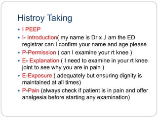
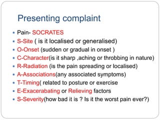
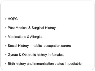
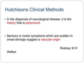
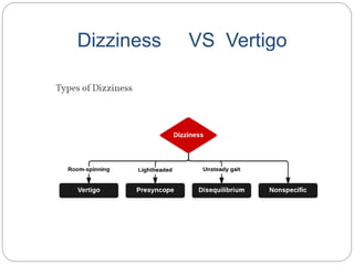
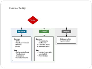
DR SHIVAJI MALLAMPATI,
MBBS,FEM,MRCEM.
EMERGENCY PHYSICIAN,
CARE HOSPITALS VIZAG.
NEUROLOGY HISTORY TAKING
Differential Diagnosis
VITAMIN CDE
 Vascular
 Inflammatory/Infectious
 Traumatic
 Autoimmune/Allergic
 Metabolic
 Idiopathic/Intoxication/Iatrogenic
 Neoplastic
 Congenital
 Degenerative/Drugs
 Endocrine
Histroy Taking
 I PEEP
 I- Introduction( my name is Dr x ,I am the ED
registrar can I confirm your name and age please
 P-Permission ( can I examine your rt knee )
 E- Explanation ( I need to examine in your rt knee
joint to see why you are in pain )
 E-Exposure ( adequately but ensuring dignity is
maintained at all times)
 P-Pain (always check if patient is in pain and offer
analgesia before starting any examination)
Presenting complaint
 Pain- SOCRATES
 S-Site ( is it localised or generalised)
 O-Onset (sudden or gradual in onset )
 C-Character(is it sharp ,aching or throbbing in nature)
 R-Radiation (is the pain spreading or localised)
 A-Associations(any associated symptoms)
 T-Timing( related to posture or exercise
 E-Exacerabating or Relieving factors
 S-Severity(how bad it is ? Is it the worst pain ever?)
 HOPC
 Past Medical & Surgical Histroy
 Medications & Allergies
 Social Histroy – habits ,occupation,carers
 Gynae & Obstretic histroy in females
 Birth history and immunization status in pediatric
Neurology History Taking
 Headache
 Seizures
 Syncope
 Altered Mental Status(AMS)
 Dizziness
 Vertigo
 Low Back ache(LBA)
 FAST
 Visual symptoms
Hutchisons Clinical Methods
 In the diagnosis of neurological disease ,it is the
history that is paramount
 Sensory or motor symptoms which are sudden in
onset strongly suggest a vascular origin
Rodney W H
Walkar
Headache
 primary headaches :when there is no underlying
cause (such as migraine or cluster headaches)
 secondary headaches : associated with an
underlying cause (such as tumor, meningitis, or
subarachnoid hemorrhage)
 Headache remains as non specific symptom in
the background histroy of fever
 Sudden onset of severe headache,thunderclap
headache or “worst headache of their life” is
suggestive of vascular cause
 Subarachnoid hemorrhage or Intracerebral
Hemorrhage
 The imaging modality of choice is CT Brain plain
 Even if the CT is normal and clinical diagnosis is
SAH
 Admit the patient in a moniter bed and do LP to
detect blood or xanthochromia in CSF
 Acute onset of a severe headache is subarachnoid
SAH
 The risk of rebleeding is greatest in the first 24 hours
and can be reduced by BP control.
 MAP of <140 mm Hg is a reasonable target while
avoiding hypotension
 DOC :is labetlol ,avoid NTG
 Vasospasm is most common 2 days to 3 weeks after
subarachnoid hemorrhage
 DOC :nimodipine, 60 milligrams PO every 4 hours
,initiated within 96 hours of symptom onset
 Headache+ Fever +Altered Mental Status is suggesttive
of
 CNS infection( meningitis ,encephalitis ,Brain abscess)
 Meningismus is an important clinical clue to the
presence of infection or hemorrhage
 95% of patients with bacterial meningitis present
with at least two of the four findings ,classic triad (fever
+neck stiffness+AMS )plus headache).
 Never delay administration of empiric antibiotic
therapy for neuroimaging or to perform LP, because
antibiotic treatment takes precedence over definitive
diagnosis.
 ceftriaxone, 2 gm IV plus vancomycin15 mg/kg IV, to
cover the common pathogens S.pneumoniae and N.
meningitides.
 The second priority is administration of steroids to
patients with presumptive pneumococcal meningitis.
 The recommended dosage of dexamethasone is 10
mg IV for adults.
 Consider adding acyclovir if herpes simplex virus
 Intracranial hemorrhage may occur with or without a
history of trauma.
 New or progressive headache, with or without
associated neurologic deficit.
 This is particularly important in the elderly, those with
chronic alcohol and substance abuse, and patients
using antiplatelet and anticoagulant agents.
 Acute headache with associated vestibular
symptoms (vertigo or ataxia) should be considered a
cerebellar hemorrhage until proven otherwise.
 Headache worsened by Valsalva maneuver.
 Headache causing awakening from sleep.
 Headache that changes with posture.
 Recent cancer diagnosis, or mental status change.
 Patients presenting with new headache symptoms,
especially in the presence of certain known risk factors.
 Hypercoagulable states such as use of oral
contraceptives, hematologic disorders, factor V Leiden
homozygous mutation, protein S or protein C deficiency,
and anti–thrombin III deficiency
 Cerebral venous thrombosis ,CVT
 Cerebral venous thrombosis is more common in women,
especially in the peripartum period, and in patients with a
recent surgical history.
 Patient presenting with severe headache, visual
changes, seizures, and encephalopathy in the
setting of marked blood pressure elevation.
 It is most common in patients undergoing active
treatment with immunesuppressive or
chemotherapeutic agents, as well as in patients with
end-stage renal disease.
 Posterior Reversible Encephalopathy Syndrome
PRES.
 New onset of headache in a elderly male >50 yrs
associated with fever jaw claudication or transient
ischemic attack symptoms, especially transient visual
loss.
 Temporal arteritis, also k/a GCA, is an inflammatory
condition affecting the small and medium-sized intra
and extracranial vessels.
 Diagnosis is made by the presence of three of the five
criteria.
 Begin treatment with prednisone, 60 milligrams PO
daily
RCVS
 This condition is one of a short list of conditions that can
mimic subarachnoid hemorrhage.
 Characterized by the occurrence of one or more
“thunderclap” headaches.
 The diagnosis should only be considered when the
evaluation for subarachnoid hemorrhage has proven
negative.
 The key diagnostic feature (multiple areas of cerebral
vasoconstriction on cerebral angiography) is most
commonly found on follow-up angiography between 2
and 3 weeks after symptom onset
Hypertensive Headache
 Uncontrolled hypertension can be associated with
headache,especially in conditions where there is a
rapid and marked rise in blood pressure. such as in
 pheochromocytoma,
 posterior reversible encephalopathy syndrome,
 hypertensive crisis,
 pre-eclampsia,
 eclampsia.
Red flags of Headache
 Patients should be assessed for clinical features that
suggest a particular cause of headache and ‘red-flag’
features.
 New onset or change in headache in patients who
are age> 50.
 Thunderclap headache.
 Abnormal neurological examination (focal and non-
focal signs).
 Headache that changes with posture.
 Change in headache frequency, characteristics, or
associated symptoms.
 Headache precipitated by physical exertion or valsalva
manoeuvre.
 Patients with risk factors for cerebral venous sinus
thrombosis.
 J aw claudication or visual disturbance.
 Neck stiff ness.
 Fever.
 New onset headache in a patient with a history of
Syncope VS Seizure
Feature SYNCOPE SEIZURE
Trigger common rare
Prodrome Presyncopal symptoms
like nausea,sweating
,pallor
Aura-unpleasant
smell,epigastric
sensation
Onset Gradual Sudden
Duration 1-30 sec 1-3 min
Colour Usually pale cyanosed
Convulsions May have movement
after LOC
Tonic Clonic
movements,automatism,
neck turned to one side
Tongue Bite Rare ,usually on the tip Common ,on the side
Post Event Rapid recovery ,N/V
afterwards,no post ictial
confusion
Post ictal
Confusion,aching
muscles,joint
Altered Mental Status (AMS)
 Consciousness requires two key components of the
central nervous system to be functioning:
 The reticular activating system RAS and at least one
cerebral hemisphere.
 Causes of failure of the reticular activating system
include:
 Brainstem stroke (ischaemic or haemorrhagic).
 Raised intracranial pressure resulting in herniation of
the brain and compression of the brainstem.
 Failure of both cerebral hemispheres may occur due to:
 Inadequate blood supply.
 Inadequate substrate for normal metabolism, e.g. oxygen
or glucose.
 Direct or indirect trauma to the cerebrum.
 Exposure of the brain to a toxic insult, e.g. infection, toxic
metabolites, or exogenous poisons.
 A stroke affecting one cerebral hemisphere does not result
in coma because the other hemisphere and the reticular
activating system are still functioning. A brainstem stroke
may lead to coma due to failure of the reticular activating
AEIOU TIPS
 Precipitating Causes: AEIOU TIPS
 A – alcohol,acidosis,arrythmias
 E – encephalopathy (hypertensive, hepatic),
electrolytes, endocrine, environmental
 I – insulin (hypoglycemia, HHNK, DKA)
 O – opiates, oxygen (hypoxia)
 U – uremia
 T – trauma, toxins
 I – infection, increased intracranial pressure
 P – psychosis, poisoning (cyanide, carbon
monoxide, etc.), porphyria
 S – stroke, shock , seizure
Dizziness VS Vertigo
Low Back Ache ( LBA )
 Acute back pain is a very common ED presentation.
 LBA & History of trauma (this may be minimal in the
elderly or those with osteoporosis),Prolonged steroid
use is S/O
 Vertebral Fracture
 LBA with Age <20 or > 50.History of malignancy,Non-
mechanical pain,Thoracic pain,Systemically unwell,
Weight loss,is S/O.
 LBA with Fever, Systemically
unwell,IVDU,Immunosuppression,
HIV. Recent bacterial infection,Non-mechanical
pain.Pain worse at night is S/O .
 Spinal Infection.
 LBA withSaddle anaesthesia,Bladder or bowel
dysfunction,Gait disturbance. Widespread or
progressive motor weakness,Bilateral sciatica is S/O .
 Cauda equina syndrome
 sudden onset of LBA in elderly male with
hemodynamic compromise& Pulsatile abdominal
mass.
 AAA
 LBA in young age <20with ,Structural deformity of
the spine, Systemically unwell.
 Ankylosing Spondylitis
RED FLAGS OF LBA
 Thoracic pain.
 Fever.
 Unexplained weight loss.
 Bladder or bowel dysfunction.
 History of carcinoma.
 Systemically unwell.
 Progressive neurological defi cit.
 Disturbed gait, saddle anaesthesia.
 Age <20 years or > 50 years.
CVA
 Sudden numbness or weakness of face, arm, or Leg
especially unilateral
 Sudden altered mental status
 Sudden aphasia
 Sudden memory deficit or spatial orientation or perception
difficulties
 Sudden visual deficit or diplopia
 Sudden dizziness, gait disturbance, or ataxia
STROKE MIMICS
 HYPOGLCEMIA
 SEIZURE : TODD” S PALSY
 TIA
 ENCEPHALOPATHY : HONK,
UREMIA,AMMONIA(HE)
 FUNCTIONAL
Timing
 To Know whether the patient is in window period .
 Should always be calculated from last seen normal.
 If window period and acute ischaemic stroke with measurable
neurological deficit :
 ED assessment should occur within 10 min.
 CT brain plain should be done within 25 min.
 CT brain to be reported within 45 min.
 IF a candidate for thrombolysis then IV rTpa with in 1 hr.
CT brain plain
 Should be normal if acute ischaemic stroke in
window period.
Neurology Histroy taking

Neurology Histroy taking

  • 1.
    DR SHIVAJI MALLAMPATI, MBBS,FEM,MRCEM. EMERGENCYPHYSICIAN, CARE HOSPITALS VIZAG. NEUROLOGY HISTORY TAKING
  • 2.
    Differential Diagnosis VITAMIN CDE Vascular  Inflammatory/Infectious  Traumatic  Autoimmune/Allergic  Metabolic  Idiopathic/Intoxication/Iatrogenic  Neoplastic  Congenital  Degenerative/Drugs  Endocrine
  • 3.
    Histroy Taking  IPEEP  I- Introduction( my name is Dr x ,I am the ED registrar can I confirm your name and age please  P-Permission ( can I examine your rt knee )  E- Explanation ( I need to examine in your rt knee joint to see why you are in pain )  E-Exposure ( adequately but ensuring dignity is maintained at all times)  P-Pain (always check if patient is in pain and offer analgesia before starting any examination)
  • 4.
    Presenting complaint  Pain-SOCRATES  S-Site ( is it localised or generalised)  O-Onset (sudden or gradual in onset )  C-Character(is it sharp ,aching or throbbing in nature)  R-Radiation (is the pain spreading or localised)  A-Associations(any associated symptoms)  T-Timing( related to posture or exercise  E-Exacerabating or Relieving factors  S-Severity(how bad it is ? Is it the worst pain ever?)
  • 5.
     HOPC  PastMedical & Surgical Histroy  Medications & Allergies  Social Histroy – habits ,occupation,carers  Gynae & Obstretic histroy in females  Birth history and immunization status in pediatric
  • 6.
    Neurology History Taking Headache  Seizures  Syncope  Altered Mental Status(AMS)  Dizziness  Vertigo  Low Back ache(LBA)  FAST  Visual symptoms
  • 7.
    Hutchisons Clinical Methods In the diagnosis of neurological disease ,it is the history that is paramount  Sensory or motor symptoms which are sudden in onset strongly suggest a vascular origin Rodney W H Walkar
  • 8.
    Headache  primary headaches:when there is no underlying cause (such as migraine or cluster headaches)  secondary headaches : associated with an underlying cause (such as tumor, meningitis, or subarachnoid hemorrhage)  Headache remains as non specific symptom in the background histroy of fever
  • 9.
     Sudden onsetof severe headache,thunderclap headache or “worst headache of their life” is suggestive of vascular cause  Subarachnoid hemorrhage or Intracerebral Hemorrhage  The imaging modality of choice is CT Brain plain  Even if the CT is normal and clinical diagnosis is SAH  Admit the patient in a moniter bed and do LP to detect blood or xanthochromia in CSF  Acute onset of a severe headache is subarachnoid
  • 10.
    SAH  The riskof rebleeding is greatest in the first 24 hours and can be reduced by BP control.  MAP of <140 mm Hg is a reasonable target while avoiding hypotension  DOC :is labetlol ,avoid NTG  Vasospasm is most common 2 days to 3 weeks after subarachnoid hemorrhage  DOC :nimodipine, 60 milligrams PO every 4 hours ,initiated within 96 hours of symptom onset
  • 11.
     Headache+ Fever+Altered Mental Status is suggesttive of  CNS infection( meningitis ,encephalitis ,Brain abscess)  Meningismus is an important clinical clue to the presence of infection or hemorrhage  95% of patients with bacterial meningitis present with at least two of the four findings ,classic triad (fever +neck stiffness+AMS )plus headache).
  • 12.
     Never delayadministration of empiric antibiotic therapy for neuroimaging or to perform LP, because antibiotic treatment takes precedence over definitive diagnosis.  ceftriaxone, 2 gm IV plus vancomycin15 mg/kg IV, to cover the common pathogens S.pneumoniae and N. meningitides.  The second priority is administration of steroids to patients with presumptive pneumococcal meningitis.  The recommended dosage of dexamethasone is 10 mg IV for adults.  Consider adding acyclovir if herpes simplex virus
  • 13.
     Intracranial hemorrhagemay occur with or without a history of trauma.  New or progressive headache, with or without associated neurologic deficit.  This is particularly important in the elderly, those with chronic alcohol and substance abuse, and patients using antiplatelet and anticoagulant agents.  Acute headache with associated vestibular symptoms (vertigo or ataxia) should be considered a cerebellar hemorrhage until proven otherwise.
  • 14.
     Headache worsenedby Valsalva maneuver.  Headache causing awakening from sleep.  Headache that changes with posture.  Recent cancer diagnosis, or mental status change.
  • 15.
     Patients presentingwith new headache symptoms, especially in the presence of certain known risk factors.  Hypercoagulable states such as use of oral contraceptives, hematologic disorders, factor V Leiden homozygous mutation, protein S or protein C deficiency, and anti–thrombin III deficiency  Cerebral venous thrombosis ,CVT  Cerebral venous thrombosis is more common in women, especially in the peripartum period, and in patients with a recent surgical history.
  • 16.
     Patient presentingwith severe headache, visual changes, seizures, and encephalopathy in the setting of marked blood pressure elevation.  It is most common in patients undergoing active treatment with immunesuppressive or chemotherapeutic agents, as well as in patients with end-stage renal disease.  Posterior Reversible Encephalopathy Syndrome PRES.
  • 17.
     New onsetof headache in a elderly male >50 yrs associated with fever jaw claudication or transient ischemic attack symptoms, especially transient visual loss.  Temporal arteritis, also k/a GCA, is an inflammatory condition affecting the small and medium-sized intra and extracranial vessels.  Diagnosis is made by the presence of three of the five criteria.  Begin treatment with prednisone, 60 milligrams PO daily
  • 18.
    RCVS  This conditionis one of a short list of conditions that can mimic subarachnoid hemorrhage.  Characterized by the occurrence of one or more “thunderclap” headaches.  The diagnosis should only be considered when the evaluation for subarachnoid hemorrhage has proven negative.  The key diagnostic feature (multiple areas of cerebral vasoconstriction on cerebral angiography) is most commonly found on follow-up angiography between 2 and 3 weeks after symptom onset
  • 19.
    Hypertensive Headache  Uncontrolledhypertension can be associated with headache,especially in conditions where there is a rapid and marked rise in blood pressure. such as in  pheochromocytoma,  posterior reversible encephalopathy syndrome,  hypertensive crisis,  pre-eclampsia,  eclampsia.
  • 21.
    Red flags ofHeadache  Patients should be assessed for clinical features that suggest a particular cause of headache and ‘red-flag’ features.  New onset or change in headache in patients who are age> 50.  Thunderclap headache.  Abnormal neurological examination (focal and non- focal signs).  Headache that changes with posture.
  • 22.
     Change inheadache frequency, characteristics, or associated symptoms.  Headache precipitated by physical exertion or valsalva manoeuvre.  Patients with risk factors for cerebral venous sinus thrombosis.  J aw claudication or visual disturbance.  Neck stiff ness.  Fever.  New onset headache in a patient with a history of
  • 23.
    Syncope VS Seizure FeatureSYNCOPE SEIZURE Trigger common rare Prodrome Presyncopal symptoms like nausea,sweating ,pallor Aura-unpleasant smell,epigastric sensation Onset Gradual Sudden Duration 1-30 sec 1-3 min Colour Usually pale cyanosed Convulsions May have movement after LOC Tonic Clonic movements,automatism, neck turned to one side Tongue Bite Rare ,usually on the tip Common ,on the side Post Event Rapid recovery ,N/V afterwards,no post ictial confusion Post ictal Confusion,aching muscles,joint
  • 25.
    Altered Mental Status(AMS)  Consciousness requires two key components of the central nervous system to be functioning:  The reticular activating system RAS and at least one cerebral hemisphere.  Causes of failure of the reticular activating system include:  Brainstem stroke (ischaemic or haemorrhagic).  Raised intracranial pressure resulting in herniation of the brain and compression of the brainstem.
  • 26.
     Failure ofboth cerebral hemispheres may occur due to:  Inadequate blood supply.  Inadequate substrate for normal metabolism, e.g. oxygen or glucose.  Direct or indirect trauma to the cerebrum.  Exposure of the brain to a toxic insult, e.g. infection, toxic metabolites, or exogenous poisons.  A stroke affecting one cerebral hemisphere does not result in coma because the other hemisphere and the reticular activating system are still functioning. A brainstem stroke may lead to coma due to failure of the reticular activating
  • 27.
    AEIOU TIPS  PrecipitatingCauses: AEIOU TIPS  A – alcohol,acidosis,arrythmias  E – encephalopathy (hypertensive, hepatic), electrolytes, endocrine, environmental  I – insulin (hypoglycemia, HHNK, DKA)  O – opiates, oxygen (hypoxia)  U – uremia  T – trauma, toxins  I – infection, increased intracranial pressure  P – psychosis, poisoning (cyanide, carbon monoxide, etc.), porphyria  S – stroke, shock , seizure
  • 28.
  • 31.
    Low Back Ache( LBA )  Acute back pain is a very common ED presentation.  LBA & History of trauma (this may be minimal in the elderly or those with osteoporosis),Prolonged steroid use is S/O  Vertebral Fracture  LBA with Age <20 or > 50.History of malignancy,Non- mechanical pain,Thoracic pain,Systemically unwell, Weight loss,is S/O.
  • 32.
     LBA withFever, Systemically unwell,IVDU,Immunosuppression, HIV. Recent bacterial infection,Non-mechanical pain.Pain worse at night is S/O .  Spinal Infection.  LBA withSaddle anaesthesia,Bladder or bowel dysfunction,Gait disturbance. Widespread or progressive motor weakness,Bilateral sciatica is S/O .  Cauda equina syndrome
  • 33.
     sudden onsetof LBA in elderly male with hemodynamic compromise& Pulsatile abdominal mass.  AAA  LBA in young age <20with ,Structural deformity of the spine, Systemically unwell.  Ankylosing Spondylitis
  • 34.
    RED FLAGS OFLBA  Thoracic pain.  Fever.  Unexplained weight loss.  Bladder or bowel dysfunction.  History of carcinoma.  Systemically unwell.  Progressive neurological defi cit.  Disturbed gait, saddle anaesthesia.  Age <20 years or > 50 years.
  • 35.
    CVA  Sudden numbnessor weakness of face, arm, or Leg especially unilateral  Sudden altered mental status  Sudden aphasia  Sudden memory deficit or spatial orientation or perception difficulties  Sudden visual deficit or diplopia  Sudden dizziness, gait disturbance, or ataxia
  • 37.
    STROKE MIMICS  HYPOGLCEMIA SEIZURE : TODD” S PALSY  TIA  ENCEPHALOPATHY : HONK, UREMIA,AMMONIA(HE)  FUNCTIONAL
  • 38.
    Timing  To Knowwhether the patient is in window period .  Should always be calculated from last seen normal.  If window period and acute ischaemic stroke with measurable neurological deficit :  ED assessment should occur within 10 min.  CT brain plain should be done within 25 min.  CT brain to be reported within 45 min.  IF a candidate for thrombolysis then IV rTpa with in 1 hr.
  • 39.
    CT brain plain Should be normal if acute ischaemic stroke in window period.