This document outlines neonatal sepsis, including:
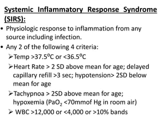
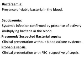
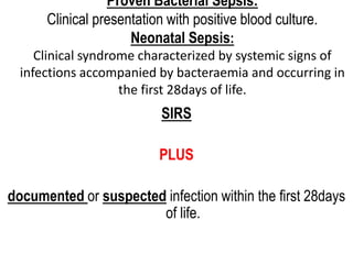
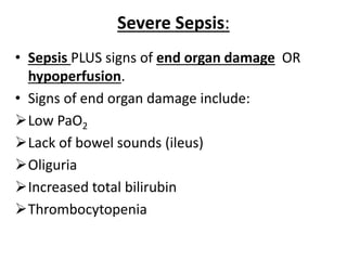
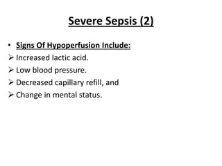
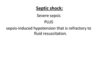
- It defines terms like sepsis, severe sepsis, and septic shock.
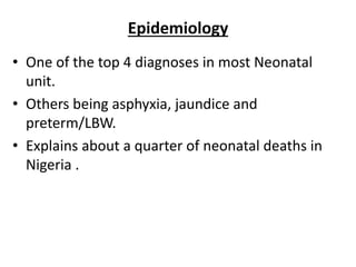
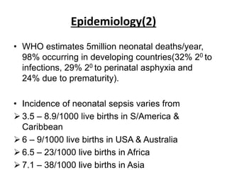
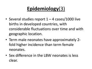
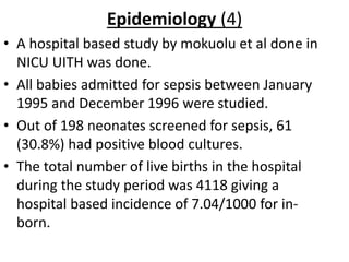
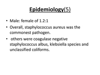
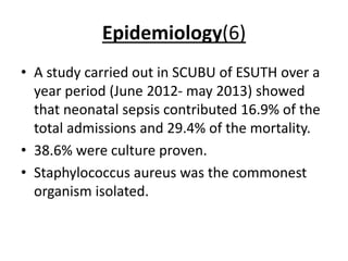
- Epidemiology shows it is one of the top causes of NICU admission and mortality, with incidence varying globally.
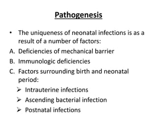
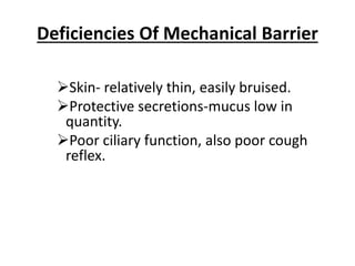
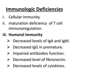
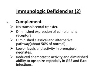
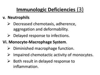
- Pathogenesis is explained by neonates' immature immune systems and barriers to infection.
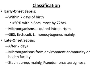
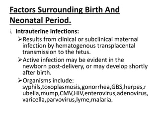
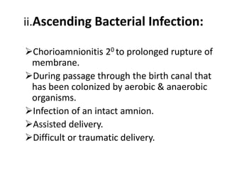
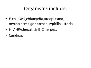
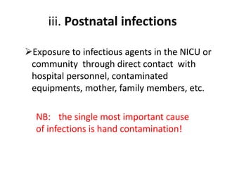
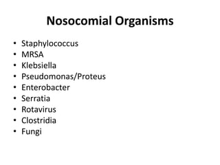
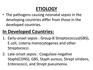
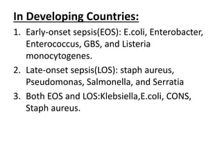
- Etiology differs between developing and developed countries.
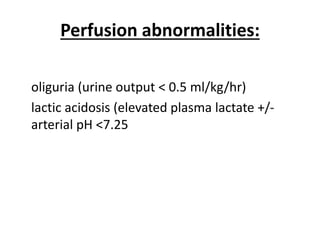
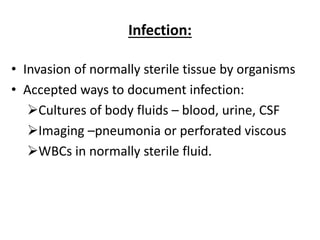
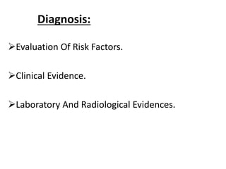
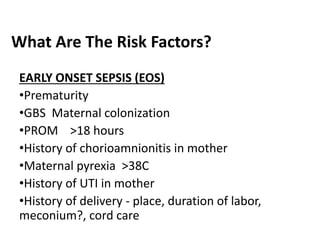
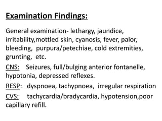
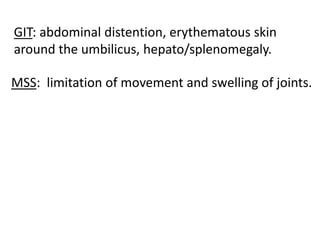
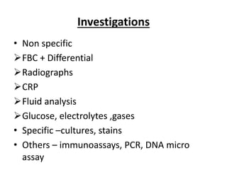
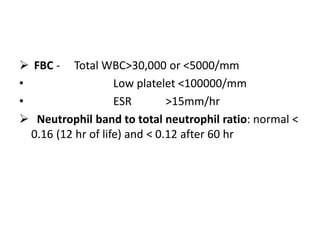
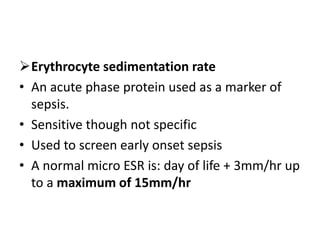
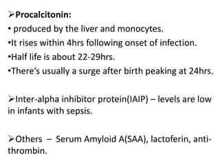
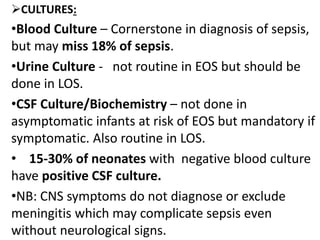
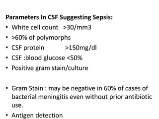
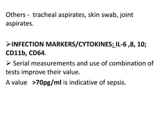
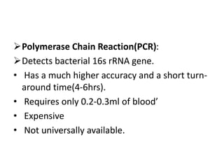
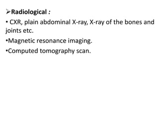
- Diagnosis involves clinical evaluation, labs like blood counts and cultures, and radiology when needed.
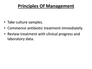
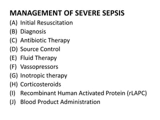
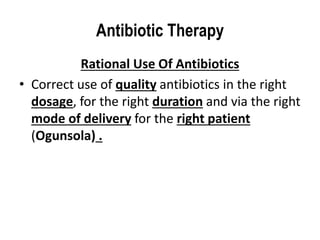
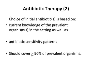
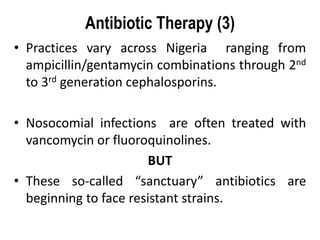
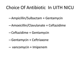
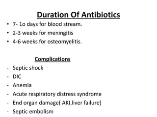
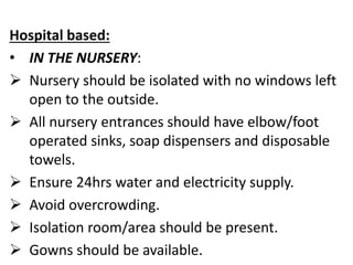
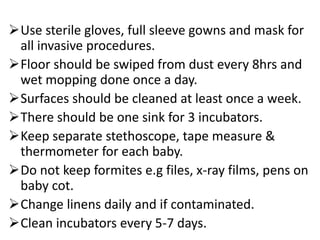
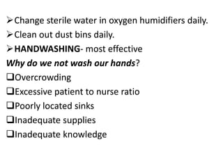
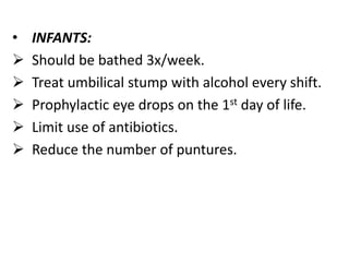
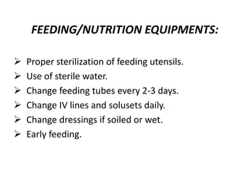
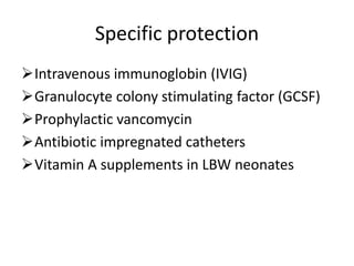
- Management involves cultures, antibiotics, fluid resuscitation, inotropes, and source control as needed for severe cases.