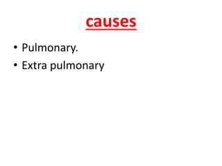
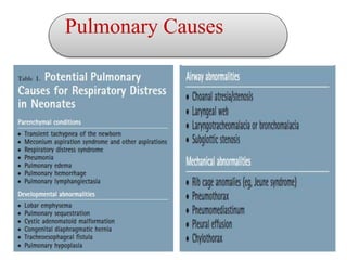
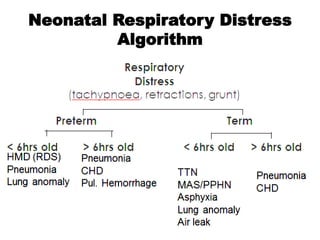
Respiratory Distress in the
Term and
Near – Term infant
by :
Ahmad Salama
(Newborn Resident)
Introduction
*One of the most common reasons for admission of
term neonates to a NICU.
*Main cause of morbidity and mortality in the
newborn period.
* The circumstances of the newborn's birth provide
important clues to the diagnosis.
Infants at Risk for
Developing Respiratory
Distress
Preterm Infants
Infants with birth asphyxia
Infants of Diabetic Mothers
Infants born by Cesarean Section
Infants born to mothers with fever, Prolonged
ROM, foul-smelling amniotic fluid.
Meconium in amniotic fluid.
Clicical Presentation
Tachypnea (RR > 60/min)
Nasal flaring
Retraction
Grunting
Delayed or decreased air entry
+/- Cyanosis
+/- Desaturation
causes
• Pulmonary.
• Extra pulmonary
Pulmonary Causes
Neonatal Respiratory Distress
Algorithm
MINIMAL
RESPIRATORY
DISEASE
• It’s transient respiratory signs persist for
less than 4 hours.
Due to:
• Hypothermia (Surfactant function is temperature-
dependent)
• Acidosis
C/P:
• The baby, near or at term
• presents within the first 2–3 hours, commonly
after being transferred to the postnatal ward
with the mother.
• The infant usually has tachypnea( respiratory
rate of up to 80–100/min)
• expiratory grunt.
• mild sternal or intercostal recession
• Cyanosis, if present, is relieved by administering
25–30% oxygen.
Ix :
 CXR … ABG… CBC
INFECTION SHOULD BE EXCLUDED
Rx :
• Antibiotics ( until culture results)
• O2 therapy
Parenchymal Diseases
1-TRANSIENT
TACHYPNEA OF
THE NEWBORN
(TTN)
o also known as RDS type 2 or Wet lung .
o Relatively benign condition
o self limited
o The most common cause of neonatal respiratory
distress.
o constituting more than 40 percent of cases
o 11 per 1,000 live births..
They are not at risk for
other illnesses.
Risk factors:
• Elective CS .
• ♂.
• Maternal Asthma .
• maternal Diabetes
Onset:
• within 2 hours after delivery
pathophysiolgy
Fetal lung Fluid
• Amount:About 20 ml/kg near term
• Rate of production :2-5 ml/kg/hr.
• The presence of lung liquid is important for normal lung
development; chronic drainage results in pulmonary hypoplasia.
**lung liquid has a high chloride but low bicarbonate and protein
Concentration.
**The dominant force mediating lung liquid secretion is the
secondary active transport of chloride ions from the interstitial space
into the lung lumen.
****During labour and delivery, the concentration of adrenaline
(epinephrine) increases and, as a consequence, lung liquid secretion
ceases and resorption begins.
Management
Most significant discriminatory findings are the
onset of the illness and the degree of distress
exhibited by the infant
Symptoms can last from a few hours to two days
TTN is a clinical diagnosis
CXR   
Rx:
• It is supportive with close observation
because the condition is usually self limited.
• Low flow supplemental oxygen may be
necessary for several hours.
(usually FIO2 < 40 %)
• Oral furosemide (Lasix) has not been shown to
significantly improve status and should not be
given
Malignant TTN
• Severe Hypoxic Respiratory Failure in LPT Infants
• LPT + ECS Pulmonary Hypertension
• Gradual increase in oxygen requirement and
subsequent evidence of PPHN
2-Respiratory
Distress
Syndrome
(RDS)
• These infants are typically 34 to 37 weeks of gestation
• RF : include maternal diabetes, multiple births ,Elective
CS , perinatal asphyxia, cold stress, and infants whose
siblings suffered from RDS.
• Because their surfactant sufficiency is borderline and
they have larger pulmonary reserves, affected infants
may be able to cope without ventilation for longer than
smaller preterm infants. Infants who have RDS may do
well with nasal continuous positive airway pressure or
may require ventilation.
3- Meconium
Aspiration
Syndrome
(MAS)
MAS is defined as respiratory distress
in an infant born through meconium-
stained amniotic fluid whose
symptoms cannot otherwise be
explained.
** POST-MATURE** ??
* 13 % MSAF  4-5 % develop MAS
**Meconium :
is composed of desquamated cells, secretions,
lanugo, water, bile pigments, pancreatic
enzymes, and amniotic fluid.
**It is sterile although when aspirated, is
locally irritative, obstructive, and a medium for
bacterial culture.
**
occurs early in the first
trimester of pregnancy . Fetal defecation slows
after 16 weeks gestation and becomes infrequent
by 20 weeks, concurrent with innervation of the
anal sphincter. From approximately 20 to 34
weeks, fetal passage of meconium remains
infrequent
• Meconium passage may represent hypoxia or
fetal distress in utero
Physiology
**The passage of meconium from the fetus into
amnion is prevented by lack of peristalsis (low
motilin level), tonic contraction of the anal
sphincter, terminal cap of viscous meconium.
**MSAF may be a natural phenomenon that
doesn’t indicate fetal distress but mature GI tract
in post term fetus with increased motilin level.
**Vagal stimulation by cord or head compression
may be associated with passage of meconium in
the absence of fetal distress.
Several investigators have suggested that most
cases of meconium aspiration occur in utero when
fetal gasping is initiated before delivery
Meconium has been found distally as far as the
alveoli in some stillborn infants and in some infants
that die within hours of delivery.
There is currently no way to distinguish between
the infant who has developed MAS by intrauterine
respiration or gasping and the infant who has
developed MAS by inhalation of meconium at the
first breaths after delivery
Risk factors for MSAF
Maternal HT
Maternal DM
Maternal heavy cigarette smoking
Maternal chronic respiratory or CV Dx
Post term pregnancy
Pre-eclampsia/eclampsia
Oligohydramnios
IUGR
Poor biophysical profile
Abnormal fetal HR pattern
Alarm of MAS
THICK MECONIUM
FETAL TACHYCARDIA
LACK OF INCREASE HEART RATE DURING
INTRA PARTUM MONITORING
LOW CORD PH
MAS must be
considered in any
infant born through
MSAF who develops
symptoms of RD.
Clinical Presentation
**Evidence of postmaturity: peeling skin, long
fingernails, and decreased vernix.
**The vernix, umbilical cord, and nails may be
meconium-stained, depending upon how long
the infant has been exposed in utero.
**In general, nails will become stained after 6
hours and vernix after 12 to 14 hours of
exposure.
**The chest typically appears barrel-shaped, with
an increased anterior-posterior diameter caused
by overinflation.
**Auscultation reveals rales and rhonchi
immediately after birth
Some patients are asymptomatic at birth and
develop worsening signs of respiratory distress as
the meconium moves from the large airways into
the lower tracheobronchial tree.
*Symptoms similar to infants with TTN, but the
presentation may suggest a more severe condition.
*Infants have greater degrees of tachypnea, retraction
and lethargy immediately after delivery.
*Some infants will have an asymptomatic period of
several hours before respiratory distress become
apparent.
*Arterial Blood Gases will reveal more acidosis,
hypercapnia and hypoxemia than in infants
with TTN.
* Hypoxia occurs because aspiration takes place in
utero
.
Complications
Partial obstruction
complete obstruction
Surfactant destruction
Chemical pneumonitis &Bacterial pneumonia
Asphyxia
PPHN
Cleary&Wiswell
The management of MAS remains a CHALLENGE.
Goals:
Increased oxygenation while minimizing the
barotrauma (may lead to air leak) by minimal MAP
and as short IT as possible.
Prevent pulmonary hypertension.
Successful transition from intrauterine to
extrauterine life with a drop in pulmonary arterial
resistance and an increase in pulmonary blood flow.
**intrapartum oropharyngeal suction before
delivery of the body in all cases of MSAF ???
**Elective intubation and tracheal suction was a
standard therapy in the past. X X
**SURFACTANT ???!! (↓↓↓ use of ECMO or
iNO )
** HFOV
Surfactant for MAS in full term/near term
infants??
Cochrane Database Syst Rev. 2007 Jul 18;(3):CD002054
El Shahed A, Dargaville P, Ohlsson A, Soll R.
CONCLUSIONS: In infants with MAS, surfactant
administration may reduce the severity of
respiratory illness and decrease the number of
infants with progressive respiratory failure requiring
support with ECMO. The relative efficacy of
surfactant therapy compared to, or in conjunction
with, other approaches to treatment including
inhaled nitric oxide, liquid ventilation, surfactant
lavage and high frequency ventilation remains to be
tested.
4-Pneumonia
Pneumonia may be acquired in utero, during
delivery (or perinatally), or postnatally in the
nursery or at home.
** Early onset or Late onset
**The definition of the pneumonia was based on the presence
of PMNL in the alveoli or interstitium ,
although the presence of bacteria was not necessary for the
definition.
Routes of Acquisition
Early-onset pneumonia generally within three
days of birth, is acquired from the mother by one of
three routes:
Intrauterine aspiration of infected amniotic fluid
Transplacental transmission
Aspiration during or after birth of infected amniotic
fluid.
Late-onset pneumonia , which occurs during
hospitalization or after discharge, generally arises from
organisms colonizing the hospitalized newborn or is
nosocomially acquired from infected individuals or
contaminated equipment..
RISK FACTORS
1-PROM >18 hrs (early onset)
2-Infants who require assisted ventilation.(LOS)
3-Anomalies of the airway (eg,TOF,CCAM)
4-Severe underlying disease
5- Prolonged hospitalization
6- Neurologic impairment resulting in aspiration
7-Nosocomial infections occasionally are traced to
poor handwashing or overcrowding
CLINICAL MANIFESTATIONS
Early-onset pneumonia commonly presents with
respiratory distress beginning at or soon after birth.
Infants may have associated lethargy, apnea,
tachycardia and poor perfusion, sometimes
progressing to septic shock. Some infants develop
pulmonary hypertension. Other signs include
temperature instability, metabolic acidosis, and
abdominal distension.
None of these signs is specific for pneumonia.
.
Late-onset pneumonia is marked by changes in
the overall condition of the newborn and can
include nonspecific signs of apnea, tachypnea,
poor feeding, abdominal distention, jaundice,
emesis, respiratory distress, and circulatory
collapse.
Ventilator-dependent infants may have
increased oxygen and ventilator requirements
or purulent tracheal secretions
Diagnosis
Because signs of pneumonia are
nonspecific, any newborn infant
with sudden onset of respiratory
distress or other signs of illness
should be evaluated for
pneumonia and/or sepsis.
1- CultureS
2-CXR :
* Bilateral alveolar densities with air
bronchograms are characteristic .
*Irregular patchy infiltrates or occasionally a
normal pattern also occur.
Congenital pneumonia is a severe disease that
frequently results in either stillbirth or death
within the first 24 hours after birth.
Management
includes oxygen therapy, ventilatory support,
antibiotics,and often vasopressor support such
as dopamine and dobutamine.
5-Congenital
Diaphragmatic
Hernia
(CDH)
CDH is a developmental abnormality of the
diaphragm resulting in a defect that permits
abdominal viscera to enter the chest.
Usually the defect occurs before the 8th week of
embryonic life.
Posterolateral segments of the diaphragm and
more often on the left side.
Triad at DR : abdomen to be scaphoid. Air entry
is reduced on the affected side, and the heart
sounds are displaced.
TUBE + NGT
Bagging with mask XXXXXXXX
(ECMO),
HFOV
, delayed surgical repair,
permissivehypercapnia,
nitric oxide,
surfactant administration,
6- Pulmonary
Air Leak
syndromes
Comprise a spectrum of diseases with the same
underlying pathophysiology.
Pneumomediastinum,
Pneumothorax,
Pulmonary interstitial emphysema (PIEs)
Pneumopericardium
Overdistension of alveolar sacs or terminal
airways leads to disruption of airway integrity,
resulting in dissection of air into surrounding
spaces.
Most commonly seen in neonates with lung
disease who are on ventilatory support but can
also occur spontaneously. The more severe the
lung disease, the higher the incidence of pulmonary
air leak.
Risk Factors for Air Leak Syndromes
Spontaneous 0.5%
Ventilatory support 15-20%
CPAP 5%
Meconium staining / aspiration
Surfactant therapy
Vigorous resuscitation (bag ventilation)
Clinical Presentation of Neonates with Air Leak
Syndromes:
Respiratory distress or sudden deterioration of
clinical course with alteration of vital signs and
worsening of blood gases.
Asymmetry of thorax is present in unilateral
cases.
7-Persistant
Pulmonary
Hypertension
of the newborn
(PPHN)
It is characterized by high resistance in the
pulmonary arteries, which produces an
obstruction of blood flow through the lungs and
right-to-left shunting through the ductus
arteriosus and/or foramen ovale.
**IDIOPATHIC (OR) SECONDARY
** Near-term, term, or post term infants.
Suspected with hypoxia
refractory to
conventional ventilation
Maladaptation of the pulmonary vascular bed—
functional pulmonary vasoconstriction with
normal structural development and anatomy
(eg, MAS, cold stress, asphyxia, sepsis)
Maldevelopment of the pulmonary
vascular bed—abnormal pulmonary
vascular structure resulting in excessive
muscularization (eg, fetal ductal closure,
congenital heart disease)
Underdevelopment of the pulmonary
vascular bed—decreased cross-sectional
area of pulmonary vascular bed secondary to
hypoplasia (eg, Potter’s syndrome,
diaphragmatic hernia)
P a t h o p h y s i o l o g y :
*The neonatal pulmonary vasculature is sensitive to changes in
arterial oxygen tension (PaO2) and pH.
With hypoxemia and acidemia, the pulmonary vasculature constricts,
resulting in increased pulmonary vascular resistance. High pulmonary
vascular resistance promotes blood flow away from the lungs
through the ductus arteriosus into the systemic system and results in
right-to-left shunting
It also maintains higher right-sided pressures in the heart. When right
atrial pressure is greater than left atrial pressure and pulmonary
artery pressure is greater than systemic pressure, blood flow follows
the path of least resistance through the foramen ovale and ductus
arteriosus, again bypassing the lungs.
This promotion of right-to-left shunting results in hypoxemia due to
venous admixture. The cycle repeats as hypoxemia increases
pulmonary vascular resistance, resulting in further intrapulmonary
shunting, hypoxemia, and pulmonary vasoconstriction.
C l i n i c a l P r e s e n t a t i o n :
Respiratory distress and cyanosis worsen despite
high concentrations of inspired oxygen.
Arterial blood gases demonstrate severe hypoxemia, normal
or mildly elevated (PaCO2), and metabolic acidosis.
The diagnostic work-up for PPHN may include a
hyperoxia/hyperventilation test and/or preductal and postductal PaO2 tests.
With the hyperoxia/hyperventilation test, the infant is placed in 100% FiO2
and hyperventilated at rates > 100 BPM. An increase in PaO2 from < 50 mm
Hg before the test to > 100 mm Hg after the test is indicative of PPHN.
Preductal and postductal blood is sampled to demonstrate a right-to-left
shunt through the ductal arteriosus.
Blood is drawn simultaneously from a preductal site (right radial or either
temporal artery) and postductal site (umbilical, femoral, or posterior tibial
artery). In the hypoxemic infant, ductal shunting is demonstrated with a
PaO2 difference > 15 to 20 mm Hg between the preductal and postductal
sites.
Pulse oximetry also demonstrates an arterial oxygen percent saturation
(SaO2) difference between the right arm and the rest of the body and
supports the diagnosis of PPHN.
Diagnosis of PPHN can be made by
demonstration of a shunt by
two-dimensional echocardiogram
Severity of PPHN
Oxygenation index (OI) can be used to measure
severity of PPHN.
Oxygenation Index:
(Mean Airway Pressure x FiO2 x 100)/Pao2
Management :
The goal of treatment is
to correct hypoxemia and
acidosis and promote
pulmonary vascular dilation.
Rx:
1- O2:
Pulm VD … avoid prolonged hyperoxiemia …
2- Assisted Ventilation :
HyperVentilation
PaCO2 in the range of 40 to 50 mmHg to minimize lung injury associated with high tidal volumes.
3-Sedation
4-Surfactant.
5-Circulatory Support ….. Vasopressors
6-Correction Of Acidosis ….. HCO3
7-iNO(OI >25)
8- ECMO (OI >40).
My presentation nicu

My presentation nicu

  • 2.
    Respiratory Distress inthe Term and Near – Term infant by : Ahmad Salama (Newborn Resident)
  • 3.
    Introduction *One of themost common reasons for admission of term neonates to a NICU. *Main cause of morbidity and mortality in the newborn period. * The circumstances of the newborn's birth provide important clues to the diagnosis.
  • 4.
    Infants at Riskfor Developing Respiratory Distress Preterm Infants Infants with birth asphyxia Infants of Diabetic Mothers Infants born by Cesarean Section Infants born to mothers with fever, Prolonged ROM, foul-smelling amniotic fluid. Meconium in amniotic fluid.
  • 5.
    Clicical Presentation Tachypnea (RR> 60/min) Nasal flaring Retraction Grunting Delayed or decreased air entry +/- Cyanosis +/- Desaturation
  • 9.
  • 10.
  • 12.
  • 15.
  • 16.
    • It’s transientrespiratory signs persist for less than 4 hours. Due to: • Hypothermia (Surfactant function is temperature- dependent) • Acidosis
  • 17.
    C/P: • The baby,near or at term • presents within the first 2–3 hours, commonly after being transferred to the postnatal ward with the mother. • The infant usually has tachypnea( respiratory rate of up to 80–100/min) • expiratory grunt. • mild sternal or intercostal recession • Cyanosis, if present, is relieved by administering 25–30% oxygen.
  • 18.
    Ix :  CXR… ABG… CBC INFECTION SHOULD BE EXCLUDED Rx : • Antibiotics ( until culture results) • O2 therapy
  • 19.
  • 20.
    o also knownas RDS type 2 or Wet lung . o Relatively benign condition o self limited o The most common cause of neonatal respiratory distress. o constituting more than 40 percent of cases o 11 per 1,000 live births.. They are not at risk for other illnesses.
  • 21.
    Risk factors: • ElectiveCS . • ♂. • Maternal Asthma . • maternal Diabetes Onset: • within 2 hours after delivery
  • 22.
    pathophysiolgy Fetal lung Fluid •Amount:About 20 ml/kg near term • Rate of production :2-5 ml/kg/hr. • The presence of lung liquid is important for normal lung development; chronic drainage results in pulmonary hypoplasia. **lung liquid has a high chloride but low bicarbonate and protein Concentration. **The dominant force mediating lung liquid secretion is the secondary active transport of chloride ions from the interstitial space into the lung lumen. ****During labour and delivery, the concentration of adrenaline (epinephrine) increases and, as a consequence, lung liquid secretion ceases and resorption begins.
  • 23.
    Management Most significant discriminatoryfindings are the onset of the illness and the degree of distress exhibited by the infant Symptoms can last from a few hours to two days TTN is a clinical diagnosis CXR   
  • 24.
    Rx: • It issupportive with close observation because the condition is usually self limited. • Low flow supplemental oxygen may be necessary for several hours. (usually FIO2 < 40 %) • Oral furosemide (Lasix) has not been shown to significantly improve status and should not be given
  • 26.
    Malignant TTN • SevereHypoxic Respiratory Failure in LPT Infants • LPT + ECS Pulmonary Hypertension • Gradual increase in oxygen requirement and subsequent evidence of PPHN
  • 27.
  • 28.
    • These infantsare typically 34 to 37 weeks of gestation • RF : include maternal diabetes, multiple births ,Elective CS , perinatal asphyxia, cold stress, and infants whose siblings suffered from RDS. • Because their surfactant sufficiency is borderline and they have larger pulmonary reserves, affected infants may be able to cope without ventilation for longer than smaller preterm infants. Infants who have RDS may do well with nasal continuous positive airway pressure or may require ventilation.
  • 29.
  • 31.
    MAS is definedas respiratory distress in an infant born through meconium- stained amniotic fluid whose symptoms cannot otherwise be explained. ** POST-MATURE** ?? * 13 % MSAF  4-5 % develop MAS
  • 32.
    **Meconium : is composedof desquamated cells, secretions, lanugo, water, bile pigments, pancreatic enzymes, and amniotic fluid. **It is sterile although when aspirated, is locally irritative, obstructive, and a medium for bacterial culture. **
  • 33.
    occurs early inthe first trimester of pregnancy . Fetal defecation slows after 16 weeks gestation and becomes infrequent by 20 weeks, concurrent with innervation of the anal sphincter. From approximately 20 to 34 weeks, fetal passage of meconium remains infrequent • Meconium passage may represent hypoxia or fetal distress in utero
  • 34.
    Physiology **The passage ofmeconium from the fetus into amnion is prevented by lack of peristalsis (low motilin level), tonic contraction of the anal sphincter, terminal cap of viscous meconium. **MSAF may be a natural phenomenon that doesn’t indicate fetal distress but mature GI tract in post term fetus with increased motilin level. **Vagal stimulation by cord or head compression may be associated with passage of meconium in the absence of fetal distress.
  • 35.
    Several investigators havesuggested that most cases of meconium aspiration occur in utero when fetal gasping is initiated before delivery Meconium has been found distally as far as the alveoli in some stillborn infants and in some infants that die within hours of delivery. There is currently no way to distinguish between the infant who has developed MAS by intrauterine respiration or gasping and the infant who has developed MAS by inhalation of meconium at the first breaths after delivery
  • 36.
    Risk factors forMSAF Maternal HT Maternal DM Maternal heavy cigarette smoking Maternal chronic respiratory or CV Dx Post term pregnancy Pre-eclampsia/eclampsia Oligohydramnios IUGR Poor biophysical profile Abnormal fetal HR pattern
  • 37.
    Alarm of MAS THICKMECONIUM FETAL TACHYCARDIA LACK OF INCREASE HEART RATE DURING INTRA PARTUM MONITORING LOW CORD PH
  • 42.
    MAS must be consideredin any infant born through MSAF who develops symptoms of RD.
  • 43.
    Clinical Presentation **Evidence ofpostmaturity: peeling skin, long fingernails, and decreased vernix. **The vernix, umbilical cord, and nails may be meconium-stained, depending upon how long the infant has been exposed in utero. **In general, nails will become stained after 6 hours and vernix after 12 to 14 hours of exposure.
  • 44.
    **The chest typicallyappears barrel-shaped, with an increased anterior-posterior diameter caused by overinflation. **Auscultation reveals rales and rhonchi immediately after birth Some patients are asymptomatic at birth and develop worsening signs of respiratory distress as the meconium moves from the large airways into the lower tracheobronchial tree.
  • 45.
    *Symptoms similar toinfants with TTN, but the presentation may suggest a more severe condition. *Infants have greater degrees of tachypnea, retraction and lethargy immediately after delivery. *Some infants will have an asymptomatic period of several hours before respiratory distress become apparent. *Arterial Blood Gases will reveal more acidosis, hypercapnia and hypoxemia than in infants with TTN. * Hypoxia occurs because aspiration takes place in utero .
  • 46.
    Complications Partial obstruction complete obstruction Surfactantdestruction Chemical pneumonitis &Bacterial pneumonia Asphyxia PPHN
  • 47.
  • 48.
    The management ofMAS remains a CHALLENGE. Goals: Increased oxygenation while minimizing the barotrauma (may lead to air leak) by minimal MAP and as short IT as possible. Prevent pulmonary hypertension. Successful transition from intrauterine to extrauterine life with a drop in pulmonary arterial resistance and an increase in pulmonary blood flow.
  • 49.
    **intrapartum oropharyngeal suctionbefore delivery of the body in all cases of MSAF ??? **Elective intubation and tracheal suction was a standard therapy in the past. X X **SURFACTANT ???!! (↓↓↓ use of ECMO or iNO ) ** HFOV
  • 50.
    Surfactant for MASin full term/near term infants?? Cochrane Database Syst Rev. 2007 Jul 18;(3):CD002054 El Shahed A, Dargaville P, Ohlsson A, Soll R. CONCLUSIONS: In infants with MAS, surfactant administration may reduce the severity of respiratory illness and decrease the number of infants with progressive respiratory failure requiring support with ECMO. The relative efficacy of surfactant therapy compared to, or in conjunction with, other approaches to treatment including inhaled nitric oxide, liquid ventilation, surfactant lavage and high frequency ventilation remains to be tested.
  • 51.
  • 52.
    Pneumonia may beacquired in utero, during delivery (or perinatally), or postnatally in the nursery or at home. ** Early onset or Late onset **The definition of the pneumonia was based on the presence of PMNL in the alveoli or interstitium , although the presence of bacteria was not necessary for the definition.
  • 53.
    Routes of Acquisition Early-onsetpneumonia generally within three days of birth, is acquired from the mother by one of three routes: Intrauterine aspiration of infected amniotic fluid Transplacental transmission Aspiration during or after birth of infected amniotic fluid. Late-onset pneumonia , which occurs during hospitalization or after discharge, generally arises from organisms colonizing the hospitalized newborn or is nosocomially acquired from infected individuals or contaminated equipment..
  • 56.
    RISK FACTORS 1-PROM >18hrs (early onset) 2-Infants who require assisted ventilation.(LOS) 3-Anomalies of the airway (eg,TOF,CCAM) 4-Severe underlying disease 5- Prolonged hospitalization 6- Neurologic impairment resulting in aspiration 7-Nosocomial infections occasionally are traced to poor handwashing or overcrowding
  • 57.
    CLINICAL MANIFESTATIONS Early-onset pneumoniacommonly presents with respiratory distress beginning at or soon after birth. Infants may have associated lethargy, apnea, tachycardia and poor perfusion, sometimes progressing to septic shock. Some infants develop pulmonary hypertension. Other signs include temperature instability, metabolic acidosis, and abdominal distension. None of these signs is specific for pneumonia. .
  • 58.
    Late-onset pneumonia ismarked by changes in the overall condition of the newborn and can include nonspecific signs of apnea, tachypnea, poor feeding, abdominal distention, jaundice, emesis, respiratory distress, and circulatory collapse. Ventilator-dependent infants may have increased oxygen and ventilator requirements or purulent tracheal secretions
  • 59.
    Diagnosis Because signs ofpneumonia are nonspecific, any newborn infant with sudden onset of respiratory distress or other signs of illness should be evaluated for pneumonia and/or sepsis.
  • 60.
    1- CultureS 2-CXR : *Bilateral alveolar densities with air bronchograms are characteristic . *Irregular patchy infiltrates or occasionally a normal pattern also occur.
  • 61.
    Congenital pneumonia isa severe disease that frequently results in either stillbirth or death within the first 24 hours after birth. Management includes oxygen therapy, ventilatory support, antibiotics,and often vasopressor support such as dopamine and dobutamine.
  • 62.
  • 63.
    CDH is adevelopmental abnormality of the diaphragm resulting in a defect that permits abdominal viscera to enter the chest. Usually the defect occurs before the 8th week of embryonic life. Posterolateral segments of the diaphragm and more often on the left side. Triad at DR : abdomen to be scaphoid. Air entry is reduced on the affected side, and the heart sounds are displaced.
  • 65.
    TUBE + NGT Baggingwith mask XXXXXXXX (ECMO), HFOV , delayed surgical repair, permissivehypercapnia, nitric oxide, surfactant administration,
  • 66.
  • 67.
    Comprise a spectrumof diseases with the same underlying pathophysiology. Pneumomediastinum, Pneumothorax, Pulmonary interstitial emphysema (PIEs) Pneumopericardium Overdistension of alveolar sacs or terminal airways leads to disruption of airway integrity, resulting in dissection of air into surrounding spaces.
  • 68.
    Most commonly seenin neonates with lung disease who are on ventilatory support but can also occur spontaneously. The more severe the lung disease, the higher the incidence of pulmonary air leak. Risk Factors for Air Leak Syndromes Spontaneous 0.5% Ventilatory support 15-20% CPAP 5% Meconium staining / aspiration Surfactant therapy Vigorous resuscitation (bag ventilation)
  • 71.
    Clinical Presentation ofNeonates with Air Leak Syndromes: Respiratory distress or sudden deterioration of clinical course with alteration of vital signs and worsening of blood gases. Asymmetry of thorax is present in unilateral cases.
  • 72.
  • 73.
    It is characterizedby high resistance in the pulmonary arteries, which produces an obstruction of blood flow through the lungs and right-to-left shunting through the ductus arteriosus and/or foramen ovale. **IDIOPATHIC (OR) SECONDARY ** Near-term, term, or post term infants. Suspected with hypoxia refractory to conventional ventilation
  • 74.
    Maladaptation of thepulmonary vascular bed— functional pulmonary vasoconstriction with normal structural development and anatomy (eg, MAS, cold stress, asphyxia, sepsis) Maldevelopment of the pulmonary vascular bed—abnormal pulmonary vascular structure resulting in excessive muscularization (eg, fetal ductal closure, congenital heart disease) Underdevelopment of the pulmonary vascular bed—decreased cross-sectional area of pulmonary vascular bed secondary to hypoplasia (eg, Potter’s syndrome, diaphragmatic hernia)
  • 76.
    P a th o p h y s i o l o g y : *The neonatal pulmonary vasculature is sensitive to changes in arterial oxygen tension (PaO2) and pH. With hypoxemia and acidemia, the pulmonary vasculature constricts, resulting in increased pulmonary vascular resistance. High pulmonary vascular resistance promotes blood flow away from the lungs through the ductus arteriosus into the systemic system and results in right-to-left shunting It also maintains higher right-sided pressures in the heart. When right atrial pressure is greater than left atrial pressure and pulmonary artery pressure is greater than systemic pressure, blood flow follows the path of least resistance through the foramen ovale and ductus arteriosus, again bypassing the lungs. This promotion of right-to-left shunting results in hypoxemia due to venous admixture. The cycle repeats as hypoxemia increases pulmonary vascular resistance, resulting in further intrapulmonary shunting, hypoxemia, and pulmonary vasoconstriction.
  • 77.
    C l in i c a l P r e s e n t a t i o n : Respiratory distress and cyanosis worsen despite high concentrations of inspired oxygen. Arterial blood gases demonstrate severe hypoxemia, normal or mildly elevated (PaCO2), and metabolic acidosis.
  • 78.
    The diagnostic work-upfor PPHN may include a hyperoxia/hyperventilation test and/or preductal and postductal PaO2 tests. With the hyperoxia/hyperventilation test, the infant is placed in 100% FiO2 and hyperventilated at rates > 100 BPM. An increase in PaO2 from < 50 mm Hg before the test to > 100 mm Hg after the test is indicative of PPHN. Preductal and postductal blood is sampled to demonstrate a right-to-left shunt through the ductal arteriosus. Blood is drawn simultaneously from a preductal site (right radial or either temporal artery) and postductal site (umbilical, femoral, or posterior tibial artery). In the hypoxemic infant, ductal shunting is demonstrated with a PaO2 difference > 15 to 20 mm Hg between the preductal and postductal sites. Pulse oximetry also demonstrates an arterial oxygen percent saturation (SaO2) difference between the right arm and the rest of the body and supports the diagnosis of PPHN. Diagnosis of PPHN can be made by demonstration of a shunt by two-dimensional echocardiogram
  • 79.
    Severity of PPHN Oxygenationindex (OI) can be used to measure severity of PPHN. Oxygenation Index: (Mean Airway Pressure x FiO2 x 100)/Pao2
  • 80.
    Management : The goalof treatment is to correct hypoxemia and acidosis and promote pulmonary vascular dilation.
  • 81.
    Rx: 1- O2: Pulm VD… avoid prolonged hyperoxiemia … 2- Assisted Ventilation : HyperVentilation PaCO2 in the range of 40 to 50 mmHg to minimize lung injury associated with high tidal volumes. 3-Sedation 4-Surfactant. 5-Circulatory Support ….. Vasopressors 6-Correction Of Acidosis ….. HCO3 7-iNO(OI >25) 8- ECMO (OI >40).