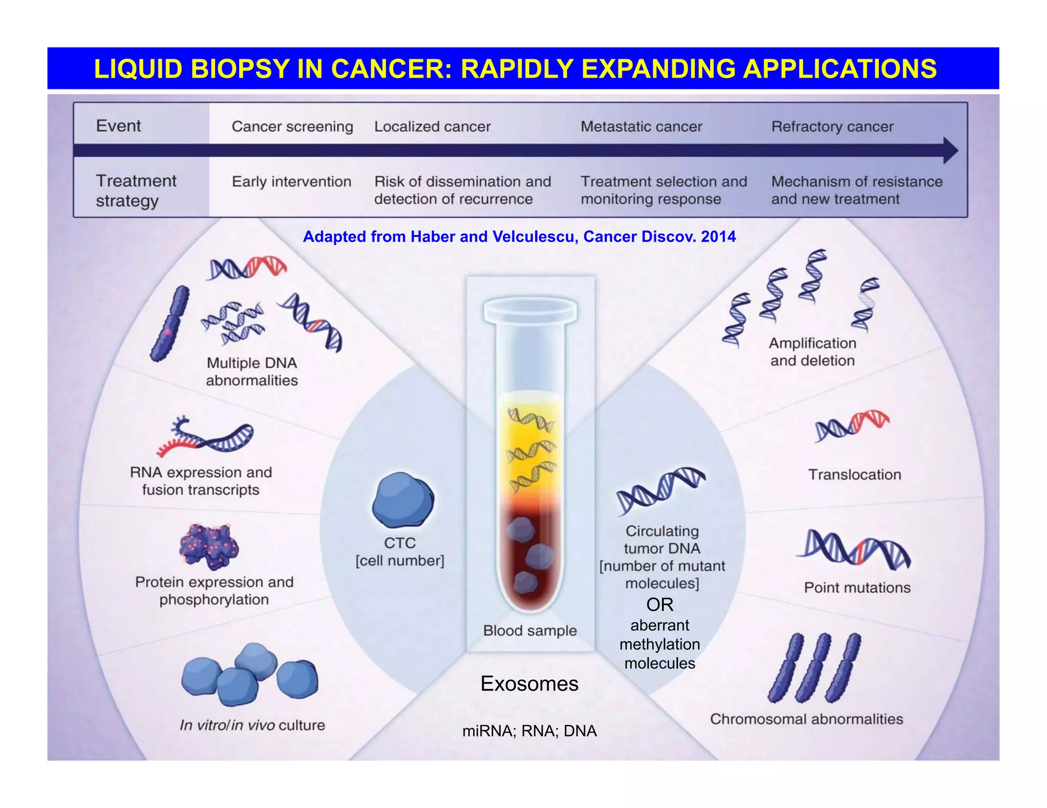
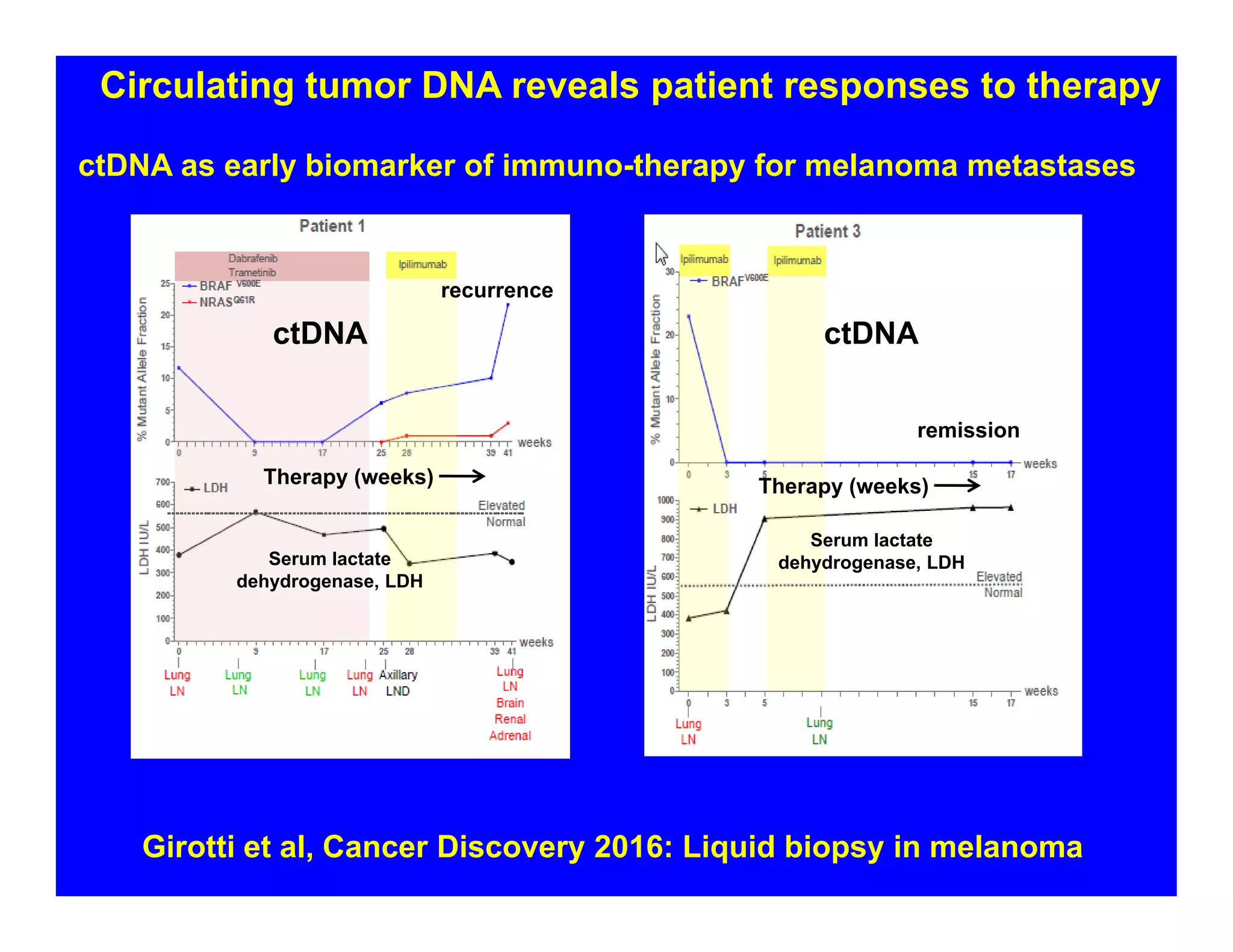
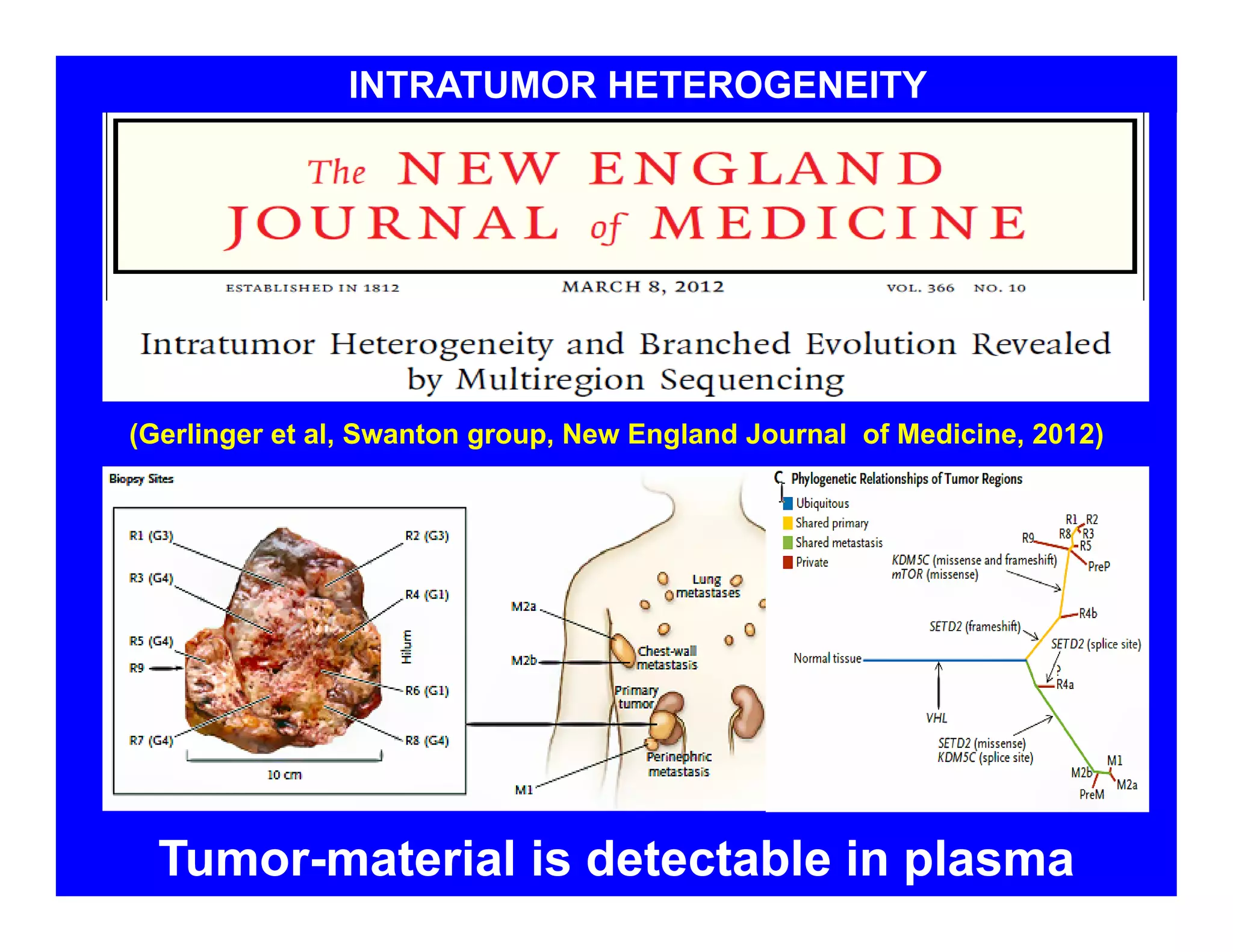
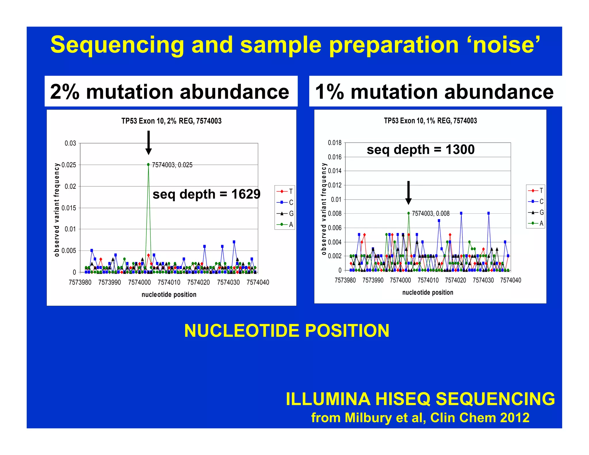
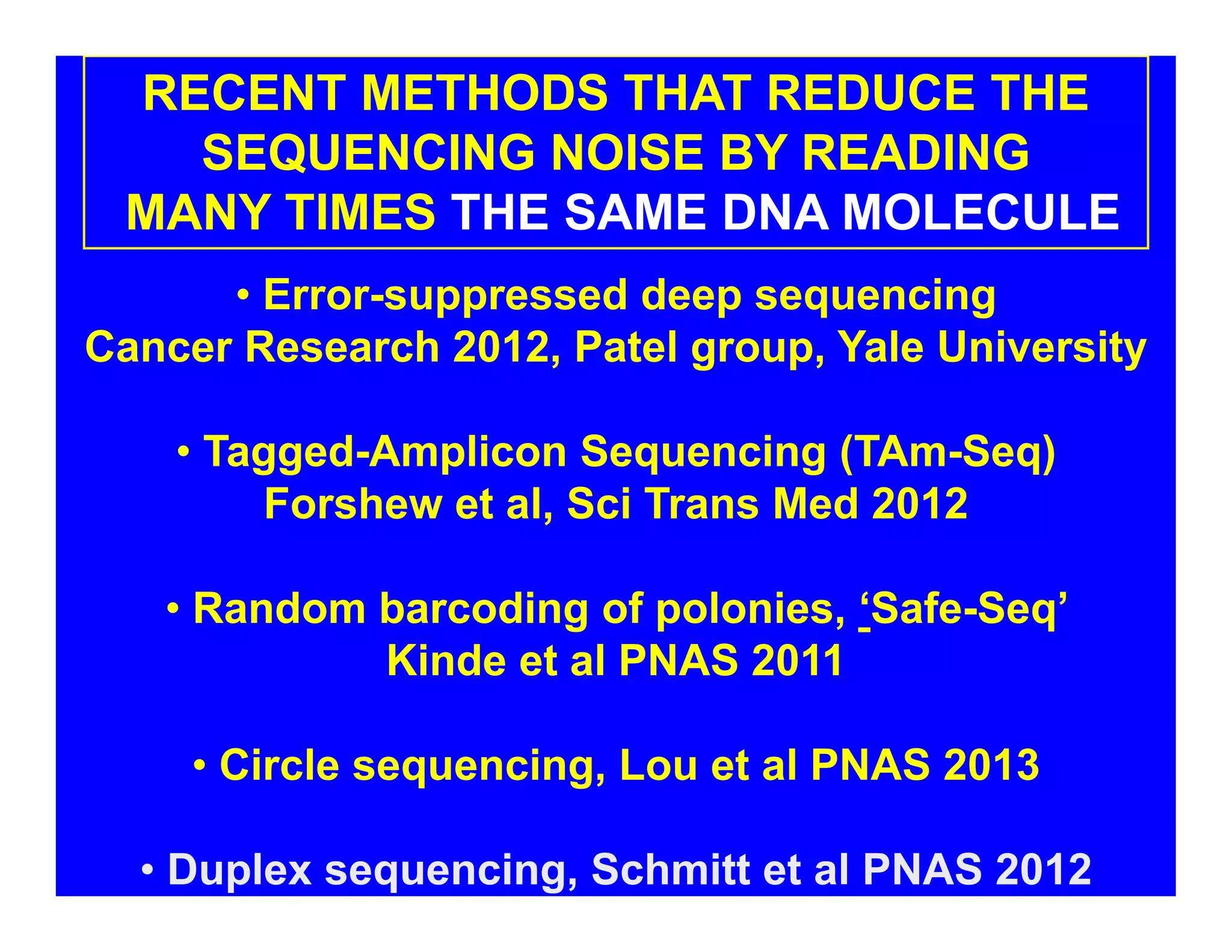
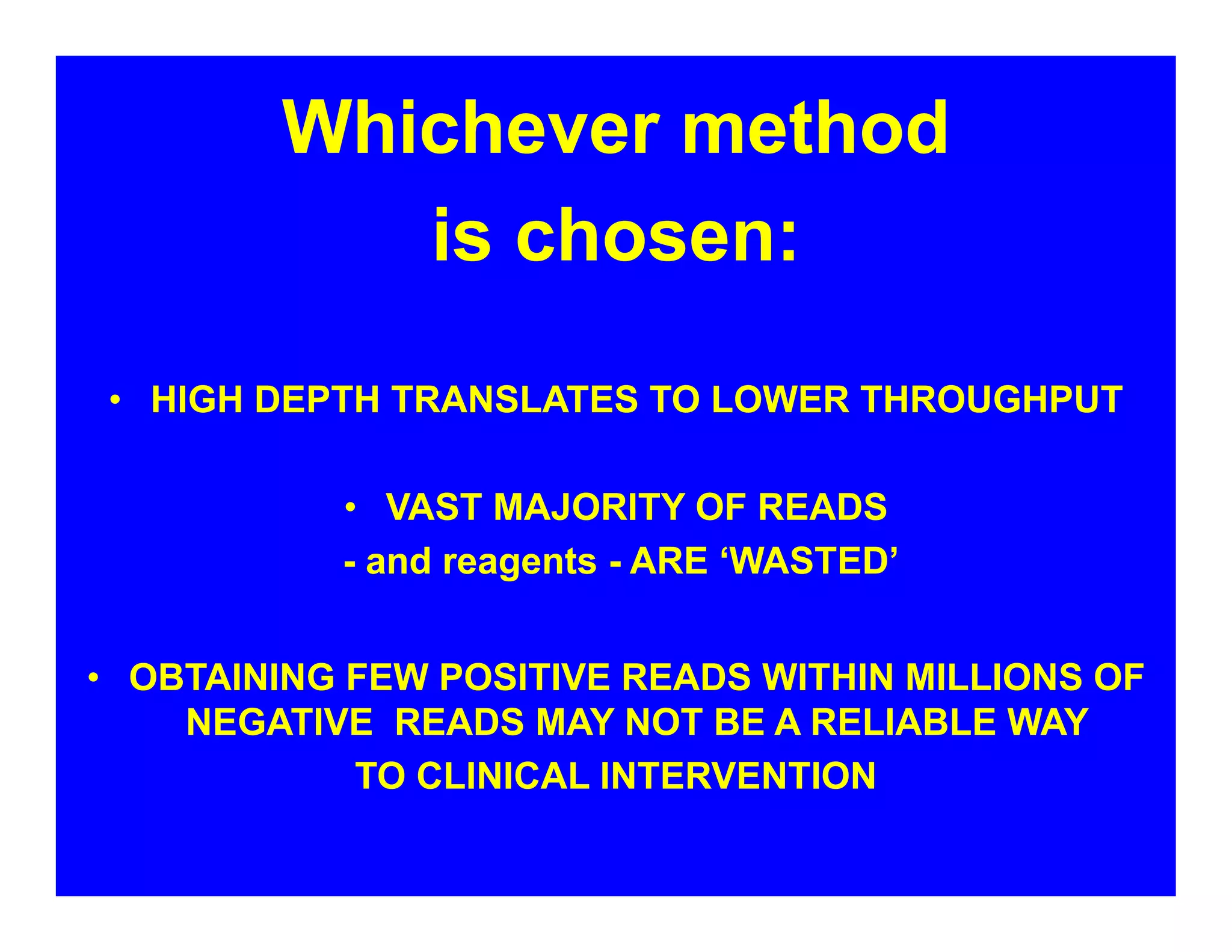
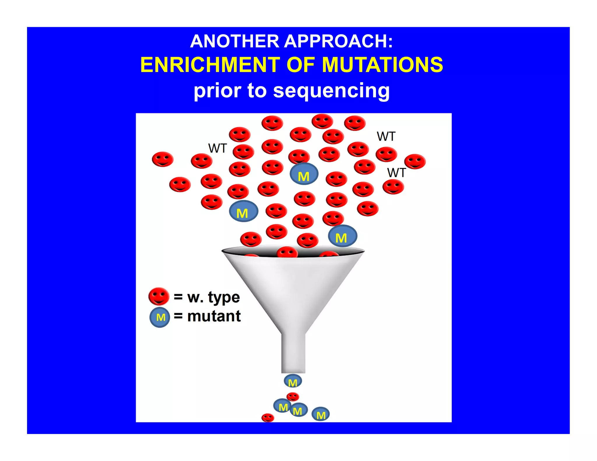
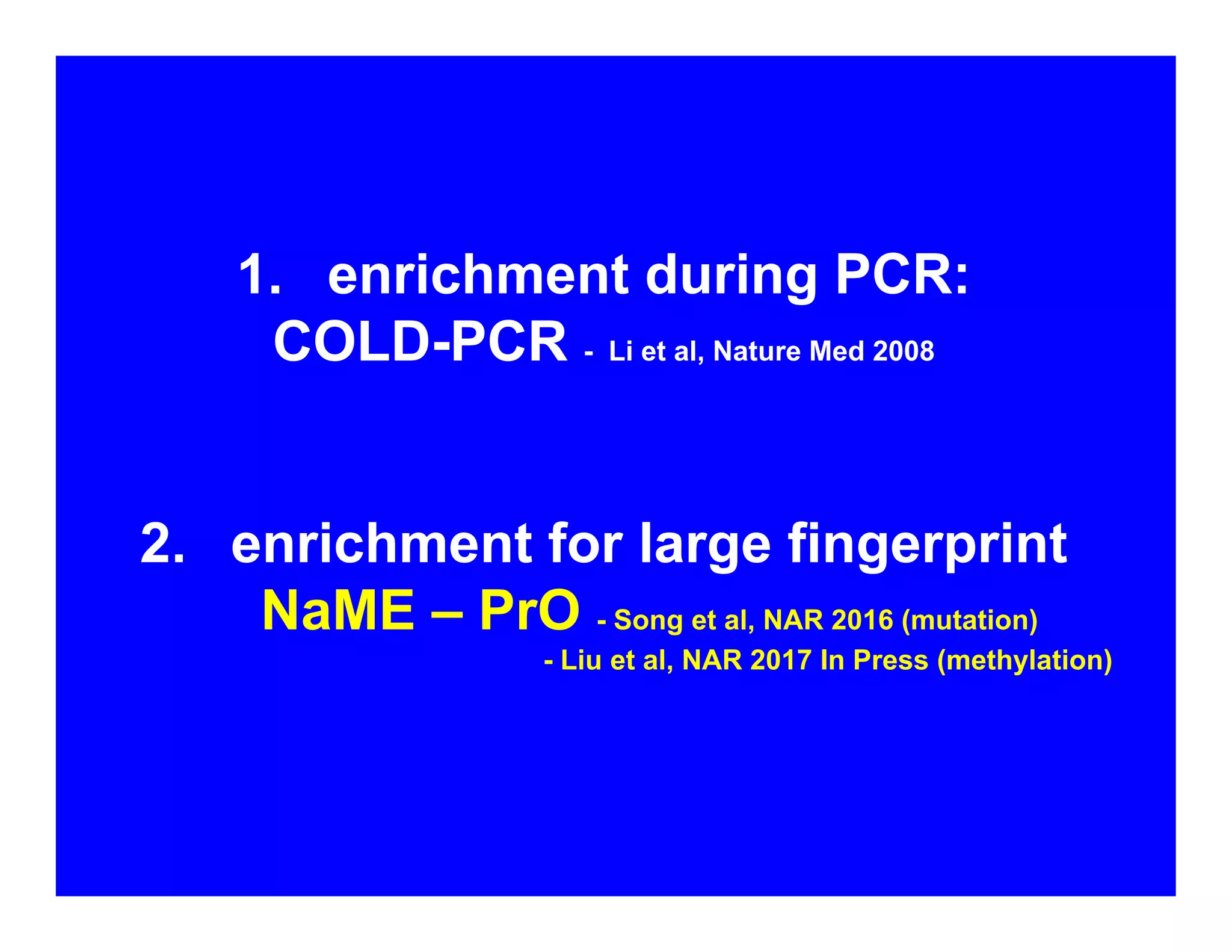
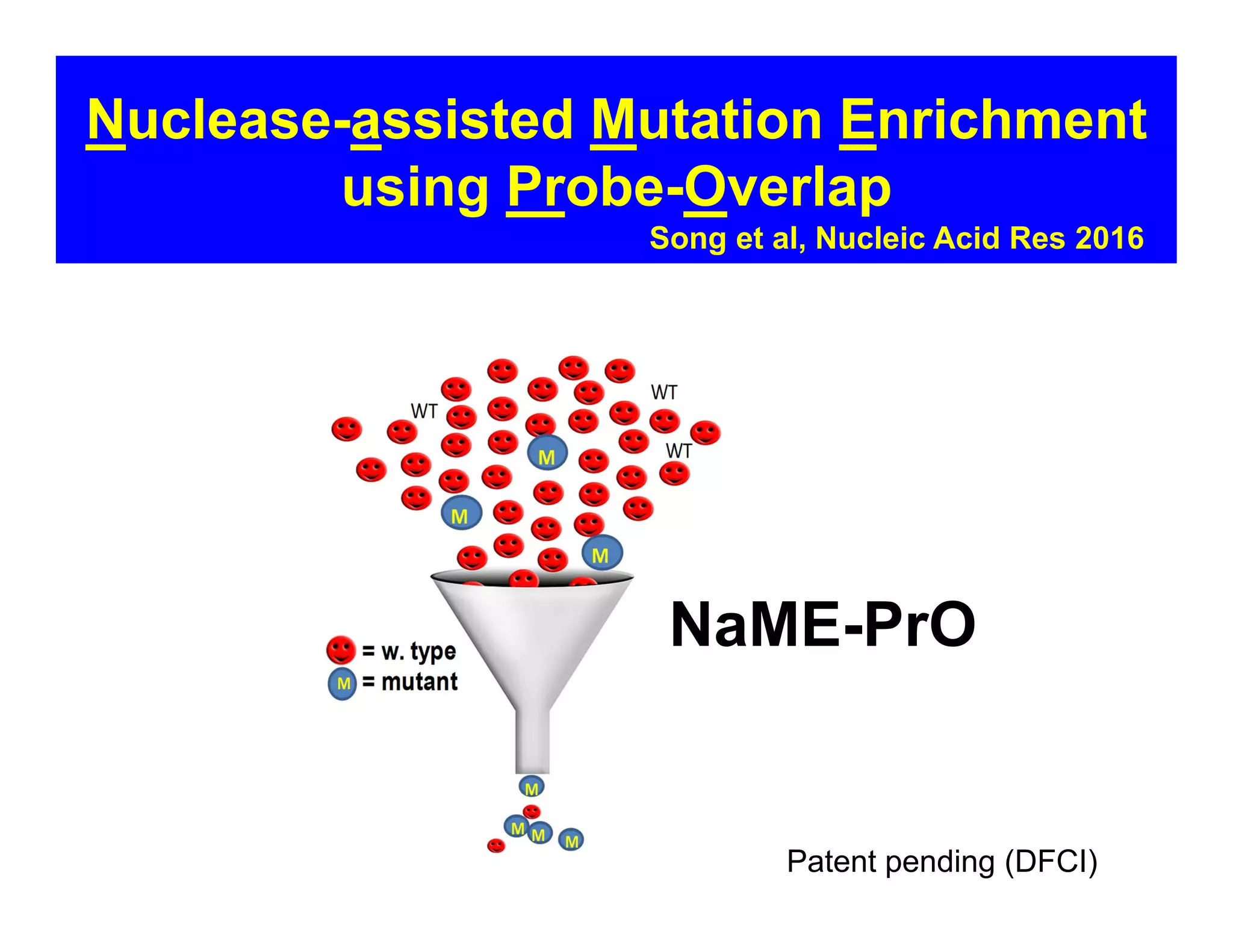
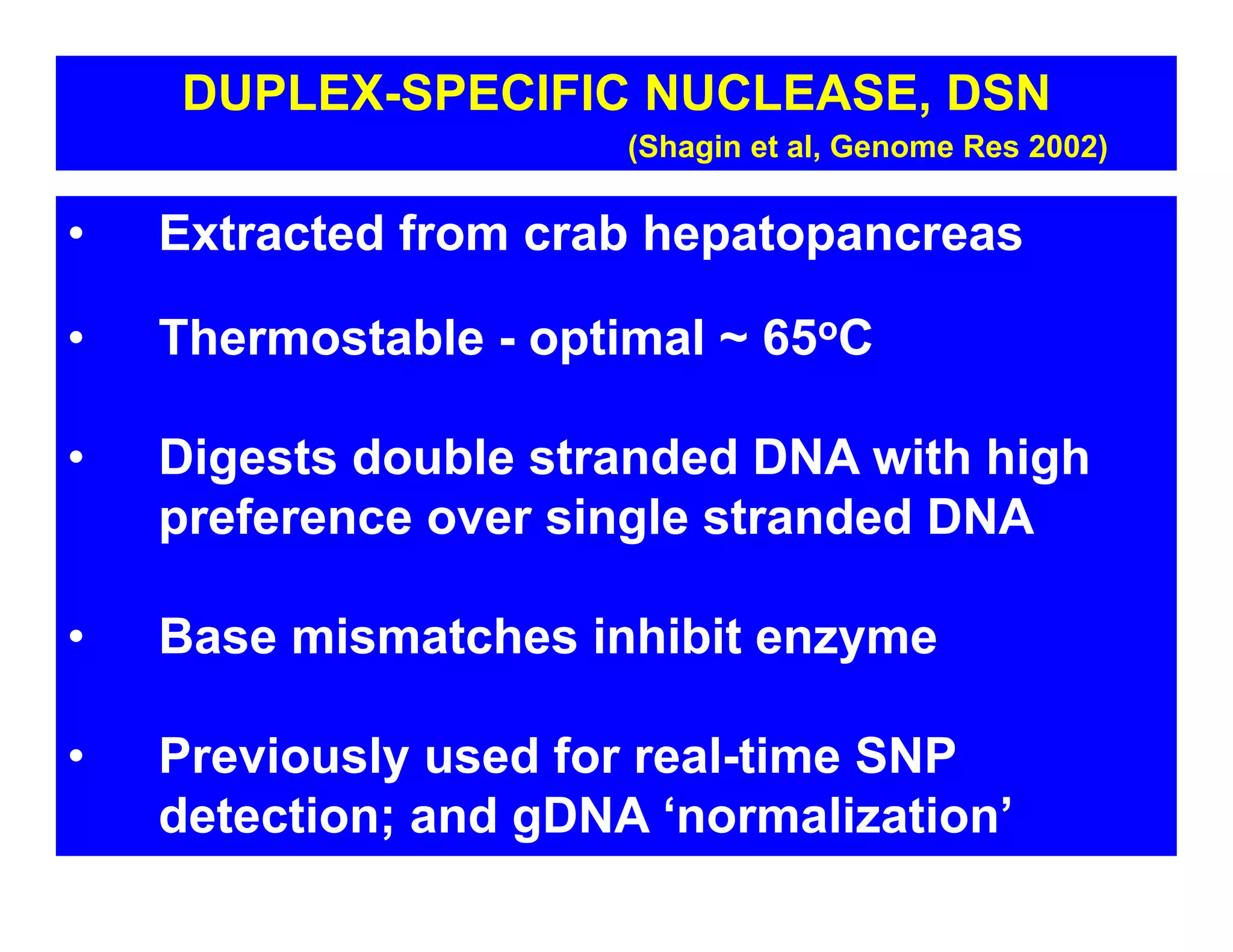
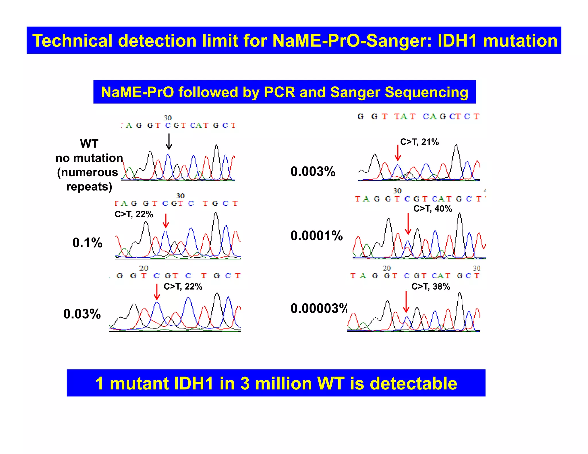
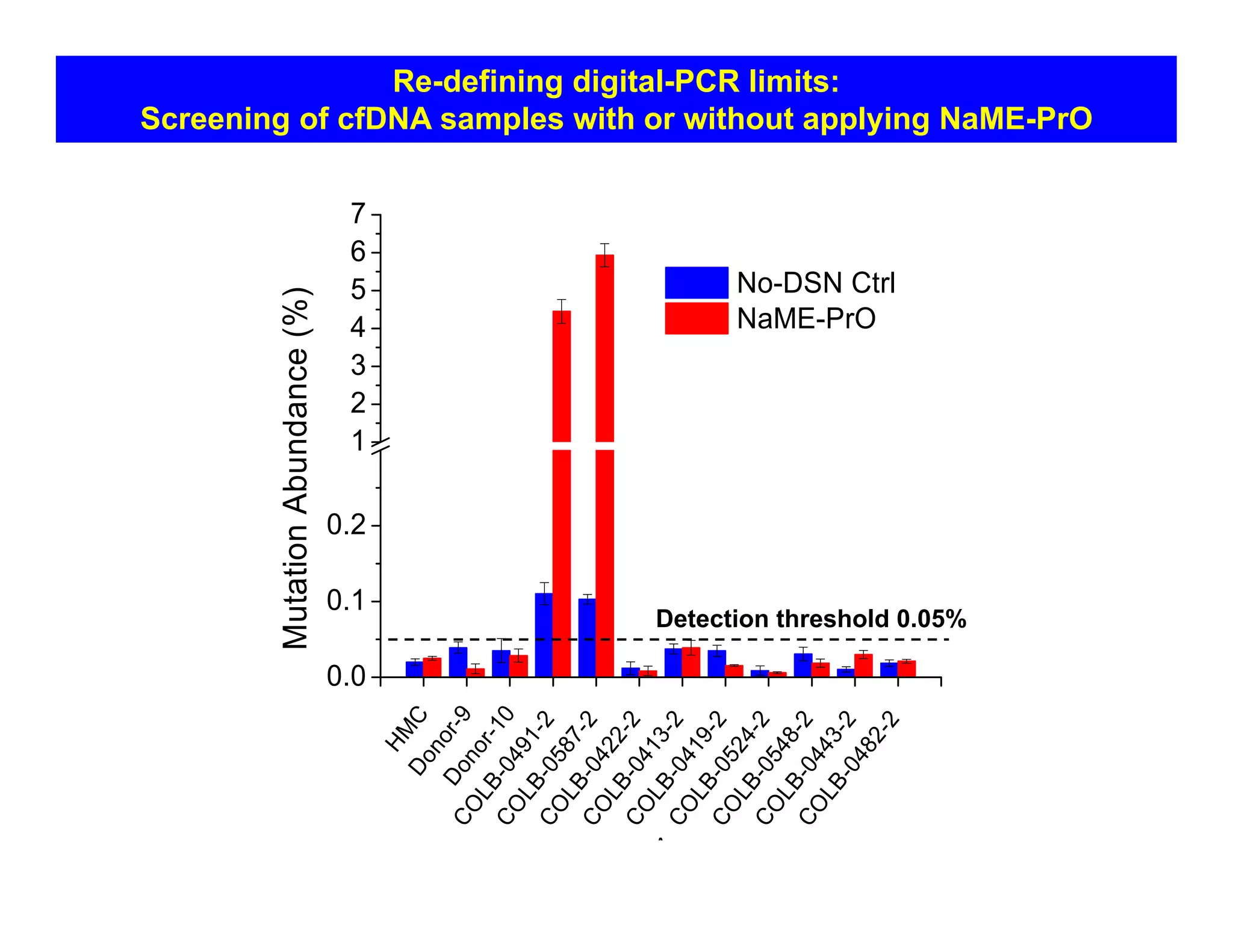
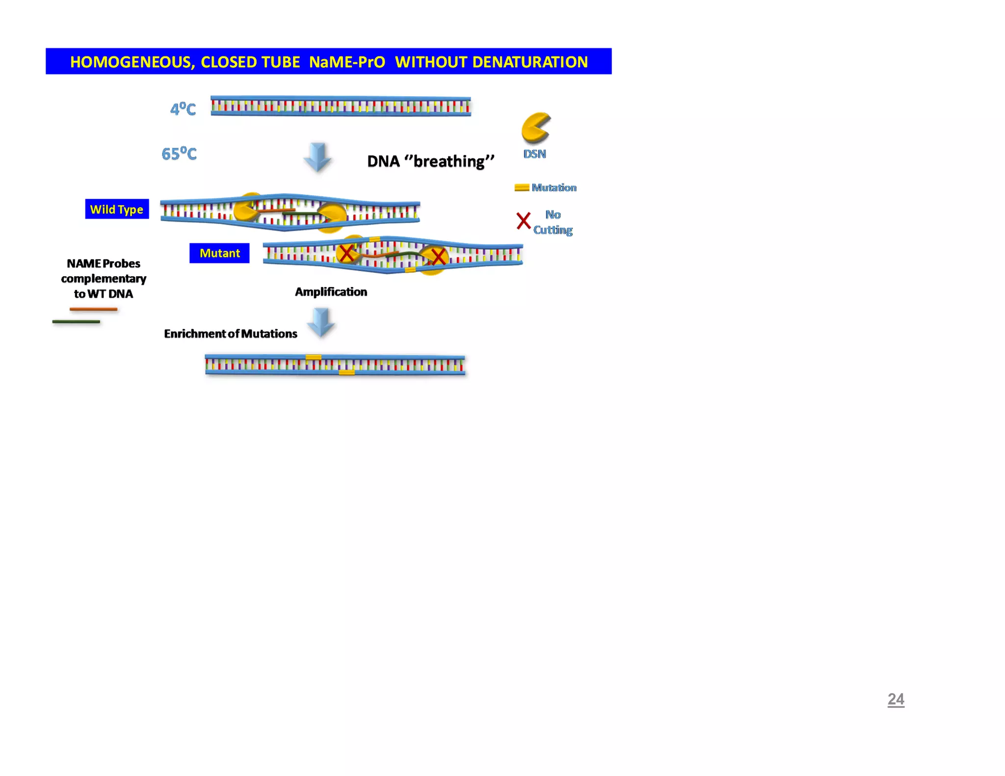
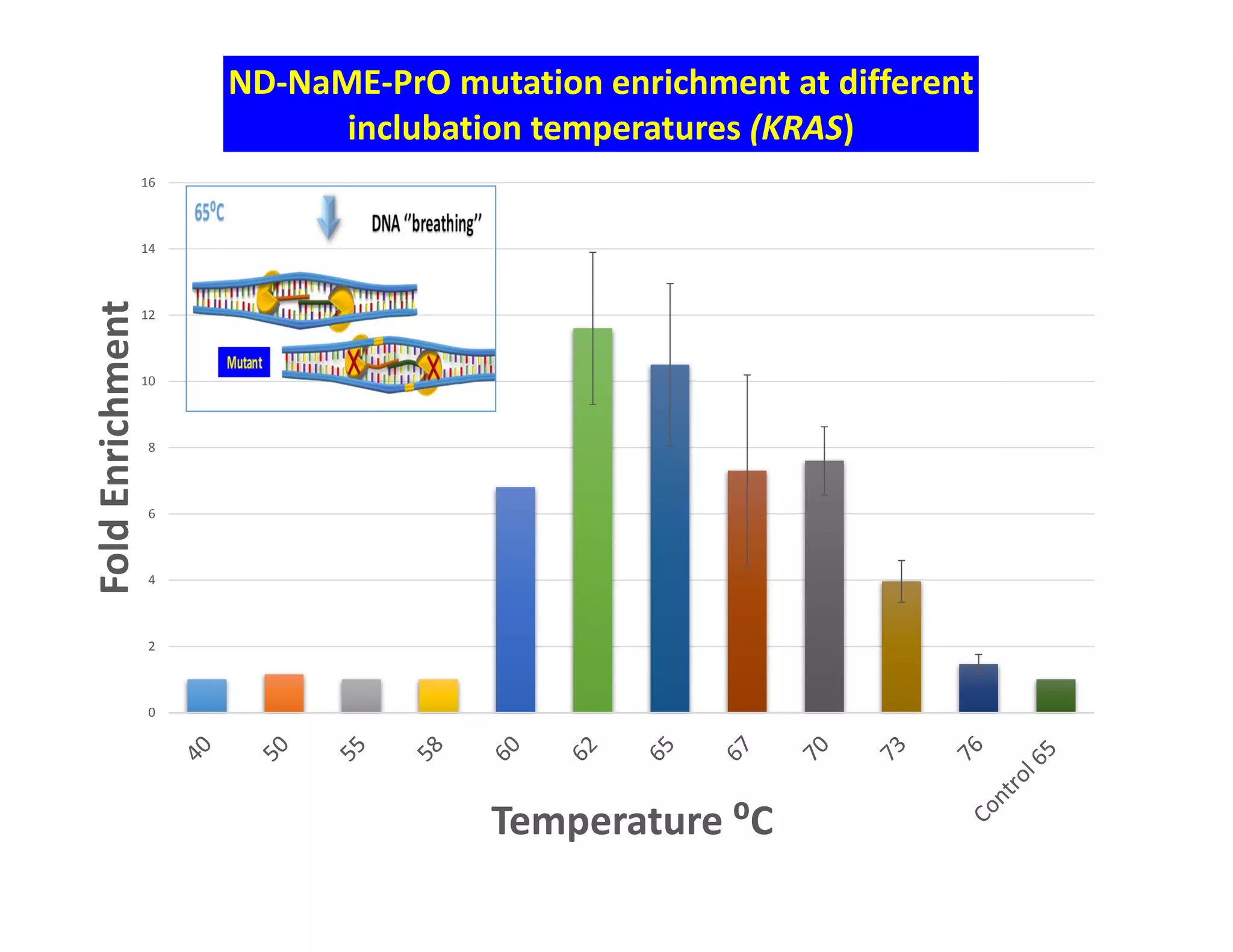
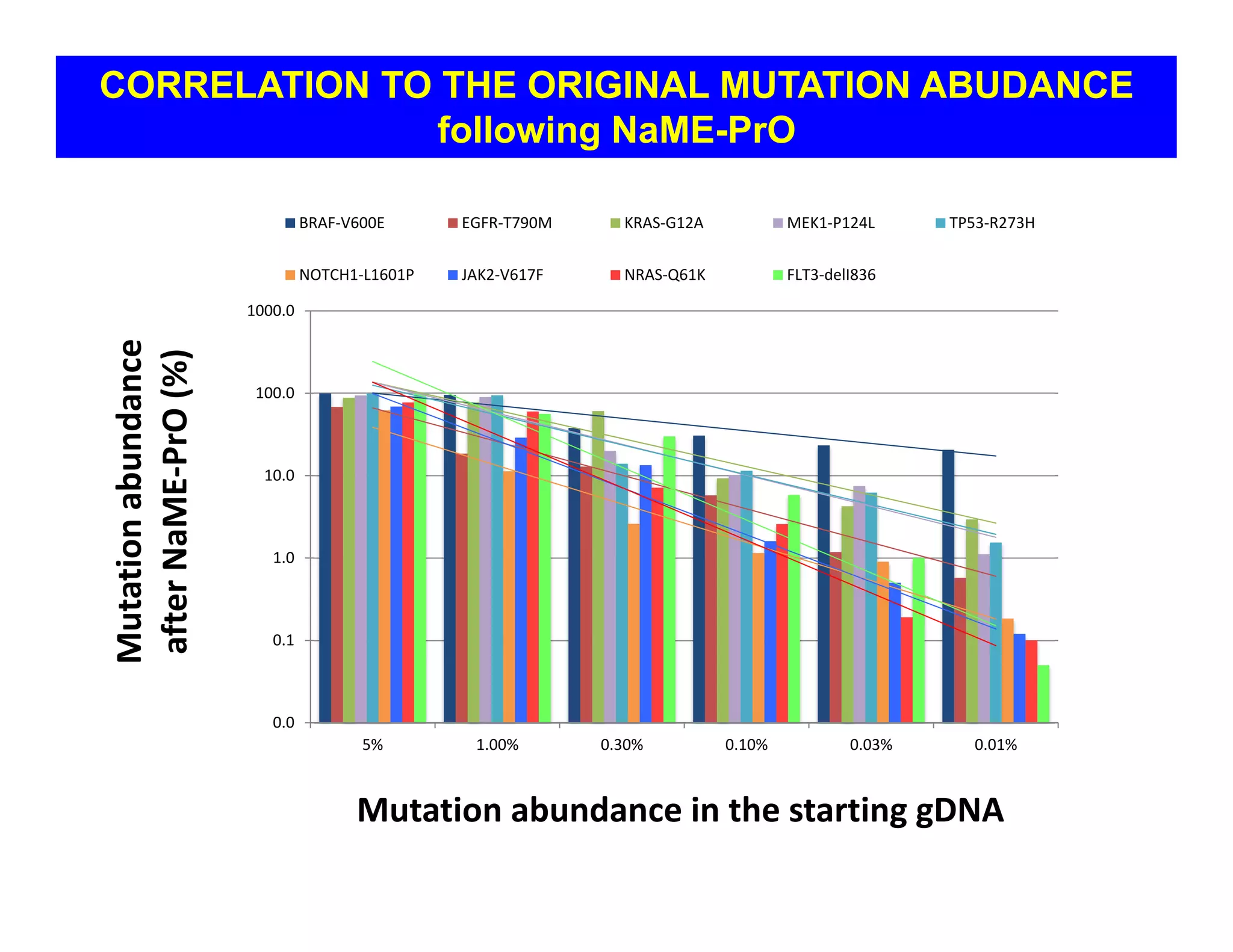
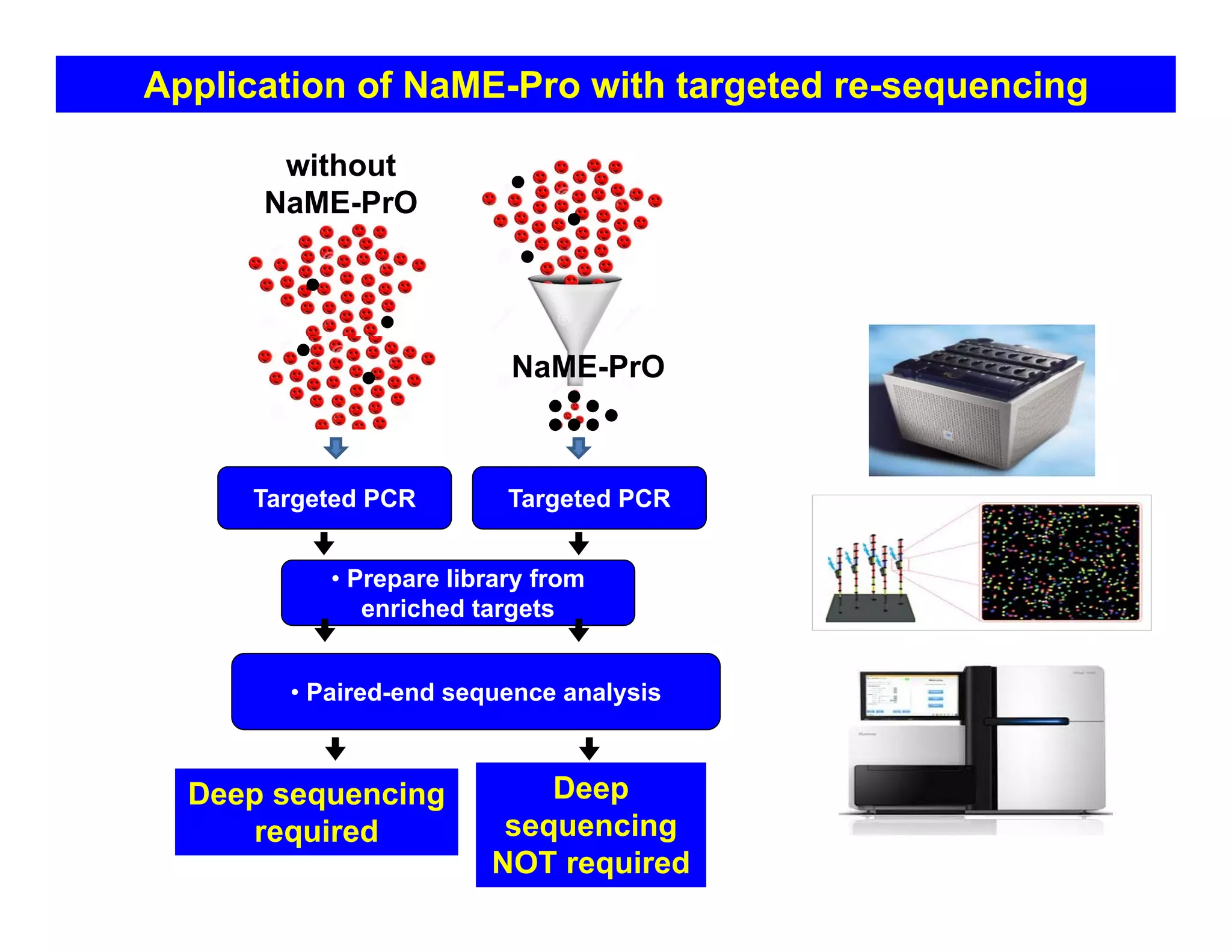
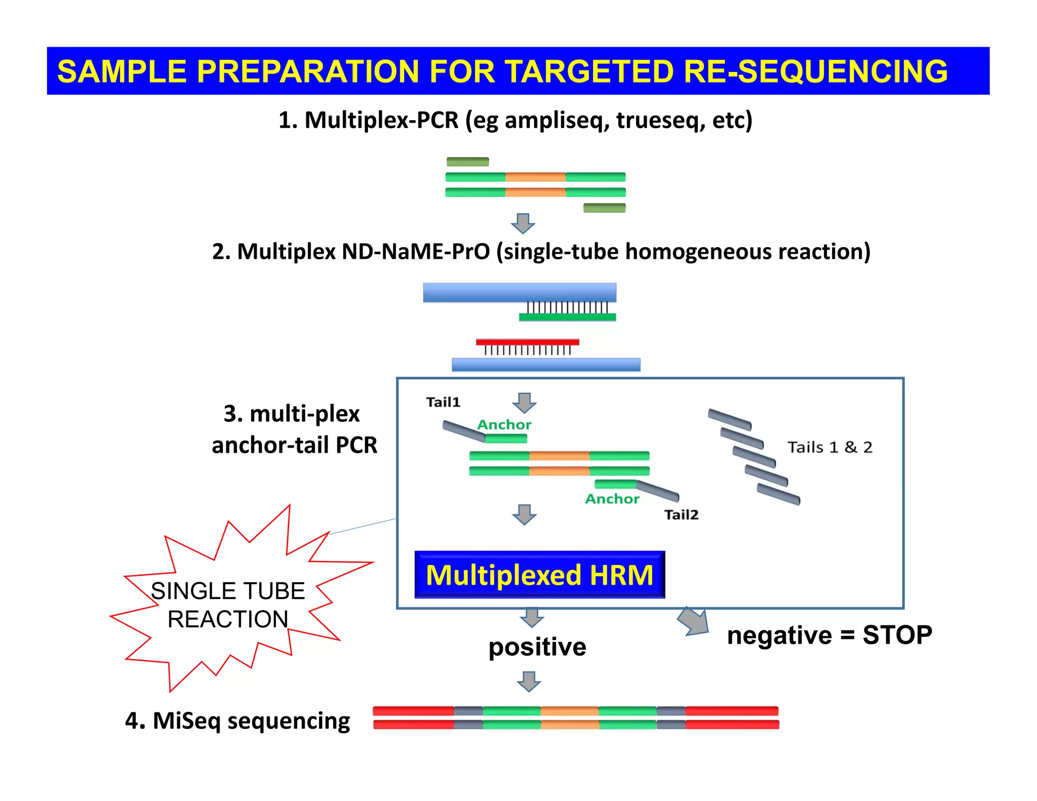
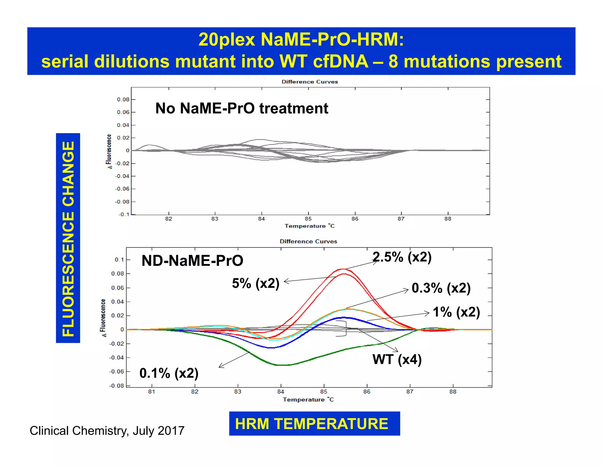
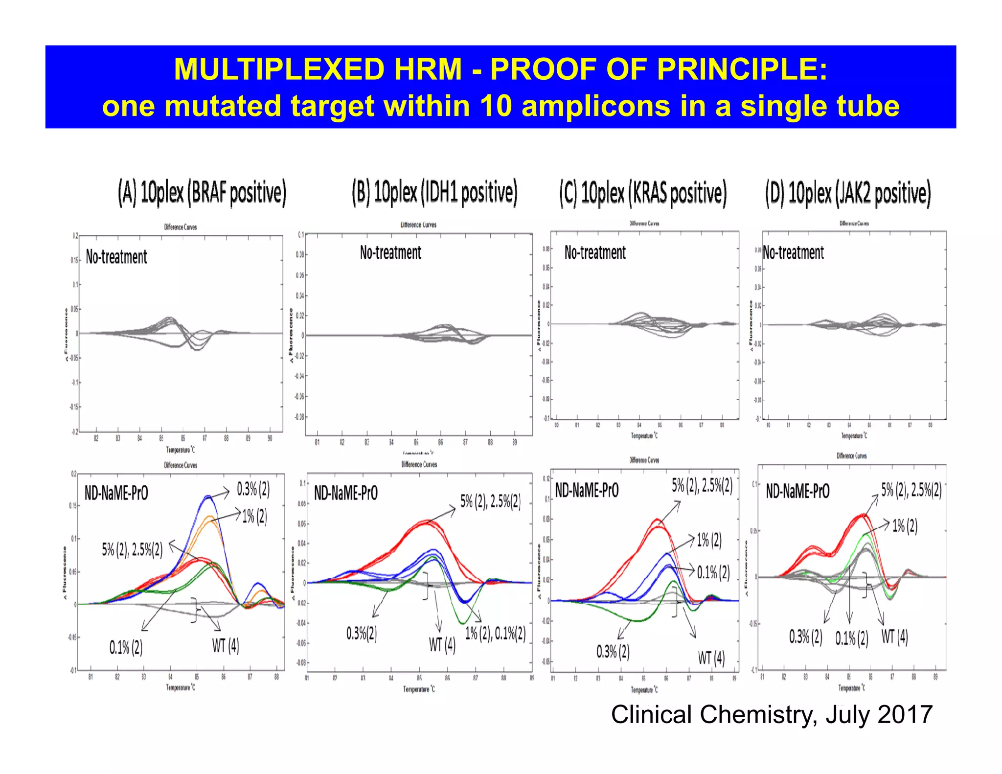
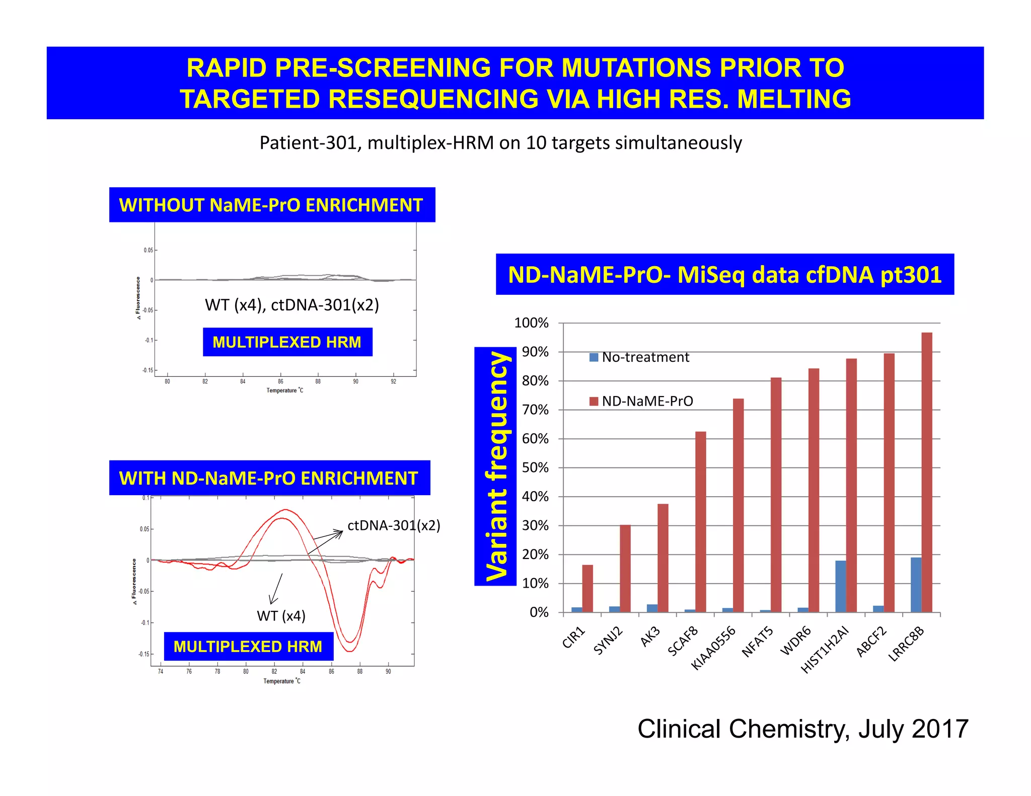
Multi-step real time PCR coupled with HRM
enables rapid mutation assessment
prior to targeted re-sequencing
G. Mike Makrigiorgos, Ph.D
Department of Radiation Oncology
Dana Farber Cancer Institute
Harvard Medical School, Boston MA
LIQUID BIOPSY IN CANCER: RAPIDLY EXPANDING APPLICATIONS
Exosomes
miRNA; RNA; DNA
OR
aberrant
methylation
molecules
Adapted from Haber and Velculescu, Cancer Discov. 2014
Girotti et al, Cancer Discovery 2016: Liquid biopsy in melanoma
Circulating tumor DNA reveals patient responses to therapy
recurrence
Therapy (weeks)
ctDNA
Serum lactate
dehydrogenase, LDH
ctDNA as early biomarker of immuno-therapy for melanoma metastases
ctDNA
Therapy (weeks)
Serum lactate
dehydrogenase, LDH
remission
INTRATUMOR HETEROGENEITY
(Gerlinger et al, Swanton group, New England Journal of Medicine, 2012)
Tumor-material is detectable in plasma
To increase sensitivity-specificity
in ccfDNA diagnostics, multiple targets
need be interrogated
• Murtaza et al, Nature 2013 Non-invasive
analysis of acquired resistance to cancer
therapy by sequencing of plasma DNA
• Newman et al, Nature Med 2014 (CAPP
sequencing) An ultrasensitive method for
quantitating circulating tumor DNA with broad
patient coverage
Next Generation Sequencing Technology:
revolutionizing personalized medicine
and tumor biology
-but good enough for detecting low-level mutations
in heterogeneous tumors or plasma??
• PROBLEM 1:
THE ‘DEPTH’
The lower the mutant fraction
the more times a sequence
needs to be sampled
CURRENTLY IT IS NOT
COST - EFFECTIVE TO SEQUENCE THE
ENTIRE GENOME MULTIPLE TIMES
e.g. HiSeq ~40 Gb/lane~4x each allele
GENOME FRACTIONATION
(targeted re-sequencing)
IS MANDATORY FOR MUTATIONS AT
LEVELS ~1% OR BELOW
•PROBLEM 2:
THE ‘NOISE’
TP53 Exon 10, 2% REG, 7574003
7574003, 0.025
0
0.005
0.01
0.015
0.02
0.025
0.03
7573980 7573990 7574000 7574010 7574020 7574030 7574040
nucleotide position
observedvariantfrequency
T
C
G
A
seq depth = 1629
2% mutation abundance
TP53 Exon 10, 1% REG, 7574003
7574003, 0.008
0
0.002
0.004
0.006
0.008
0.01
0.012
0.014
0.016
0.018
7573980 7573990 7574000 7574010 7574020 7574030 7574040
nucleotide position
observedvariantfrequency
T
C
G
A
seq depth = 1300
1% mutation abundance
ILLUMINA HISEQ SEQUENCING
from Milbury et al, Clin Chem 2012
Sequencing and sample preparation ‘noise’
NUCLEOTIDE POSITION
RECENT METHODS THAT REDUCE THE
SEQUENCING NOISE BY READING
MANY TIMES THE SAME DNA MOLECULE
• Error-suppressed deep sequencing
Cancer Research 2012, Patel group, Yale University
• Tagged-Amplicon Sequencing (TAm-Seq)
Forshew et al, Sci Trans Med 2012
• Random barcoding of polonies, ‘Safe-Seq’
Kinde et al PNAS 2011
• Circle sequencing, Lou et al PNAS 2013
• Duplex sequencing, Schmitt et al PNAS 2012
Whichever method
is chosen:
• HIGH DEPTH TRANSLATES TO LOWER THROUGHPUT
• VAST MAJORITY OF READS
- and reagents - ARE ‘WASTED’
• OBTAINING FEW POSITIVE READS WITHIN MILLIONS OF
NEGATIVE READS MAY NOT BE A RELIABLE WAY
TO CLINICAL INTERVENTION
ANOTHER APPROACH:
ENRICHMENT OF MUTATIONS
prior to sequencing
1. enrichment during PCR:
COLD-PCR - Li et al, Nature Med 2008
2. enrichment for large fingerprint
NaME – PrO - Song et al, NAR 2016 (mutation)
- Liu et al, NAR 2017 In Press (methylation)
Nuclease-assisted Mutation Enrichment
using Probe-Overlap
Song et al, Nucleic Acid Res 2016
NaME-PrO
Patent pending (DFCI)
• Extracted from crab hepatopancreas
• Thermostable - optimal ~ 65oC
• Digests double stranded DNA with high
preference over single stranded DNA
• Base mismatches inhibit enzyme
• Previously used for real-time SNP
detection; and gDNA ‘normalization’
DUPLEX-SPECIFIC NUCLEASE, DSN
(Shagin et al, Genome Res 2002)
Double strand
specific nuclease
(DSN, optimal Tm ~ 65oC)
TOP STRAND PROBES
BOTTOM STRAND PROBES
Partially overlapping probes
Target region
Nuclease-assisted Mutation Enrichment
using Probe Overlap (NaME-PrO)
Mutant sequenceWild type sequence
4oC
denature
95oC
PCR
NaME-PrO
65oC
lower temperature, add oligos, add DSN
no cutcut
Song et al, NAR 2016
Single-target NaME-PrO on KRAS exon 2
0.92 0.52
4.72
83.75
0.69 0.50
0
10
20
30
40
50
60
70
80
90
Mutation Abundance (%)
KRAS, p.G12V, c.35G>T
NaME-PrO PCR
KRAS
ddPCR ddPCR
mutation
enrichment
Wild type KRAS
Mutant KRAS
original
mutation
abundance
~0.5%
0.0
20.0
40.0
60.0
80.0
100.0
120.0
Mutation Abundance (%)
No‐DSN +DSN gDNA ctrl
9-plex-NaME 50plex-NaME
NaME-PrO multiplex assay (1% original mutation abundance)
0
20
40
60
80
100
120
Mutation Abundance (%)
No‐
DSN
+DSN
WT
no mutation
(numerous
repeats)
0.1%
C>T, 22%
0.03%
C>T, 22%
0.003%
C>T, 21%
0.0001%
C>T, 40%
0.00003%
C>T, 38%
Technical detection limit for NaME-PrO-Sanger: IDH1 mutation
NaME-PrO followed by PCR and Sanger Sequencing
1 mutant IDH1 in 3 million WT is detectable
HM
CDonor-9Donor-10
COLB-0491-2
CO
LB-0587-2
CO
LB-0422-2
COLB-0413-2
COLB-0419-2
CO
LB-0524-2
CO
LB-0548-2
COLB-0443-2
COLB-0482-2
0.0
0.1
0.2
1
2
3
4
5
6
7
MutationAbundance(%)
A
No-DSN Ctrl
NaME-PrO
Detection threshold 0.05%
Re-defining digital-PCR limits:
Screening of cfDNA samples with or without applying NaME-PrO
HOMOGENOUS,
CLOSED TUBE NaME-PrO
Clinical Chemistry, July 2017
24
0
2
4
6
8
10
12
14
16
Fold Enrichment
Temperature ⁰C
ND‐NaME‐PrO mutation enrichment at different 
inclubation temperatures (KRAS)
0.0
0.1
1.0
10.0
100.0
1000.0
5% 1.00% 0.30% 0.10% 0.03% 0.01%
Mutation abundance 
after NaME‐PrO(%)
Mutation abundance in the starting gDNA
BRAF‐V600E EGFR‐T790M KRAS‐G12A MEK1‐P124L TP53‐R273H
NOTCH1‐L1601P JAK2‐V617F NRAS‐Q61K FLT3‐delI836
CORRELATION TO THE ORIGINAL MUTATION ABUDANCE
following NaME-PrO
Application of NaME-Pro with targeted re-sequencing
Targeted PCR
• Prepare library from
enriched targets
• Paired-end sequence analysis
Deep sequencing
required
Targeted PCR
NaME-PrO
Deep
sequencing
NOT required
without
NaME-PrO
PRE-SCREENING VIA
Multiplexed-PCR-HRM
Clinical Chemistry, July 2017
(advanced online publication)
SAMPLE PREPARATION FOR TARGETED RE-SEQUENCING
1. Multiplex‐PCR (eg ampliseq, trueseq, etc)
Multiplexed HRM
4. MiSeq sequencing
positive negative = STOP
3. multi‐plex
anchor‐tail PCR
2. Multiplex ND‐NaME‐PrO (single‐tube homogeneous reaction)
SINGLE TUBE
REACTION
No NaME-PrO treatment
ND-NaME-PrO
WT (x4)
5% (x2)
2.5% (x2)
0.3% (x2)
1% (x2)
0.1% (x2)
20plex NaME-PrO-HRM:
serial dilutions mutant into WT cfDNA – 8 mutations present
Clinical Chemistry, July 2017
FLUORESCENCECHANGE
HRM TEMPERATURE
MULTIPLEXED HRM - PROOF OF PRINCIPLE:
one mutated target within 10 amplicons in a single tube
Clinical Chemistry, July 2017
0%
10%
20%
30%
40%
50%
60%
70%
80%
90%
100%
Variant frequency
No‐treatment
ND‐NaME‐PrO
ND‐NaME‐PrO‐ MiSeq data cfDNA pt301
Patient‐301, multiplex‐HRM on 10 targets simultaneously
WITHOUT NaME‐PrO ENRICHMENT
WT (x4), ctDNA‐301(x2)
WITH ND‐NaME‐PrO ENRICHMENT
WT (x4)
ctDNA‐301(x2)
RAPID PRE-SCREENING FOR MUTATIONS PRIOR TO
TARGETED RESEQUENCING VIA HIGH RES. MELTING
MULTIPLEXED HRM
MULTIPLEXED HRM
Clinical Chemistry, July 2017
CONCLUSIONS
• Use of circulating DNA as a biomarker for multiple clinical
endpoints in oncology is increasing rapidly
• Next generation sequencing is preferred for multiple markers
• Digital PCR and qPCR are preferred for single markers
• Sample preparation for NGS remains long and expensive process
• Mutation enrichment can greatly simplify NGS sample preparation
• NaME-PrO and COLD-PCR blend well with NGS sample
preparation, or with qPCR-only detection of cfDNA biomarkers
DFCI-BWH-MGH
Heather Parsons, MD
Keith Flaherty, MD
Ryan Sullivan, MD
Harvey Mamon, MD, Ph.D
Matthew Kulke, MD
John Quackenbush, Ph.D
Benjamin Ebert, MD, PhD
Rafael Bejar, MD, PhD
Contributors and Collaborators
Funding: NIH / NCI IMAT Program; Bridge Foundation (DFHCC)
THANK YOU FOR YOUR ATTENTION
BROAD INSTITUTE
Viktor Adalsteinsson Ph.D
MIT
Yaping Liu PhD
MAKRIGIORGOS LAB
Mariana Fitarelli-Kiehl, Ph.D
Ioannis Ladas, Ph.D
Fangyan Wu, Ph.D
Ravina Ashtaputre, BA
Carrie Leong, Ph.D

Multi-step real time PCR coupled with HRM enables rapid mutation assessment prior to targeted re-sequencing

  • 1.
    Multi-step real timePCR coupled with HRM enables rapid mutation assessment prior to targeted re-sequencing G. Mike Makrigiorgos, Ph.D Department of Radiation Oncology Dana Farber Cancer Institute Harvard Medical School, Boston MA
  • 2.
    LIQUID BIOPSY INCANCER: RAPIDLY EXPANDING APPLICATIONS Exosomes miRNA; RNA; DNA OR aberrant methylation molecules Adapted from Haber and Velculescu, Cancer Discov. 2014
  • 3.
    Girotti et al,Cancer Discovery 2016: Liquid biopsy in melanoma Circulating tumor DNA reveals patient responses to therapy recurrence Therapy (weeks) ctDNA Serum lactate dehydrogenase, LDH ctDNA as early biomarker of immuno-therapy for melanoma metastases ctDNA Therapy (weeks) Serum lactate dehydrogenase, LDH remission
  • 4.
    INTRATUMOR HETEROGENEITY (Gerlinger etal, Swanton group, New England Journal of Medicine, 2012) Tumor-material is detectable in plasma
  • 5.
    To increase sensitivity-specificity inccfDNA diagnostics, multiple targets need be interrogated • Murtaza et al, Nature 2013 Non-invasive analysis of acquired resistance to cancer therapy by sequencing of plasma DNA • Newman et al, Nature Med 2014 (CAPP sequencing) An ultrasensitive method for quantitating circulating tumor DNA with broad patient coverage
  • 6.
    Next Generation SequencingTechnology: revolutionizing personalized medicine and tumor biology -but good enough for detecting low-level mutations in heterogeneous tumors or plasma??
  • 7.
    • PROBLEM 1: THE‘DEPTH’ The lower the mutant fraction the more times a sequence needs to be sampled
  • 8.
    CURRENTLY IT ISNOT COST - EFFECTIVE TO SEQUENCE THE ENTIRE GENOME MULTIPLE TIMES e.g. HiSeq ~40 Gb/lane~4x each allele GENOME FRACTIONATION (targeted re-sequencing) IS MANDATORY FOR MUTATIONS AT LEVELS ~1% OR BELOW
  • 9.
  • 10.
    TP53 Exon 10,2% REG, 7574003 7574003, 0.025 0 0.005 0.01 0.015 0.02 0.025 0.03 7573980 7573990 7574000 7574010 7574020 7574030 7574040 nucleotide position observedvariantfrequency T C G A seq depth = 1629 2% mutation abundance TP53 Exon 10, 1% REG, 7574003 7574003, 0.008 0 0.002 0.004 0.006 0.008 0.01 0.012 0.014 0.016 0.018 7573980 7573990 7574000 7574010 7574020 7574030 7574040 nucleotide position observedvariantfrequency T C G A seq depth = 1300 1% mutation abundance ILLUMINA HISEQ SEQUENCING from Milbury et al, Clin Chem 2012 Sequencing and sample preparation ‘noise’ NUCLEOTIDE POSITION
  • 11.
    RECENT METHODS THATREDUCE THE SEQUENCING NOISE BY READING MANY TIMES THE SAME DNA MOLECULE • Error-suppressed deep sequencing Cancer Research 2012, Patel group, Yale University • Tagged-Amplicon Sequencing (TAm-Seq) Forshew et al, Sci Trans Med 2012 • Random barcoding of polonies, ‘Safe-Seq’ Kinde et al PNAS 2011 • Circle sequencing, Lou et al PNAS 2013 • Duplex sequencing, Schmitt et al PNAS 2012
  • 12.
    Whichever method is chosen: •HIGH DEPTH TRANSLATES TO LOWER THROUGHPUT • VAST MAJORITY OF READS - and reagents - ARE ‘WASTED’ • OBTAINING FEW POSITIVE READS WITHIN MILLIONS OF NEGATIVE READS MAY NOT BE A RELIABLE WAY TO CLINICAL INTERVENTION
  • 13.
    ANOTHER APPROACH: ENRICHMENT OFMUTATIONS prior to sequencing
  • 14.
    1. enrichment duringPCR: COLD-PCR - Li et al, Nature Med 2008 2. enrichment for large fingerprint NaME – PrO - Song et al, NAR 2016 (mutation) - Liu et al, NAR 2017 In Press (methylation)
  • 15.
    Nuclease-assisted Mutation Enrichment usingProbe-Overlap Song et al, Nucleic Acid Res 2016 NaME-PrO Patent pending (DFCI)
  • 16.
    • Extracted fromcrab hepatopancreas • Thermostable - optimal ~ 65oC • Digests double stranded DNA with high preference over single stranded DNA • Base mismatches inhibit enzyme • Previously used for real-time SNP detection; and gDNA ‘normalization’ DUPLEX-SPECIFIC NUCLEASE, DSN (Shagin et al, Genome Res 2002)
  • 17.
    Double strand specific nuclease (DSN,optimal Tm ~ 65oC) TOP STRAND PROBES BOTTOM STRAND PROBES Partially overlapping probes Target region Nuclease-assisted Mutation Enrichment using Probe Overlap (NaME-PrO)
  • 18.
    Mutant sequenceWild typesequence 4oC denature 95oC PCR NaME-PrO 65oC lower temperature, add oligos, add DSN no cutcut Song et al, NAR 2016
  • 19.
    Single-target NaME-PrO onKRAS exon 2 0.92 0.52 4.72 83.75 0.69 0.50 0 10 20 30 40 50 60 70 80 90 Mutation Abundance (%) KRAS, p.G12V, c.35G>T NaME-PrO PCR KRAS ddPCR ddPCR mutation enrichment Wild type KRAS Mutant KRAS original mutation abundance ~0.5%
  • 20.
    0.0 20.0 40.0 60.0 80.0 100.0 120.0 Mutation Abundance (%) No‐DSN +DSN gDNA ctrl 9-plex-NaME50plex-NaME NaME-PrO multiplex assay (1% original mutation abundance) 0 20 40 60 80 100 120 Mutation Abundance (%) No‐ DSN +DSN
  • 21.
    WT no mutation (numerous repeats) 0.1% C>T, 22% 0.03% C>T,22% 0.003% C>T, 21% 0.0001% C>T, 40% 0.00003% C>T, 38% Technical detection limit for NaME-PrO-Sanger: IDH1 mutation NaME-PrO followed by PCR and Sanger Sequencing 1 mutant IDH1 in 3 million WT is detectable
  • 22.
  • 23.
  • 24.
  • 25.
  • 26.
    0.0 0.1 1.0 10.0 100.0 1000.0 5% 1.00% 0.30%0.10% 0.03% 0.01% Mutation abundance  after NaME‐PrO(%) Mutation abundance in the starting gDNA BRAF‐V600E EGFR‐T790M KRAS‐G12A MEK1‐P124L TP53‐R273H NOTCH1‐L1601P JAK2‐V617F NRAS‐Q61K FLT3‐delI836 CORRELATION TO THE ORIGINAL MUTATION ABUDANCE following NaME-PrO
  • 27.
    Application of NaME-Prowith targeted re-sequencing Targeted PCR • Prepare library from enriched targets • Paired-end sequence analysis Deep sequencing required Targeted PCR NaME-PrO Deep sequencing NOT required without NaME-PrO
  • 28.
    PRE-SCREENING VIA Multiplexed-PCR-HRM Clinical Chemistry,July 2017 (advanced online publication)
  • 29.
    SAMPLE PREPARATION FORTARGETED RE-SEQUENCING 1. Multiplex‐PCR (eg ampliseq, trueseq, etc) Multiplexed HRM 4. MiSeq sequencing positive negative = STOP 3. multi‐plex anchor‐tail PCR 2. Multiplex ND‐NaME‐PrO (single‐tube homogeneous reaction) SINGLE TUBE REACTION
  • 30.
    No NaME-PrO treatment ND-NaME-PrO WT(x4) 5% (x2) 2.5% (x2) 0.3% (x2) 1% (x2) 0.1% (x2) 20plex NaME-PrO-HRM: serial dilutions mutant into WT cfDNA – 8 mutations present Clinical Chemistry, July 2017 FLUORESCENCECHANGE HRM TEMPERATURE
  • 31.
    MULTIPLEXED HRM -PROOF OF PRINCIPLE: one mutated target within 10 amplicons in a single tube Clinical Chemistry, July 2017
  • 32.
    0% 10% 20% 30% 40% 50% 60% 70% 80% 90% 100% Variant frequency No‐treatment ND‐NaME‐PrO ND‐NaME‐PrO‐ MiSeq data cfDNApt301 Patient‐301, multiplex‐HRM on 10 targets simultaneously WITHOUT NaME‐PrO ENRICHMENT WT (x4), ctDNA‐301(x2) WITH ND‐NaME‐PrO ENRICHMENT WT (x4) ctDNA‐301(x2) RAPID PRE-SCREENING FOR MUTATIONS PRIOR TO TARGETED RESEQUENCING VIA HIGH RES. MELTING MULTIPLEXED HRM MULTIPLEXED HRM Clinical Chemistry, July 2017
  • 33.
    CONCLUSIONS • Use ofcirculating DNA as a biomarker for multiple clinical endpoints in oncology is increasing rapidly • Next generation sequencing is preferred for multiple markers • Digital PCR and qPCR are preferred for single markers • Sample preparation for NGS remains long and expensive process • Mutation enrichment can greatly simplify NGS sample preparation • NaME-PrO and COLD-PCR blend well with NGS sample preparation, or with qPCR-only detection of cfDNA biomarkers
  • 34.
    DFCI-BWH-MGH Heather Parsons, MD KeithFlaherty, MD Ryan Sullivan, MD Harvey Mamon, MD, Ph.D Matthew Kulke, MD John Quackenbush, Ph.D Benjamin Ebert, MD, PhD Rafael Bejar, MD, PhD Contributors and Collaborators Funding: NIH / NCI IMAT Program; Bridge Foundation (DFHCC) THANK YOU FOR YOUR ATTENTION BROAD INSTITUTE Viktor Adalsteinsson Ph.D MIT Yaping Liu PhD MAKRIGIORGOS LAB Mariana Fitarelli-Kiehl, Ph.D Ioannis Ladas, Ph.D Fangyan Wu, Ph.D Ravina Ashtaputre, BA Carrie Leong, Ph.D