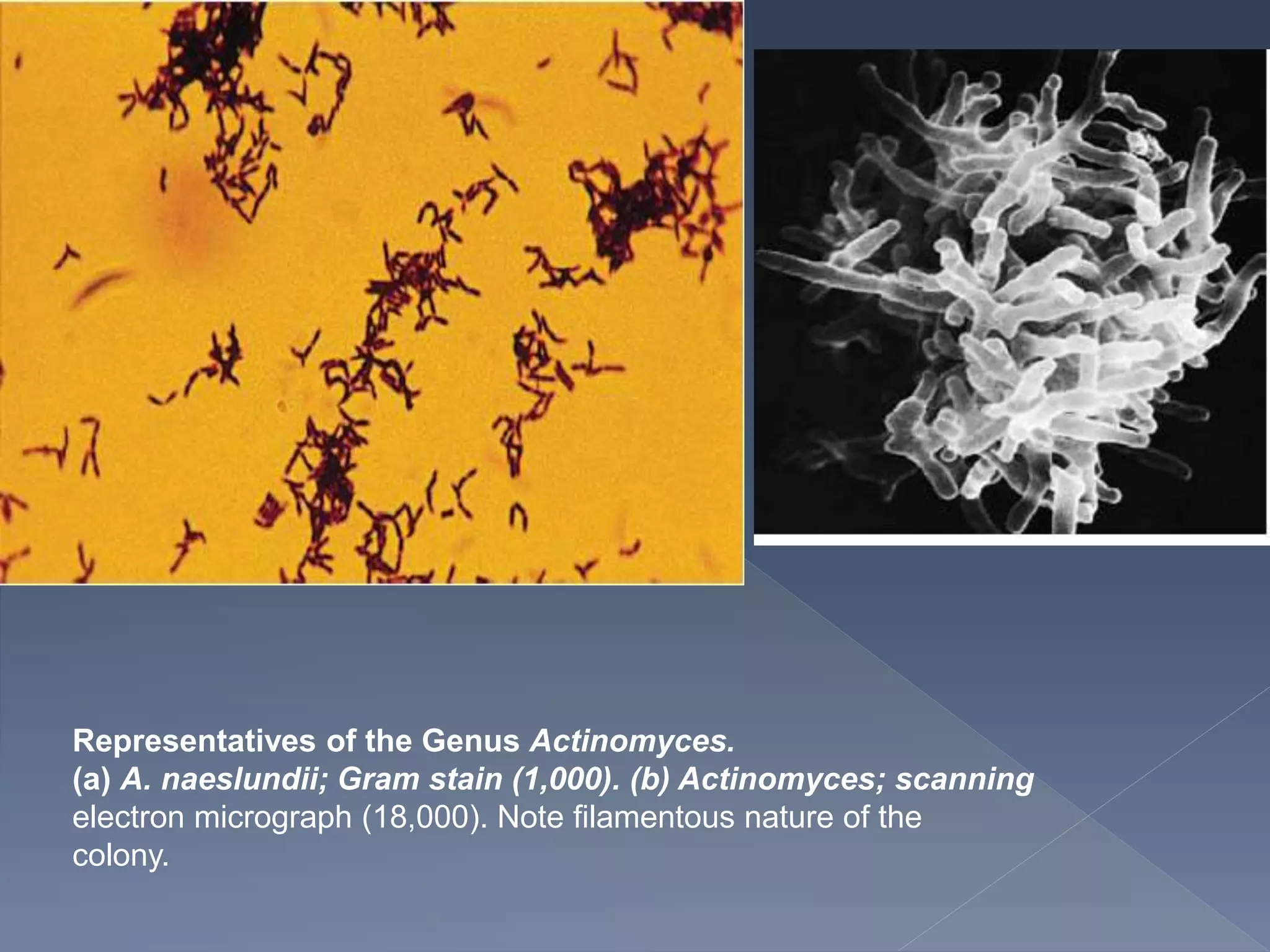
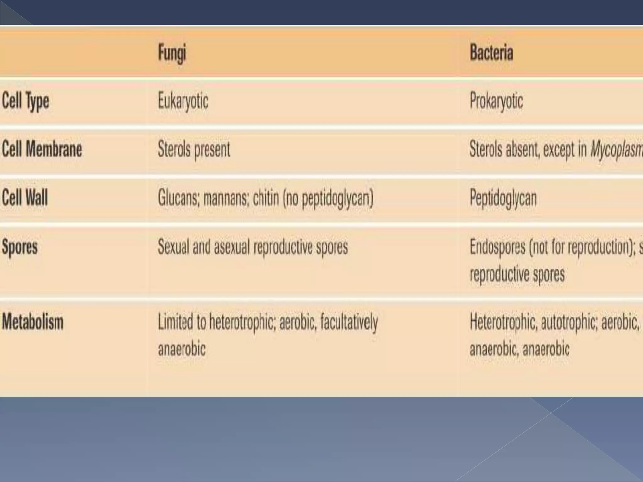
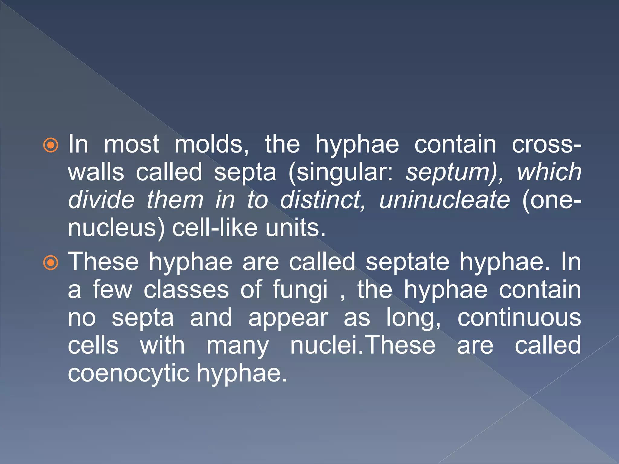
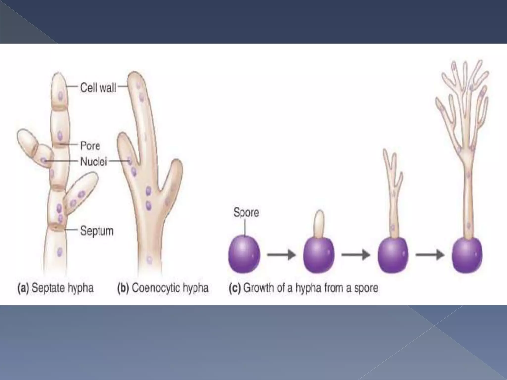
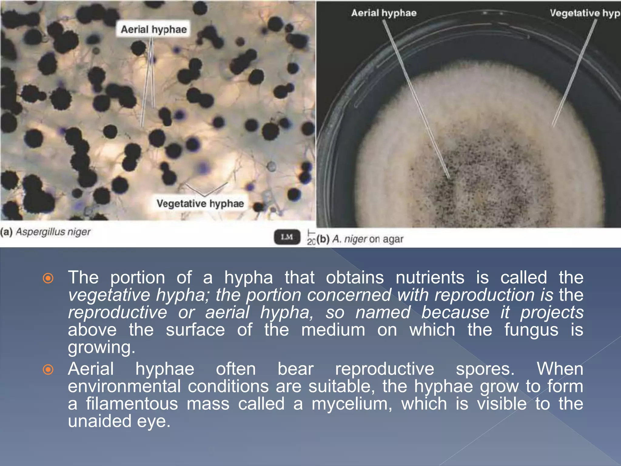
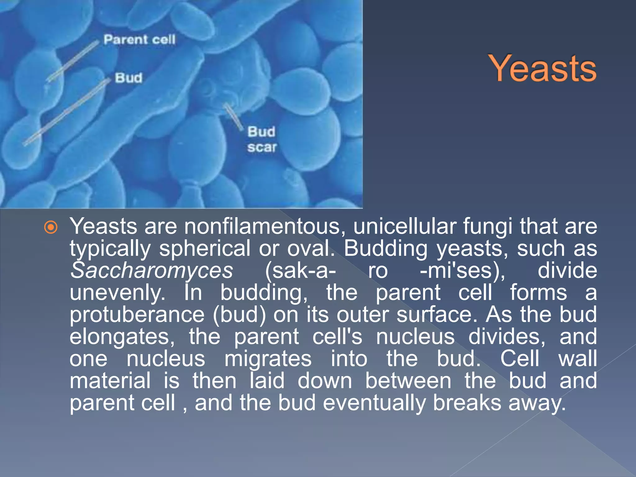
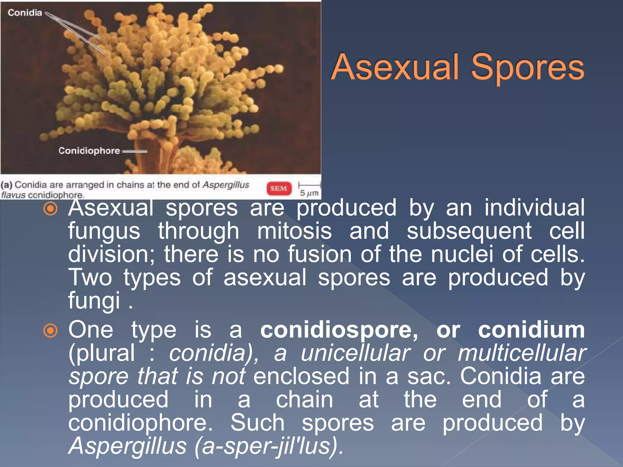
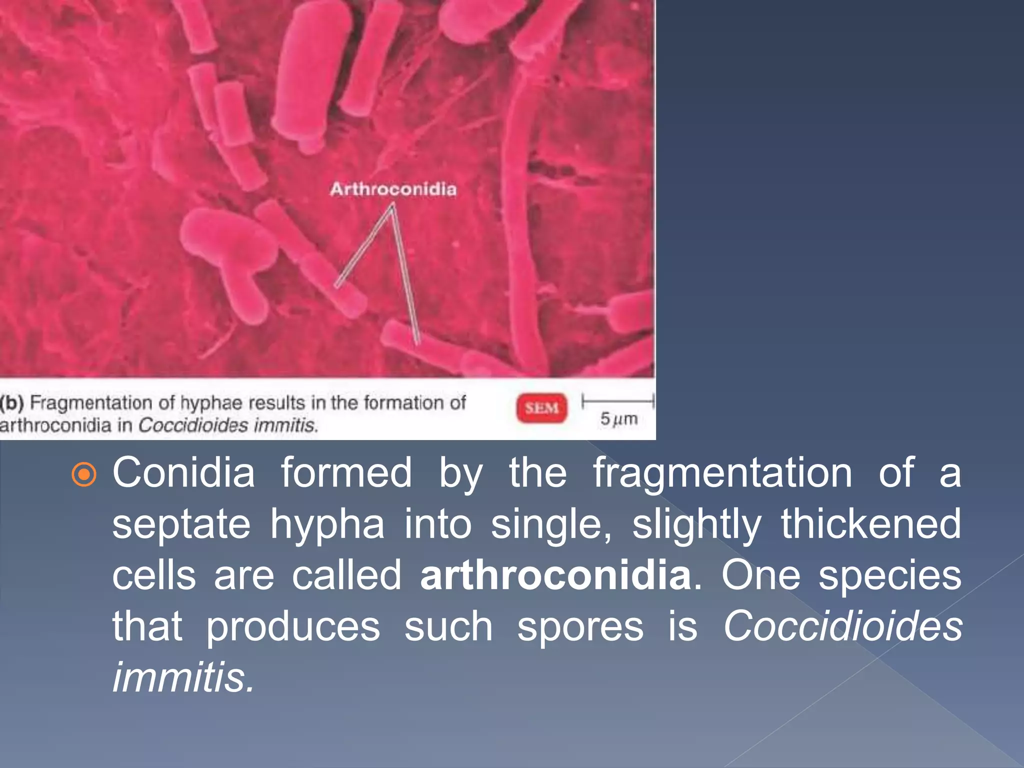
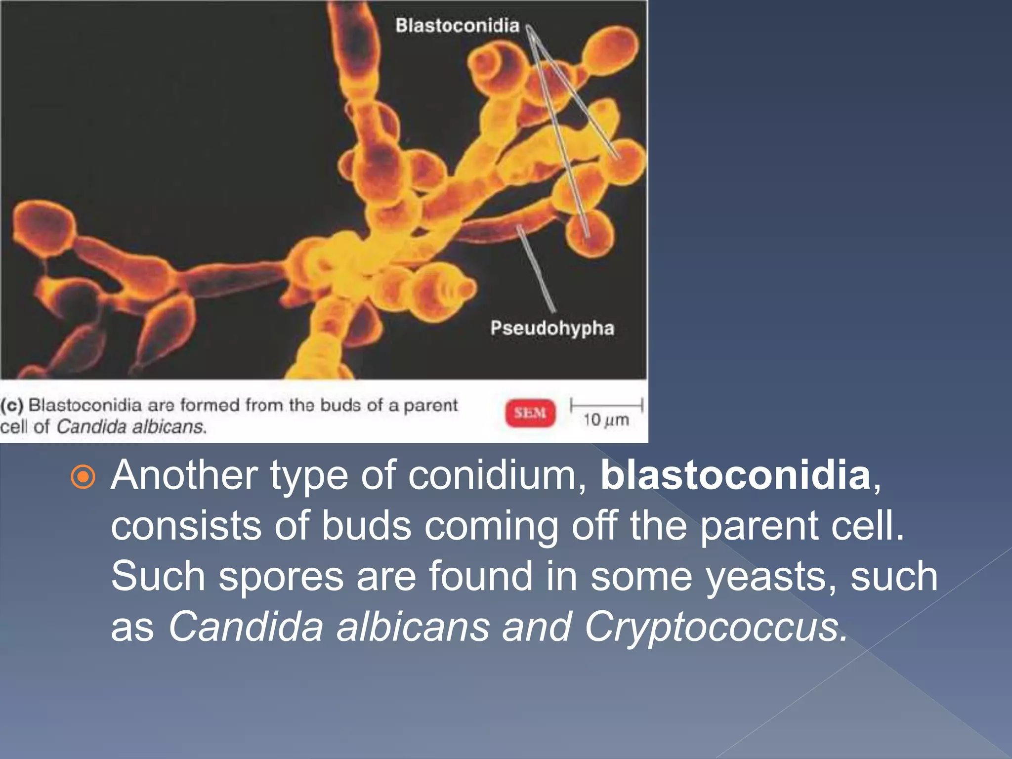
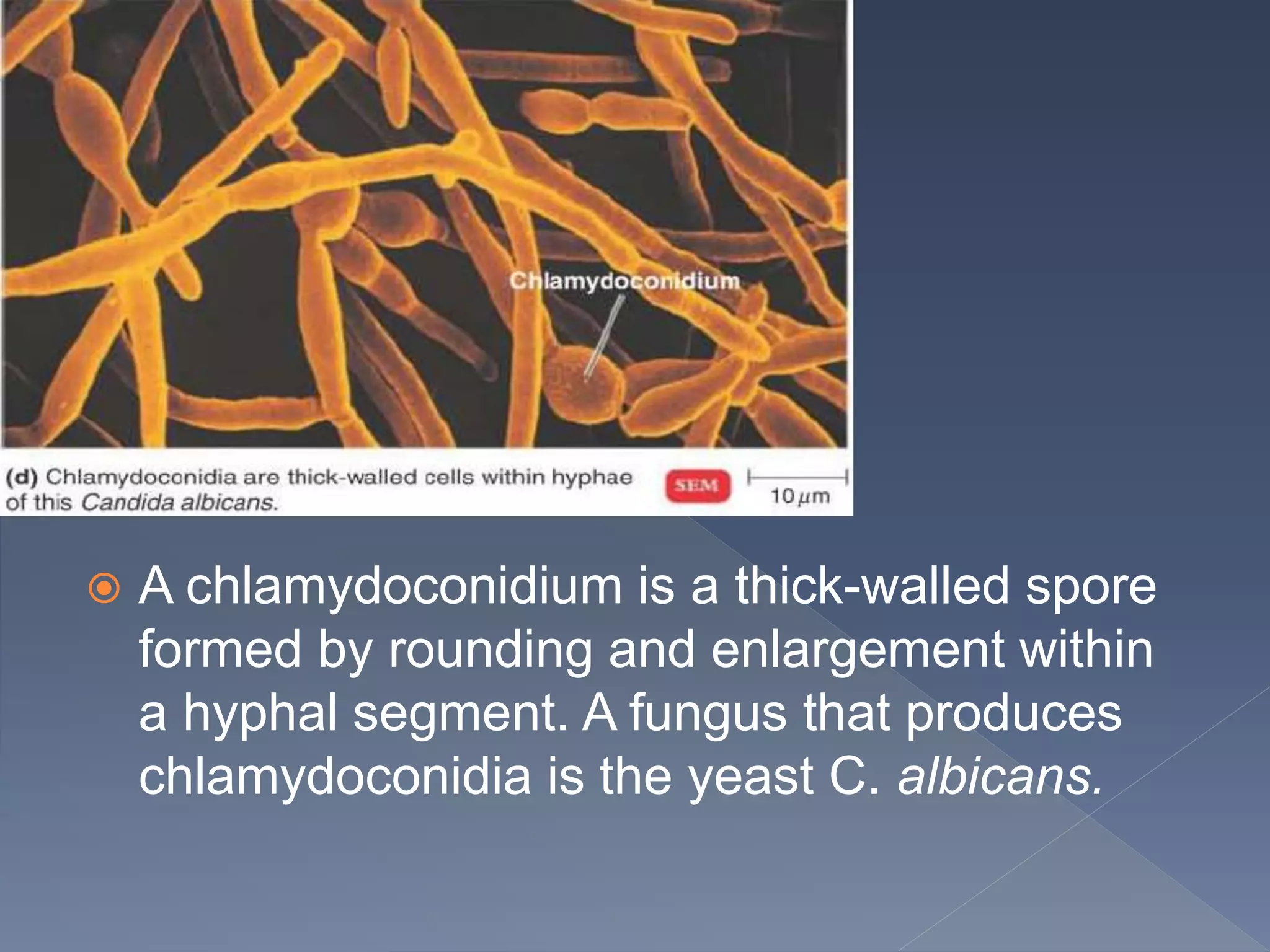
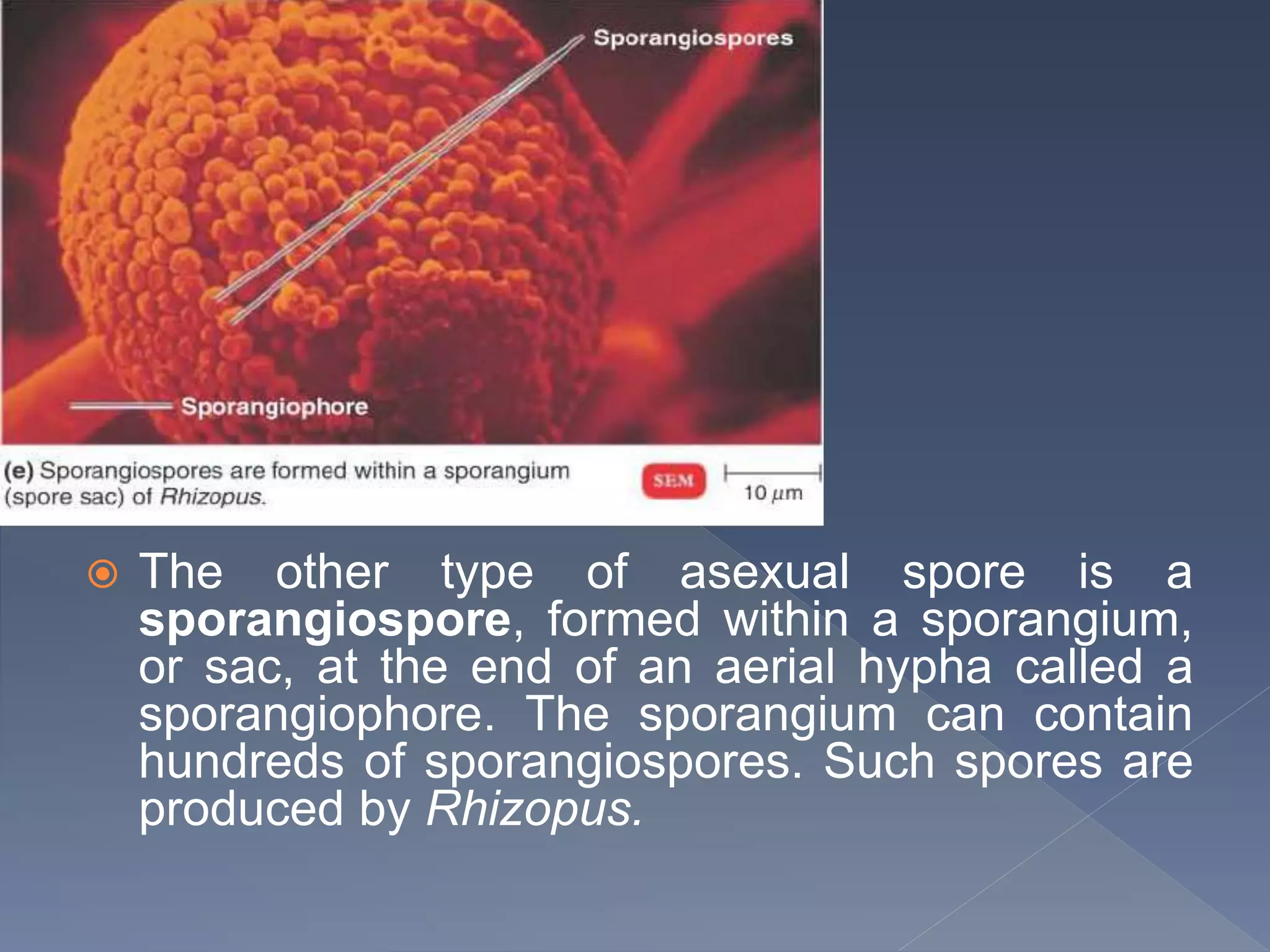
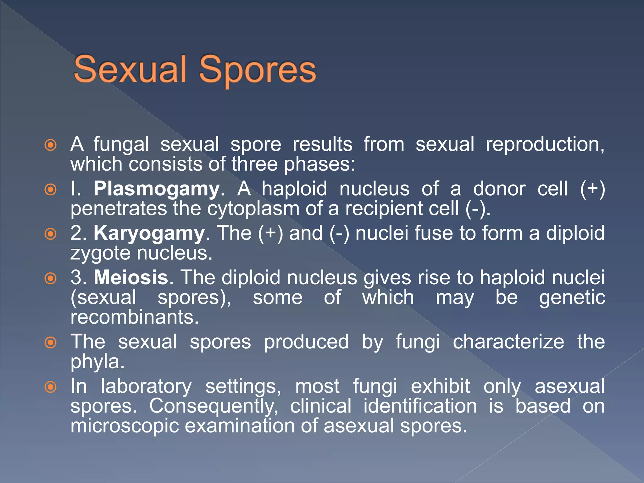
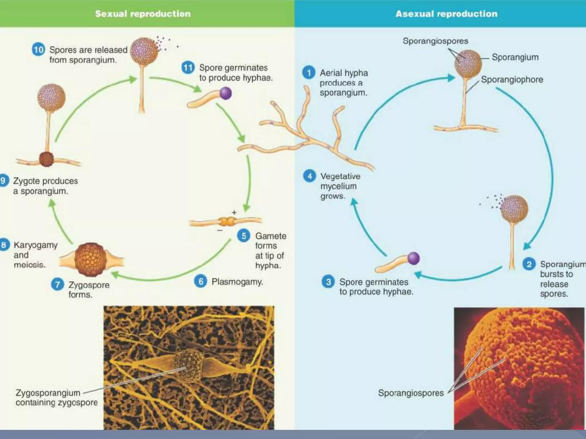
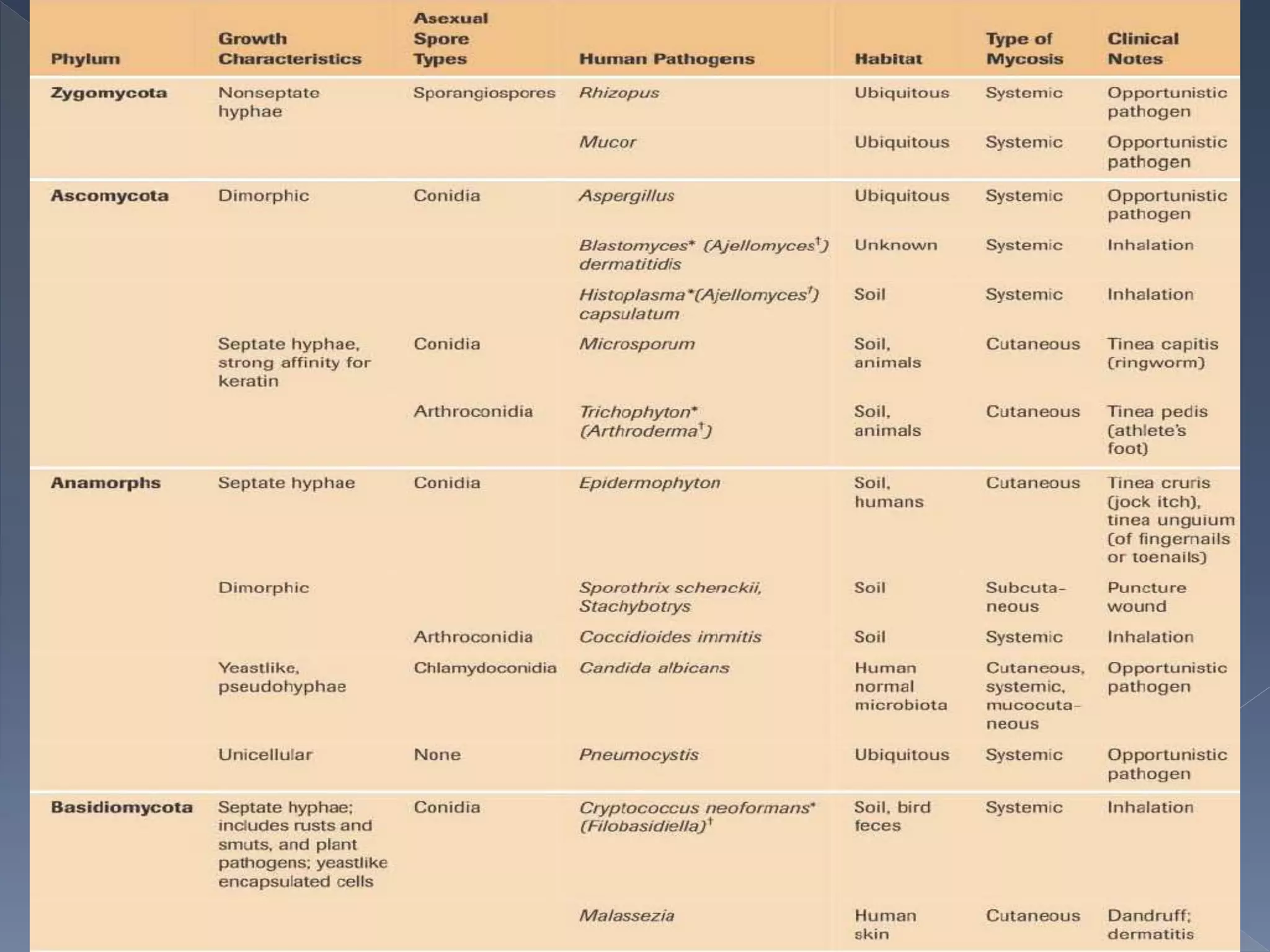
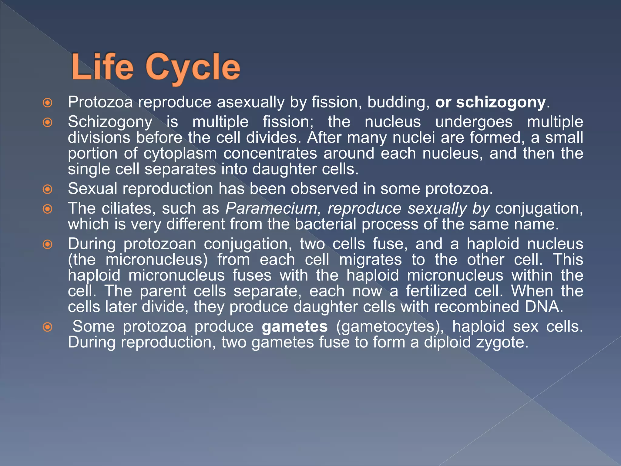
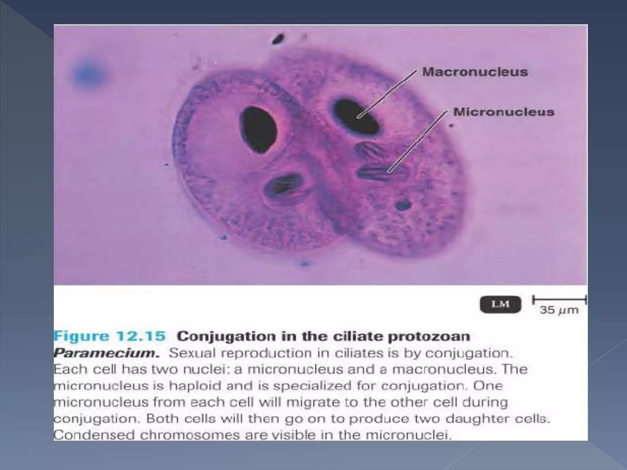
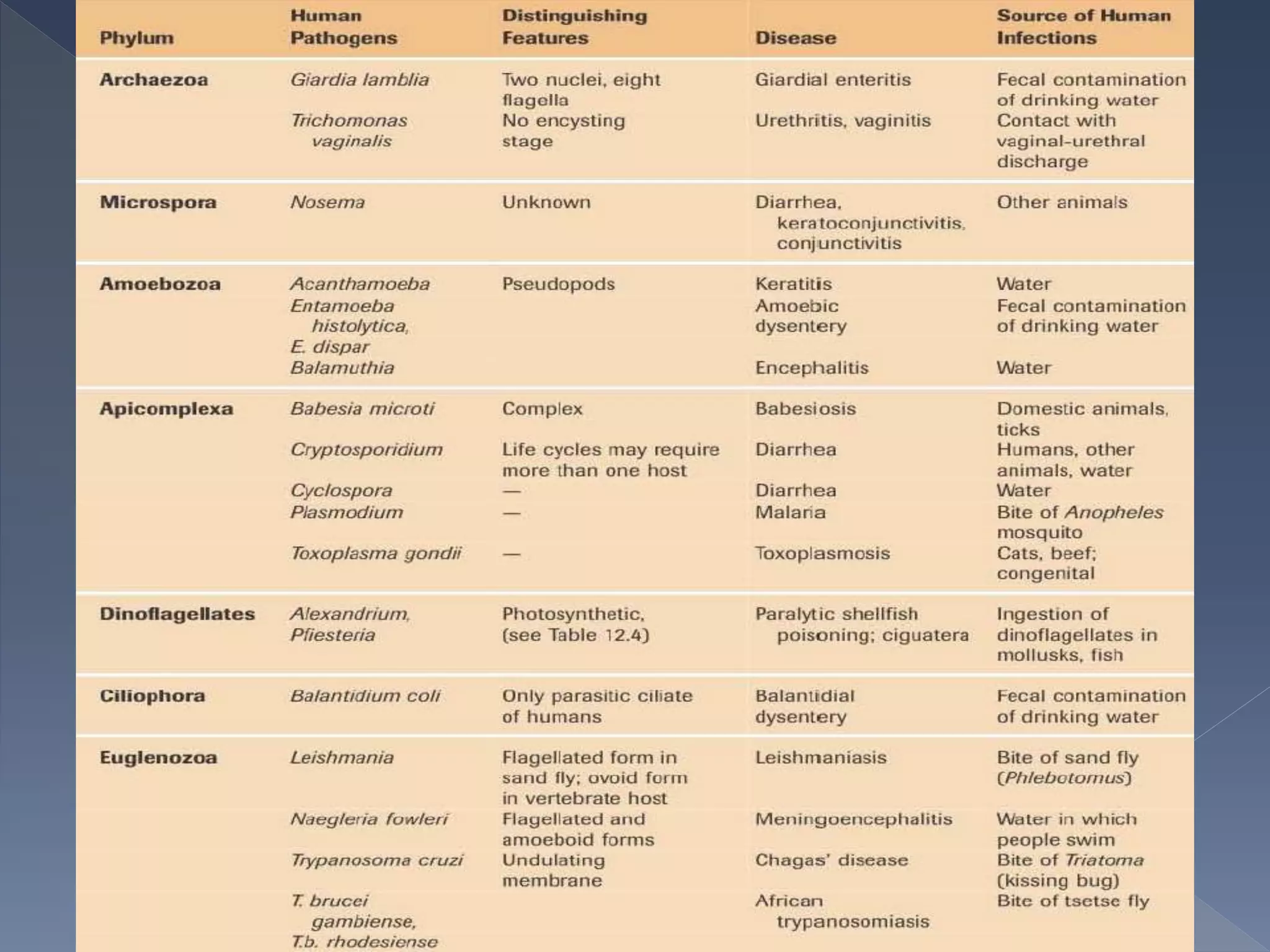
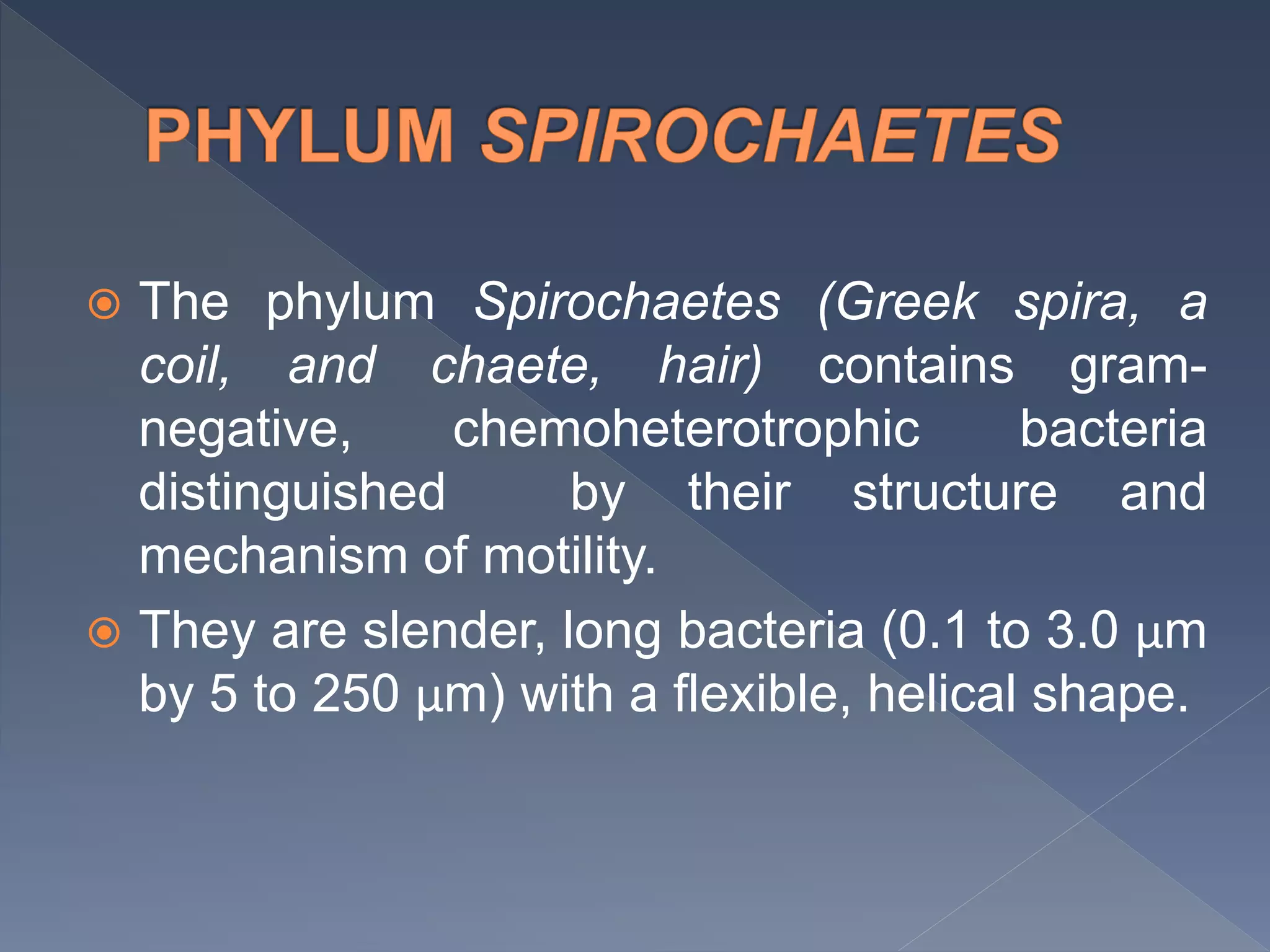
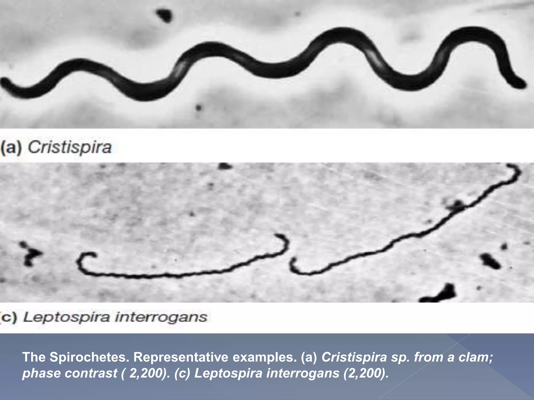
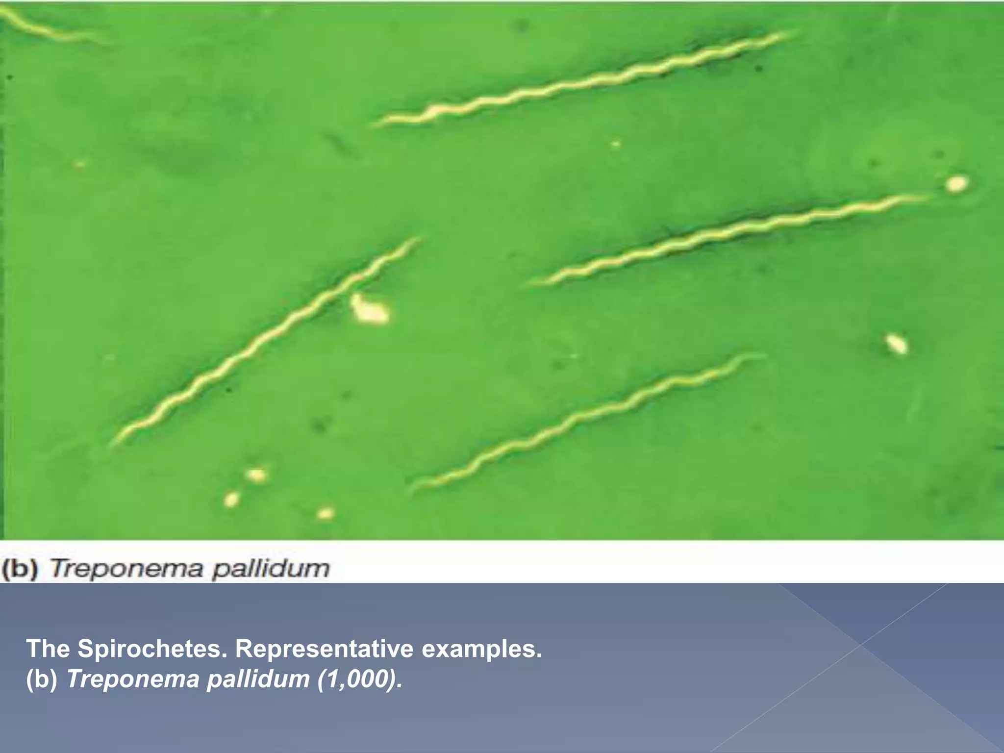
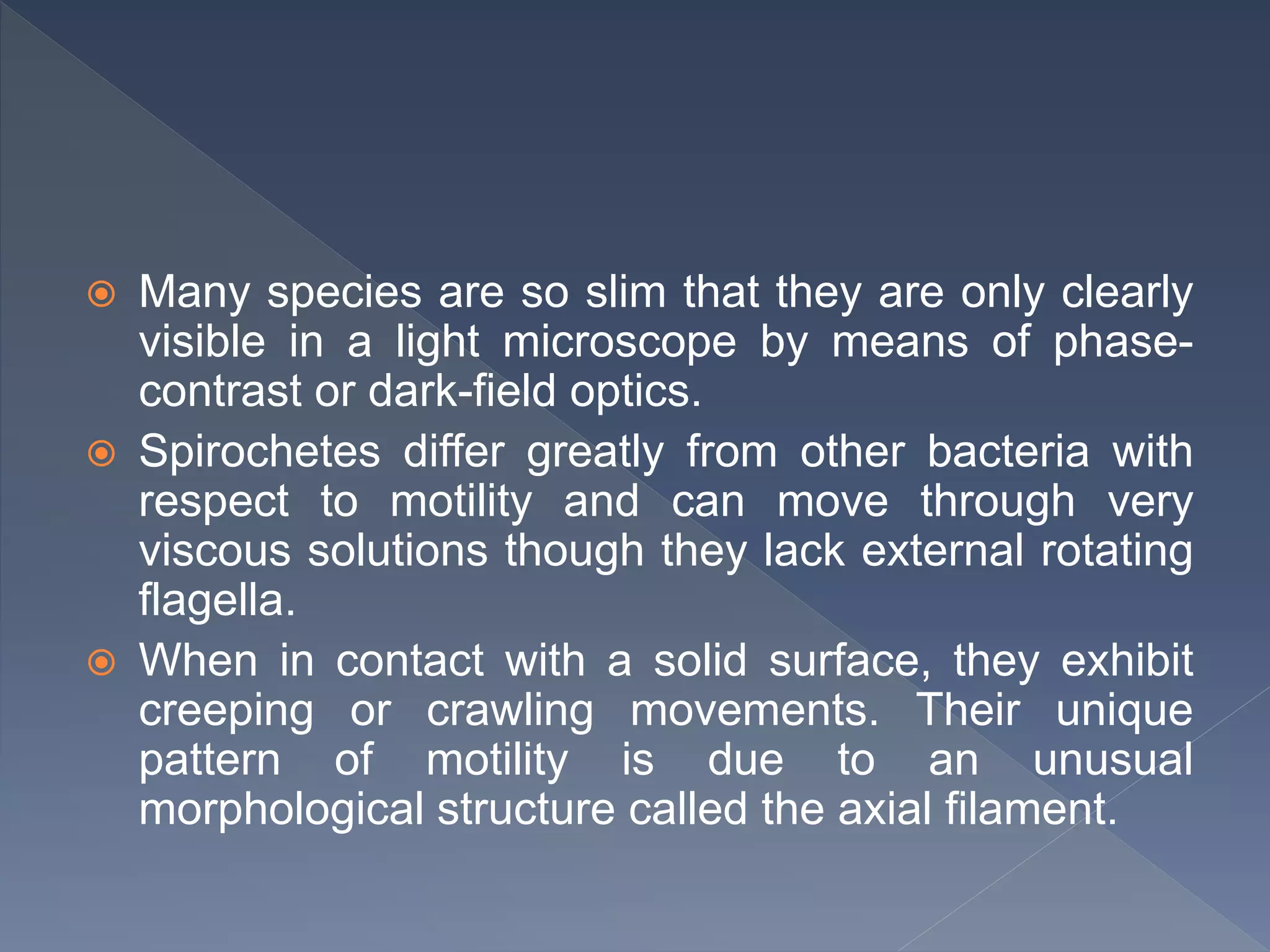
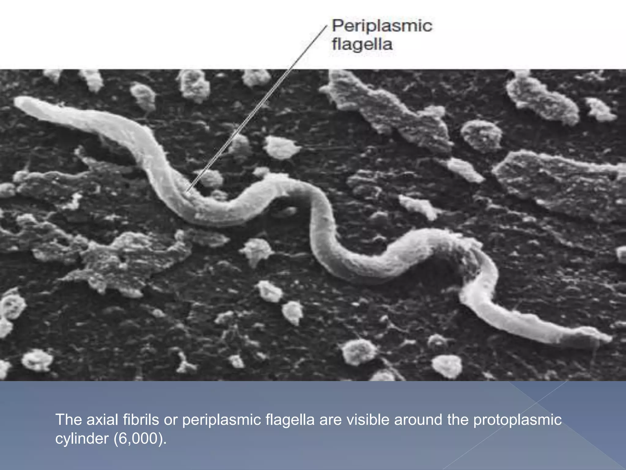
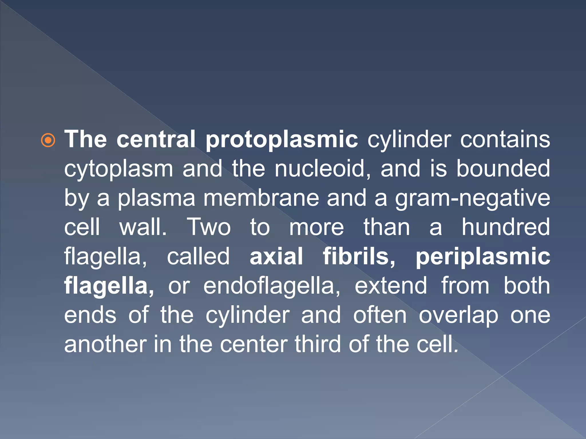
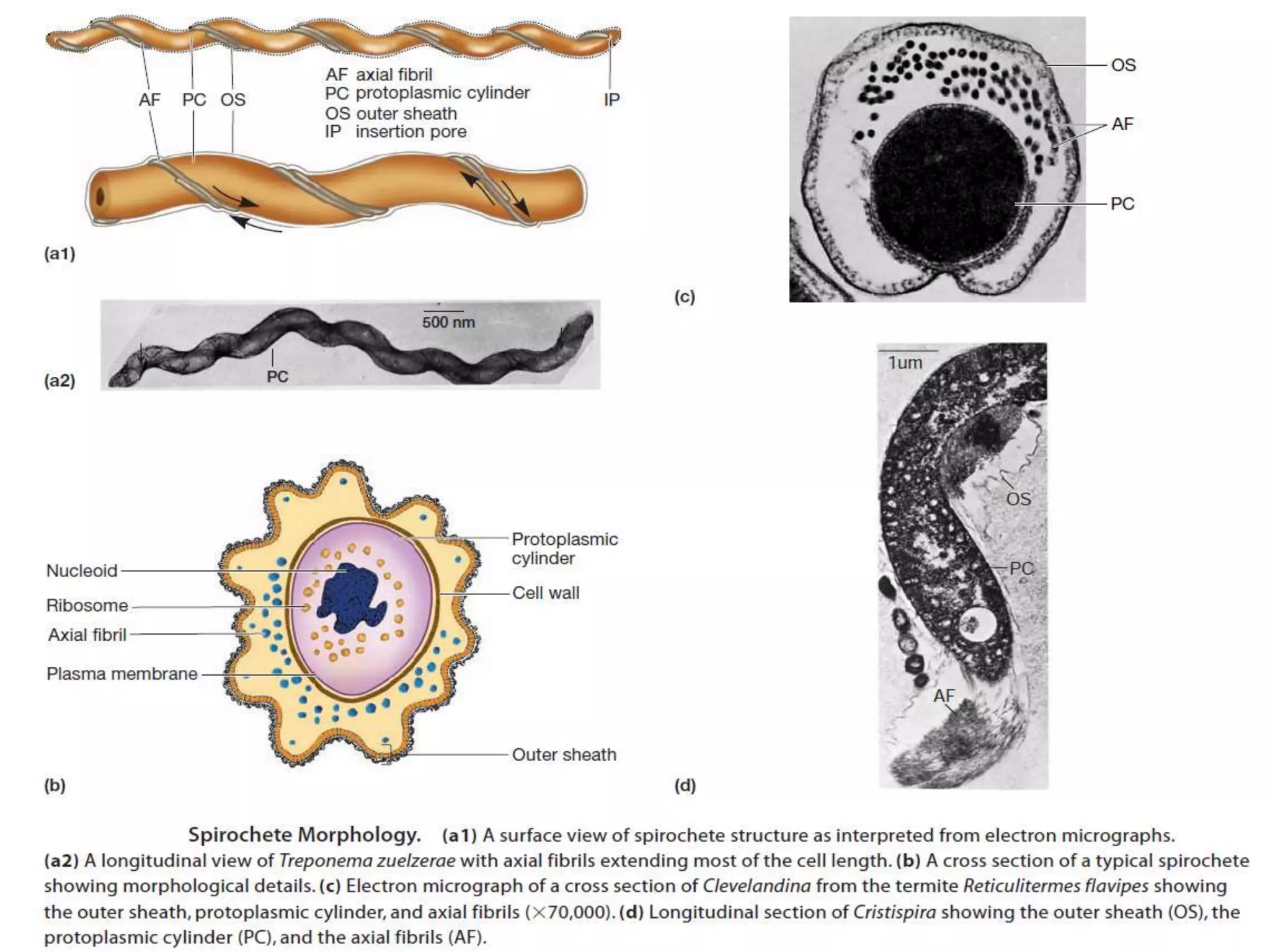
The document discusses various groups of bacteria, fungi, and protozoa, focusing on their characteristics, reproductive methods, and pathogenic potential. It details unique features of spirochetes, rickettsias, mycoplasmas, chlamydiae, actinomycetes, and fungi, highlighting their structures and the diseases they cause. Additionally, it covers the diversity within protozoa, including their reproductive strategies and modes of nutrition.
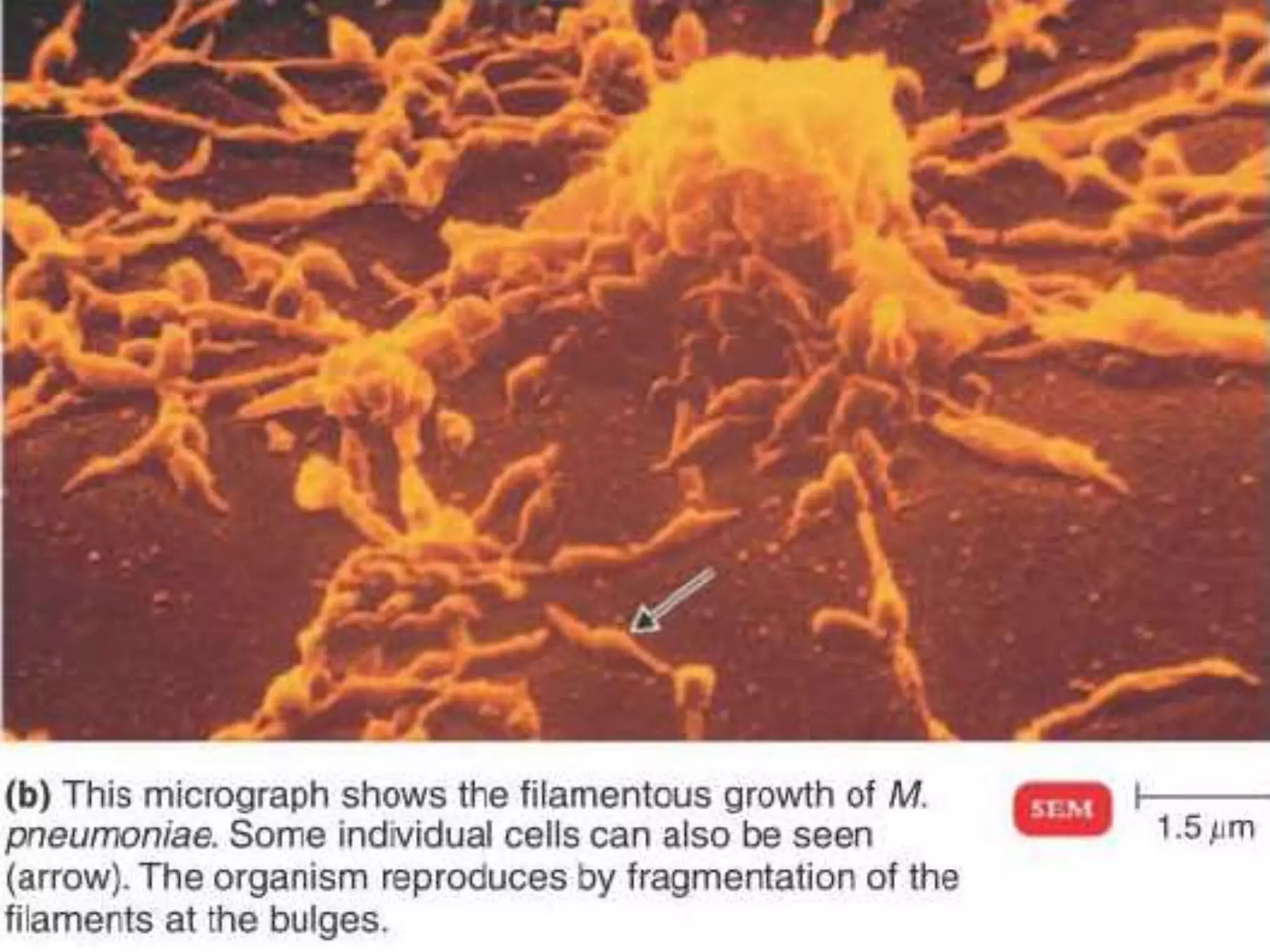
![ Mycoplasma pneumoniae. Bacteria such as M.
pneumonrae have no cell walls, and their
morphology is irregular (pleomorphic]](https://image.slidesharecdn.com/morphologystr-221106194932-5d043b26/75/morphology-structure-of-spirochete-fungi-protozoa-20-2048.jpg)