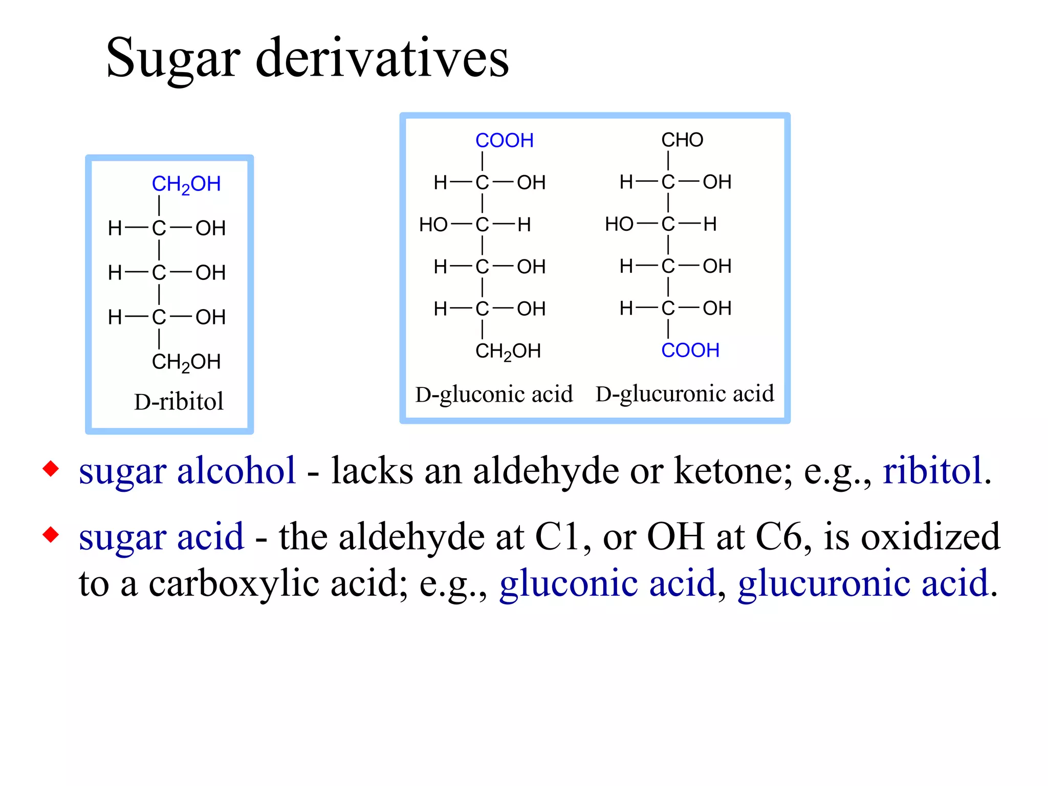
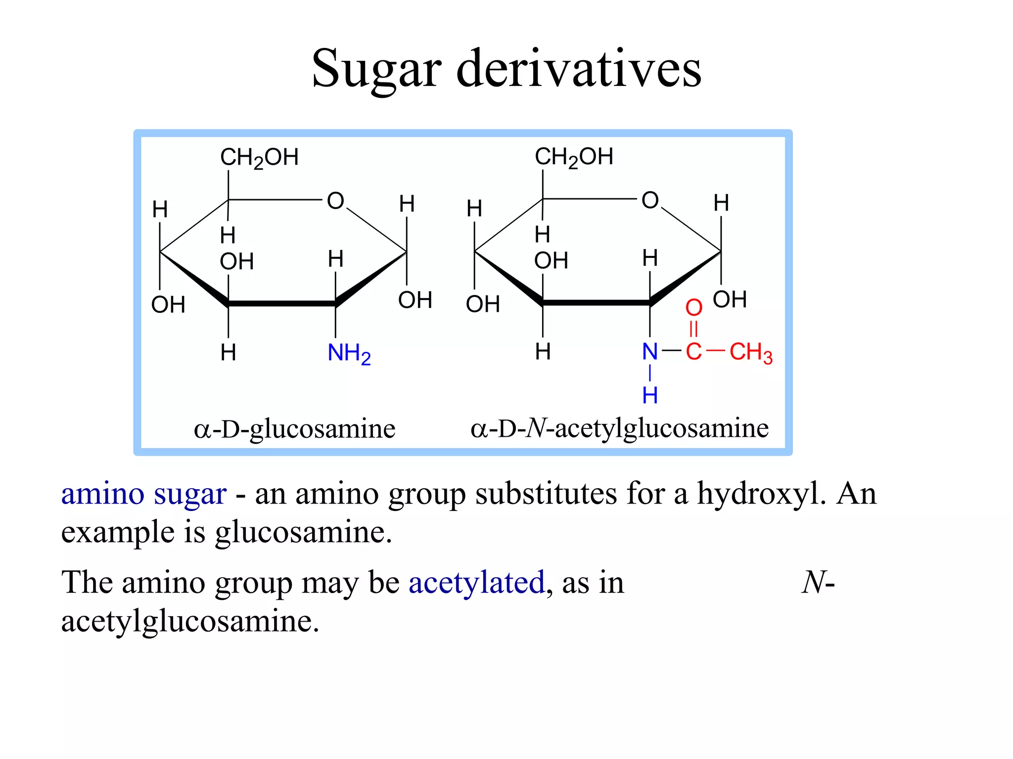
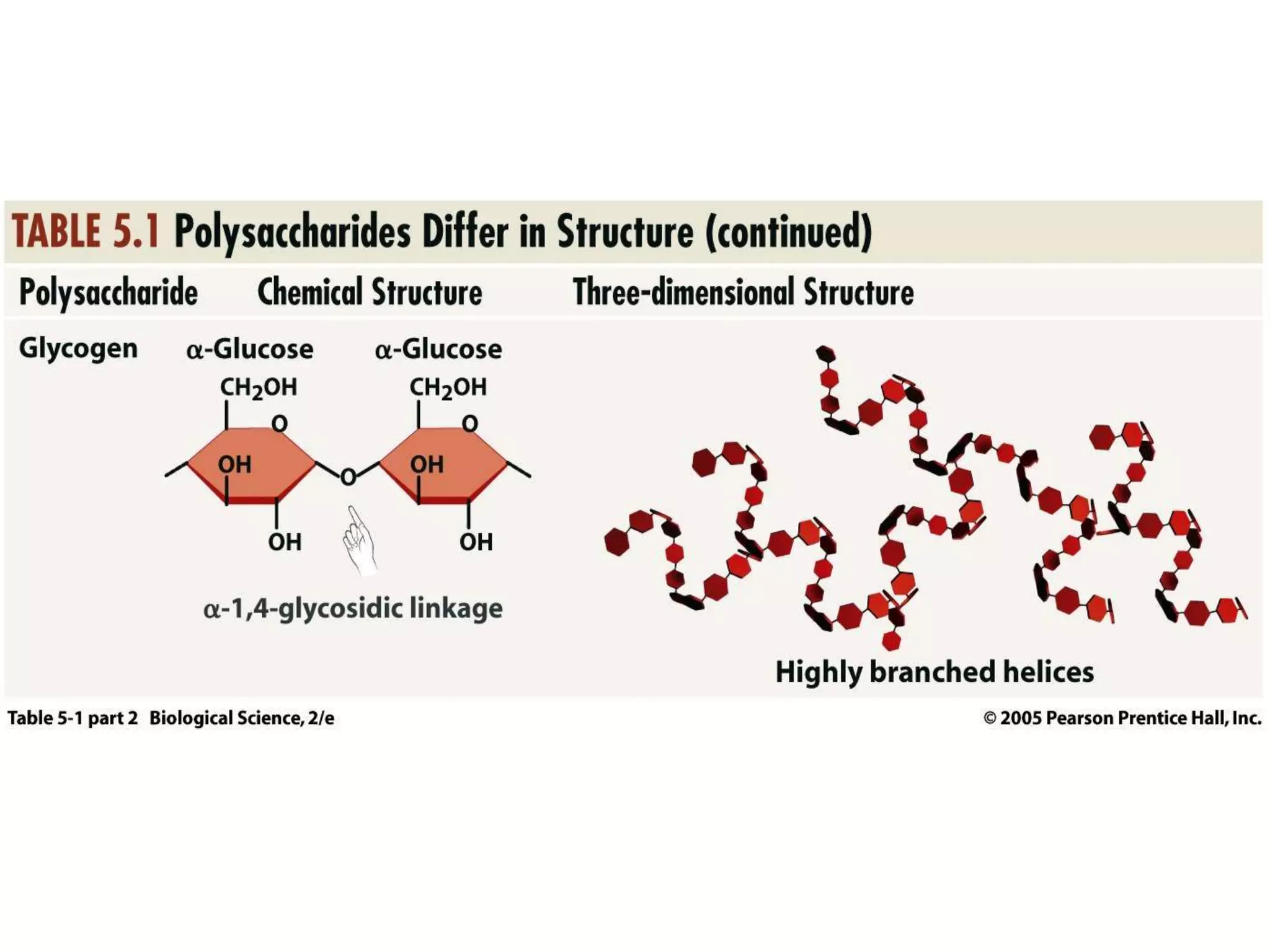
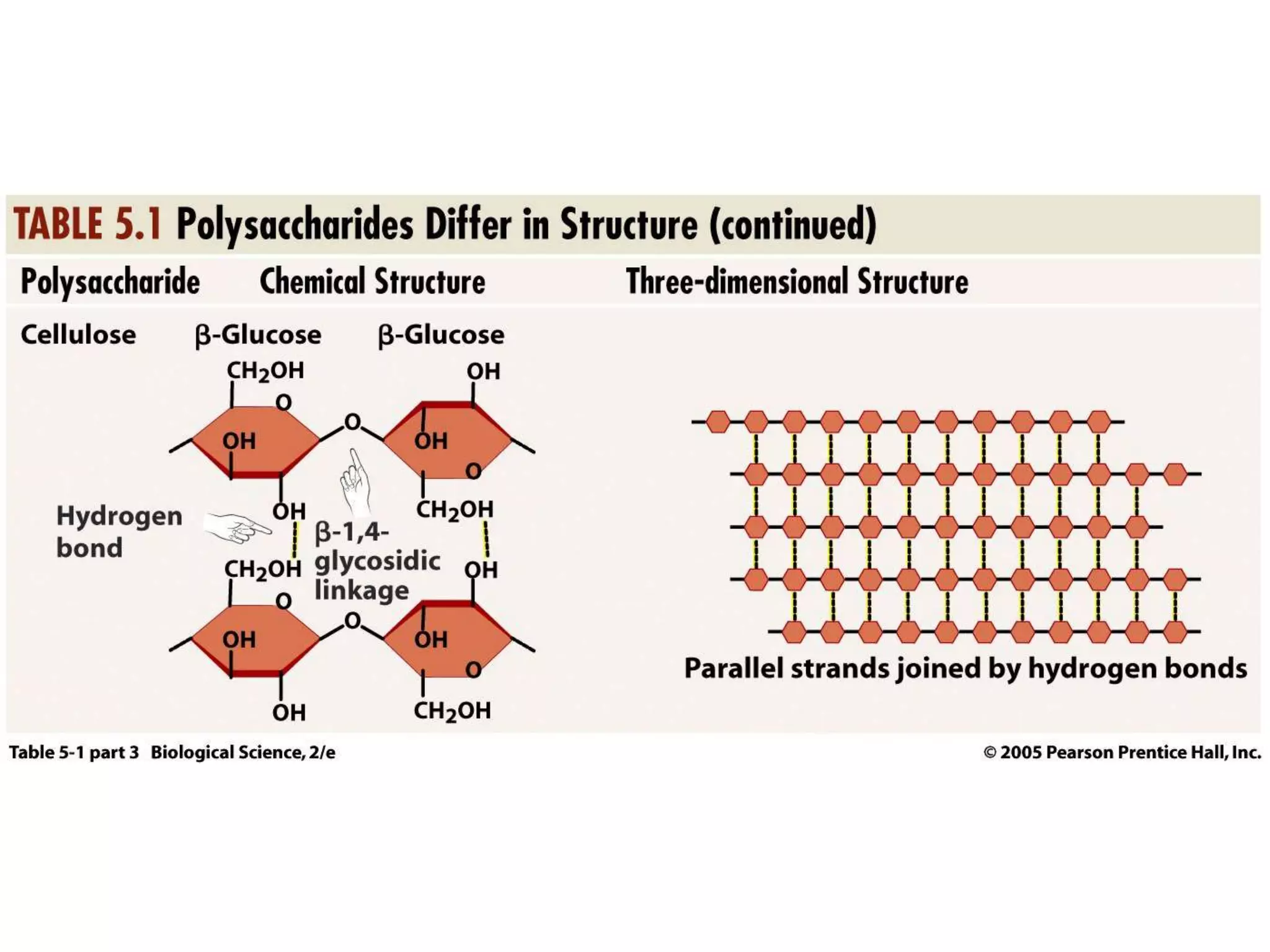
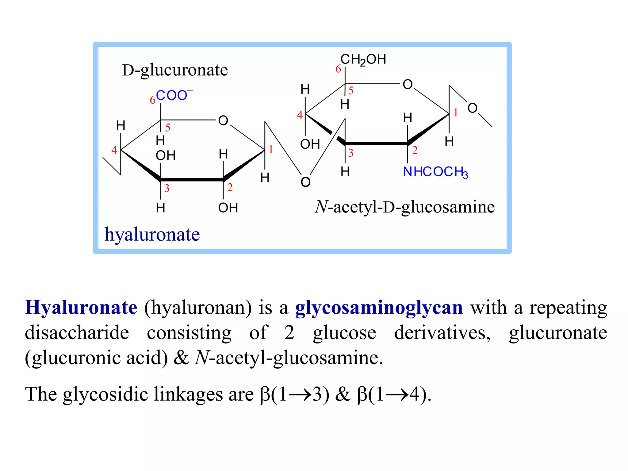
Modification of
Carbohydrates
Sugar derivatives
 sugar alcohol - lacks an aldehyde or ketone; e.g., ribitol.
 sugar acid - the aldehyde at C1, or OH at C6, is oxidized
to a carboxylic acid; e.g., gluconic acid, glucuronic acid.
CH2OH
C
C
C
CH2OH
H OH
H OH
H OH
D-ribitol
COOH
C
C
C
C
H OH
HO H
H OH
D-gluconic acid D-glucuronic acid
CH2OH
OH
H
CHO
C
C
C
C
H OH
HO H
H OH
COOH
OH
H
Sugar derivatives
amino sugar - an amino group substitutes for a hydroxyl. An
example is glucosamine.
The amino group may be acetylated, as in N-
acetylglucosamine.
H O
OH
H
OH
H
NH2
H
OH
CH2OH
H
-D-glucosamine
H O
OH
H
OH
H
N
H
OH
CH2OH
H
-D-N-acetylglucosamine
C CH3
O
H
N-acetylneuraminate (N-acetylneuraminic acid, also called sialic
acid) is often found as a terminal residue of oligosaccharide
chains of glycoproteins.
Sialic acid imparts negative charge to glycoproteins, because its
carboxyl group tends to dissociate a proton at physiological pH,
as shown here.
NH O
H
COO
OH
H
H
OH
H
H
R
C
H3C
O
HC
HC
CH2OH
OH
OH
N-acetylneuraminate (sialic acid)
R =
Glycosidic Bonds
The anomeric hydroxyl and a hydroxyl of another sugar or some
other compound can join together, splitting out water to form a
glycosidic bond:
R-OH + HO-R'  R-O-R' + H2O
E.g., methanol reacts with the anomeric OH on glucose to form
methyl glucoside (methyl-glucopyranose).
O
H
HO
H
HO
H
OH
OH
H
H
OH
-D-glucopyranose
O
H
HO
H
HO
H
OCH3
OH
H
H
OH
methyl--D-glucopyranose
CH3-OH
+
methanol
H2O
Cellobiose, a product of cellulose breakdown, is the otherwise
equivalent b anomer (O on C1 points up).
The b(1 4) glycosidic linkage is represented as a zig-zag, but one
glucose is actually flipped over relative to the other.
H O
OH
H
OH
H
OH
CH2OH
H
O H
OH
H
OH
H
OH
CH2OH
H
O
H
H
1
2
3
5
4
6
1
2
3
4
5
6
maltose
H O
OH
H
OH
H
OH
CH2OH
H
O OH
H
H
OH
H
OH
CH2OH
H
H
H
O
1
2
3
4
5
6
1
2
3
4
5
6
cellobiose
Disaccharides:
Maltose, a cleavage
product of starch (e.g.,
amylose), is a
disaccharide with an
(1 4) glycosidic link
between C1 - C4 OH of
2 glucoses.
It is the  anomer (C1 O
points down).
Other disaccharides include:
 Sucrose, common table sugar, has a glycosidic bond linking the
anomeric hydroxyls of glucose & fructose.
Because the configuration at the anomeric C of glucose is  (O
points down from ring), the linkage is (12).
The full name of sucrose is -D-glucopyranosyl-(12)-b-D-
fructopyranose.)
 Lactose, milk sugar, is composed of galactose & glucose, with
b(14) linkage from the anomeric OH of galactose. Its full name
is b-D-galactopyranosyl-(1 4)--D-glucopyranose
Polysaccharides:
Plants store glucose as amylose or amylopectin, glucose polymers
collectively called starch.
Glucose storage in polymeric form minimizes osmotic effects.
Amylose is a glucose polymer with (14) linkages.
The end of the polysaccharide with an anomeric C1 not involved in a
glycosidic bond is called the reducing end.
H O
OH
H
OH
H
OH
CH2OH
H
O H
H
OH
H
OH
CH2OH
H
O
H
H H O
O
H
OH
H
OH
CH2OH
H
H H O
H
OH
H
OH
CH2OH
H
OH
H
H O
O
H
OH
H
OH
CH2OH
H
O
H
1
6
5
4
3
1
2
amylose
Amylopectin is a glucose polymer with mainly (14) linkages, but
it also has branches formed by (16) linkages. Branches are
generally longer than shown above.
The branches produce a compact structure & provide multiple chain
ends at which enzymatic cleavage can occur.
H O
OH
H
OH
H
OH
CH2OH
H
O H
H
OH
H
OH
CH2OH
H
O
H
H H O
O
H
OH
H
OH
CH2
H
H H O
H
OH
H
OH
CH2OH
H
OH
H
H O
O
H
OH
H
OH
CH2OH
H
O
H
O
1 4
6
H O
H
OH
H
OH
CH2OH
H
H H O
H
OH
H
OH
CH2OH
H
H
O
1
OH
3
4
5
2
amylopectin
Glycogen, the glucose storage polymer in animals, is similar in
structure to amylopectin.
But glycogen has more (16) branches.
The highly branched structure permits rapid glucose release from
glycogen stores, e.g., in muscle during exercise.
The ability to rapidly mobilize glucose is more essential to animals
than to plants.
H O
OH
H
OH
H
OH
CH2OH
H
O H
H
OH
H
OH
CH2OH
H
O
H
H H O
O
H
OH
H
OH
CH2
H
H H O
H
OH
H
OH
CH2OH
H
OH
H
H O
O
H
OH
H
OH
CH2OH
H
O
H
O
1 4
6
H O
H
OH
H
OH
CH2OH
H
H H O
H
OH
H
OH
CH2OH
H
H
O
1
OH
3
4
5
2
glycogen
Cellulose, a major constituent of plant cell walls, consists of long
linear chains of glucose with b(14) linkages.
Every other glucose is flipped over, due to b linkages.
This promotes intra-chain and inter-chain H-bonds and
cellulose
H O
OH
H
OH
H
OH
CH2OH
H
O
H
OH
H
OH
CH2OH
H
O
H H O
O H
OH
H
OH
CH2OH
H
H O
H
OH
H
OH
CH2OH
H
H
OH
H O
O H
OH
H
OH
CH2OH
H
O
H H H H
1
6
5
4
3
1
2
van der Waals interactions, that
cause cellulose chains to be straight
& rigid, and pack with a crystalline
arrangement in thick bundles -
microfibrils. Schematic of arrangement of
cellulose chains in a microfibril.
Multisubunit Cellulose Synthase complexes in the plasma
membrane spin out from the cell surface microfibrils consisting of
36 parallel, interacting cellulose chains.
These microfibrils are very strong.
The role of cellulose is to impart strength and rigidity to plant cell
walls, which can withstand high hydrostatic pressure gradients.
Osmotic swelling is prevented.
Explore and compare structures of amylose & cellulose using
Chime.
cellulose
H O
OH
H
OH
H
OH
CH2OH
H
O
H
OH
H
OH
CH2OH
H
O
H H O
O H
OH
H
OH
CH2OH
H
H O
H
OH
H
OH
CH2OH
H
H
OH
H O
O H
OH
H
OH
CH2OH
H
O
H H H H
1
6
5
4
3
1
2
Glycosaminoglycans (mucopolysaccharides) are linear polymers of
repeating disaccharides.
The constituent monosaccharides tend to be modified, with acidic
groups, amino groups, sulfated hydroxyl and amino groups, etc.
Glycosaminoglycans tend to be negatively charged, because of the
prevalence of acidic groups.
H O
H
H
OH
H
OH
COO
H
H O
OH H
H
NHCOCH3
H
CH2OH
H
O
O
D-glucuronate
O
1
2
3
4
5
6
1
2
3
4
5
6
N-acetyl-D-glucosamine
hyaluronate
Hyaluronate (hyaluronan) is a glycosaminoglycan with a repeating
disaccharide consisting of 2 glucose derivatives, glucuronate
(glucuronic acid) & N-acetyl-glucosamine.
The glycosidic linkages are b(13) & b(14).
H O
H
H
OH
H
OH
COO
H
H O
OH H
H
NHCOCH3
H
CH2OH
H
O
O
D-glucuronate
O
1
2
3
4
5
6
1
2
3
4
5
6
N-acetyl-D-glucosamine
hyaluronate
Proteoglycans are glycosaminoglycans that are covalently
linked to serine residues of specific core proteins.
The glycosaminoglycan chain is synthesized by sequential
addition of sugar residues to the core protein.
heparan sulfate
glycosaminoglycan
cytosol
core
protein
transmembrane
-helix
Some proteoglycans of the extracellular matrix bind non-
covalently to hyaluronate via protein domains called link modules.
E.g.:
• Multiple copies of the aggrecan proteoglycan associate with
hyaluronate in cartilage to form large complexes.
• Versican, another proteoglycan, binds hyaluronate in the
extracellular matrix of loose connective tissues.
H O
H
H
OH
H
OH
COO
H
H O
OH H
H
NHCOCH3
H
CH2OH
H
O
O
D-glucuronate
O
1
2
3
4
5
6
1
2
3
4
5
6
N-acetyl-D-glucosamine
hyaluronate
Heparan sulfate is initially synthesized on a membrane-
embedded core protein as a polymer of alternating
N-acetylglucosamine and glucuronate residues.
Later, in segments of the polymer, glucuronate residues
may be converted to the sulfated sugar iduronic acid,
while N-acetylglucosamine residues may be deacetylated
and/or sulfated.
H O
H
OSO3

H
OH
H
COO
O H
H
NHSO3

H
OH
CH2OSO3

H
H
H
O
O
heparin or heparan sulfate - examples of residues
iduronate-2-sulfate N-sulfo-glucosamine-6-sulfate
Heparin, a soluble glycosaminoglycan
found in granules of mast cells, has a
structure similar to that of heparan
sulfates, but is more highly sulfated.
When released into the blood, it
inhibits clot formation by interacting
with the protein antithrombin.
Heparin has an extended helical
conformation.
heparin: (IDS-SGN)5
PDB 1RID
C O N S
Charge repulsion by the many negatively charged groups
may contribute to this conformation.
Heparin shown has 10 residues, alternating IDS (iduronate-
2-sulfate) & SGN (N-sulfo-glucosamine-6-sulfate).
 The core protein of a syndecan heparan sulfate
proteoglycan includes a single transmembrane -helix,
as in the simplified diagram above.
 The core protein of a glypican heparan sulfate
proteoglycan is attached to the outer surface of the
plasma membrane via covalent linkage to a modified
phosphatidylinositol lipid.
heparan sulfate
glycosaminoglycan
cytosol
core
protein
transmembrane
-helix
Some cell surface heparan
sulfate glycosaminoglycans
remain covalently linked to
core proteins embedded in
the plasma membrane.
Proteins involved in signaling & adhesion at the cell
surface recognize & bind heparan sulfate chains.
E.g., binding of some growth factors (small proteins) to
cell surface receptors is enhanced by their binding also to
heparan sulfates.
Regulated cell surface Sulf enzymes may remove sulfate
groups at particular locations on heparan sulfate chains to
alter affinity
for signal
proteins, e.g.,
growth factors. H O
H
OSO3

H
OH
H
COO
O H
H
NHSO3

H
OH
CH2OSO3

H
H
H
O
O
heparin or heparan sulfate - examples of residues
iduronate-2-sulfate N-sulfo-glucosamine-6-sulfate
Diagram
by Kirkpatrick
& Selleck.
O-linked oligosaccharide chains of glycoproteins vary
in complexity.
They link to a protein via a glycosidic bond between a
sugar residue & a serine or threonine OH.
O-linked oligosaccharides have roles in recognition,
interaction, and enzyme regulation.
H O
OH
O
H
HN
H
OH
CH2OH
H
C CH3
O
b-D-N-acetylglucosamine
CH2 CH
C
NH
O
H
serine
residue
Oligosaccharides
that are covalently
attached to proteins
or to membrane
lipids may be linear
or branched chains.
H O
OH
O
H
HN
H
OH
CH2OH
H
C CH3
O
b-D-N-acetylglucosamine
CH2 CH
C
NH
O
H
serine
residue
N-acetylglucosamine (GlcNAc) is a common O-linked
glycosylation of protein serine or threonine residues.
Many cellular proteins, including enzymes & transcription
factors, are regulated by reversible GlcNAc attachment.
Often attachment of GlcNAc to a protein OH alternates with
phosphorylation, with these 2 modifications having opposite
regulatory effects (stimulation or inhibition).
N-linked oligosaccharides of glycoproteins tend to be
complex and branched.
First N-acetylglucosamine is linked to a protein via the
side-chain N of an asparagine residue in a particular
3-amino acid sequence.
H O
OH
HN
H
H
HN
H
OH
CH2OH
H
C CH3
O
C CH2 CH
O HN
C
HN
O
HC
C
HN
HC
R
O
C
R
O
Asn
X
Ser or Thr
N-acetylglucosamine
Initial sugar in N-linked
glycoprotein oligosaccharide
Additional monosaccharides are added, and the N-linked
oligosaccharide chain is modified by removal and addition
of residues, to yield a characteristic branched structure.
NAN
Gal
NAG
Man
NAG
Gal
NAN
Man
Man
NAG
Gal
NAN
NAG
NAG
Asn
Fuc
N-linked oligosaccharide
Key:
NAN = N-acetylneuraminate
Gal = galactose
NAG = N-acetylglucosamine
Man = mannose
Fuc = fucose
Many proteins secreted by cells have attached N-linked
oligosaccharide chains.
Genetic diseases have been attributed to deficiency of
particular enzymes involved in synthesizing or modifying
oligosaccharide chains of these glycoproteins.
Such diseases, and gene knockout studies in mice, have
been used to define pathways of modification of
oligosaccharide chains of glycoproteins and glycolipids.
Carbohydrate chains of plasma membrane glycoproteins
and glycolipids usually face the outside of the cell.
They have roles in cell-cell interaction and signaling, and
in forming a protective layer on the surface of some cells.
Lectins are glycoproteins that recognize and bind to
specific oligosaccharides.
Concanavalin A & wheat germ agglutinin are plant
lectins that have been useful research tools.
The C-type lectin-like domain is a Ca++-binding
carbohydrate recognition domain in many animal lectins.
Recognition/binding of CHO moieties of glycoproteins,
glycolipids & proteoglycans by animal lectins is a factor in:
• cell-cell recognition
• adhesion of cells to the extracellular matrix
• interaction of cells with chemokines and growth factors
• recognition of disease-causing microorganisms
• initiation and control of inflammation.
Examples of animal lectins:
Mannan-binding lectin (MBL) is a glycoprotein found
in blood plasma.
It binds cell surface carbohydrates of disease-causing
microorganisms & promotes phagocytosis of these
organisms as part of the immune response.
A cleavage site just outside the transmembrane -helix
provides a mechanism for regulated release of some lectins
from the cell surface.
A cytosolic domain participates in regulated interaction
with the actin cytoskeleton.
transmembrane
-helix
lectin domain
selectin
cytoskeleton
binding domain
cytosol
outside
Selectins are integral proteins
of mammalian cell plasma
membranes with roles in
cell-cell recognition & binding.
The C-type lectin-like domain
is at the end of a multi-domain
extracellular segment extending
out from the cell surface.

Modification of Carbohydrates.ppt

  • 1.
  • 2.
    Sugar derivatives  sugaralcohol - lacks an aldehyde or ketone; e.g., ribitol.  sugar acid - the aldehyde at C1, or OH at C6, is oxidized to a carboxylic acid; e.g., gluconic acid, glucuronic acid. CH2OH C C C CH2OH H OH H OH H OH D-ribitol COOH C C C C H OH HO H H OH D-gluconic acid D-glucuronic acid CH2OH OH H CHO C C C C H OH HO H H OH COOH OH H
  • 3.
    Sugar derivatives amino sugar- an amino group substitutes for a hydroxyl. An example is glucosamine. The amino group may be acetylated, as in N- acetylglucosamine. H O OH H OH H NH2 H OH CH2OH H -D-glucosamine H O OH H OH H N H OH CH2OH H -D-N-acetylglucosamine C CH3 O H
  • 4.
    N-acetylneuraminate (N-acetylneuraminic acid,also called sialic acid) is often found as a terminal residue of oligosaccharide chains of glycoproteins. Sialic acid imparts negative charge to glycoproteins, because its carboxyl group tends to dissociate a proton at physiological pH, as shown here. NH O H COO OH H H OH H H R C H3C O HC HC CH2OH OH OH N-acetylneuraminate (sialic acid) R =
  • 5.
    Glycosidic Bonds The anomerichydroxyl and a hydroxyl of another sugar or some other compound can join together, splitting out water to form a glycosidic bond: R-OH + HO-R'  R-O-R' + H2O E.g., methanol reacts with the anomeric OH on glucose to form methyl glucoside (methyl-glucopyranose). O H HO H HO H OH OH H H OH -D-glucopyranose O H HO H HO H OCH3 OH H H OH methyl--D-glucopyranose CH3-OH + methanol H2O
  • 6.
    Cellobiose, a productof cellulose breakdown, is the otherwise equivalent b anomer (O on C1 points up). The b(1 4) glycosidic linkage is represented as a zig-zag, but one glucose is actually flipped over relative to the other. H O OH H OH H OH CH2OH H O H OH H OH H OH CH2OH H O H H 1 2 3 5 4 6 1 2 3 4 5 6 maltose H O OH H OH H OH CH2OH H O OH H H OH H OH CH2OH H H H O 1 2 3 4 5 6 1 2 3 4 5 6 cellobiose Disaccharides: Maltose, a cleavage product of starch (e.g., amylose), is a disaccharide with an (1 4) glycosidic link between C1 - C4 OH of 2 glucoses. It is the  anomer (C1 O points down).
  • 7.
    Other disaccharides include: Sucrose, common table sugar, has a glycosidic bond linking the anomeric hydroxyls of glucose & fructose. Because the configuration at the anomeric C of glucose is  (O points down from ring), the linkage is (12). The full name of sucrose is -D-glucopyranosyl-(12)-b-D- fructopyranose.)  Lactose, milk sugar, is composed of galactose & glucose, with b(14) linkage from the anomeric OH of galactose. Its full name is b-D-galactopyranosyl-(1 4)--D-glucopyranose
  • 8.
    Polysaccharides: Plants store glucoseas amylose or amylopectin, glucose polymers collectively called starch. Glucose storage in polymeric form minimizes osmotic effects. Amylose is a glucose polymer with (14) linkages. The end of the polysaccharide with an anomeric C1 not involved in a glycosidic bond is called the reducing end. H O OH H OH H OH CH2OH H O H H OH H OH CH2OH H O H H H O O H OH H OH CH2OH H H H O H OH H OH CH2OH H OH H H O O H OH H OH CH2OH H O H 1 6 5 4 3 1 2 amylose
  • 9.
    Amylopectin is aglucose polymer with mainly (14) linkages, but it also has branches formed by (16) linkages. Branches are generally longer than shown above. The branches produce a compact structure & provide multiple chain ends at which enzymatic cleavage can occur. H O OH H OH H OH CH2OH H O H H OH H OH CH2OH H O H H H O O H OH H OH CH2 H H H O H OH H OH CH2OH H OH H H O O H OH H OH CH2OH H O H O 1 4 6 H O H OH H OH CH2OH H H H O H OH H OH CH2OH H H O 1 OH 3 4 5 2 amylopectin
  • 10.
    Glycogen, the glucosestorage polymer in animals, is similar in structure to amylopectin. But glycogen has more (16) branches. The highly branched structure permits rapid glucose release from glycogen stores, e.g., in muscle during exercise. The ability to rapidly mobilize glucose is more essential to animals than to plants. H O OH H OH H OH CH2OH H O H H OH H OH CH2OH H O H H H O O H OH H OH CH2 H H H O H OH H OH CH2OH H OH H H O O H OH H OH CH2OH H O H O 1 4 6 H O H OH H OH CH2OH H H H O H OH H OH CH2OH H H O 1 OH 3 4 5 2 glycogen
  • 11.
    Cellulose, a majorconstituent of plant cell walls, consists of long linear chains of glucose with b(14) linkages. Every other glucose is flipped over, due to b linkages. This promotes intra-chain and inter-chain H-bonds and cellulose H O OH H OH H OH CH2OH H O H OH H OH CH2OH H O H H O O H OH H OH CH2OH H H O H OH H OH CH2OH H H OH H O O H OH H OH CH2OH H O H H H H 1 6 5 4 3 1 2 van der Waals interactions, that cause cellulose chains to be straight & rigid, and pack with a crystalline arrangement in thick bundles - microfibrils. Schematic of arrangement of cellulose chains in a microfibril.
  • 12.
    Multisubunit Cellulose Synthasecomplexes in the plasma membrane spin out from the cell surface microfibrils consisting of 36 parallel, interacting cellulose chains. These microfibrils are very strong. The role of cellulose is to impart strength and rigidity to plant cell walls, which can withstand high hydrostatic pressure gradients. Osmotic swelling is prevented. Explore and compare structures of amylose & cellulose using Chime. cellulose H O OH H OH H OH CH2OH H O H OH H OH CH2OH H O H H O O H OH H OH CH2OH H H O H OH H OH CH2OH H H OH H O O H OH H OH CH2OH H O H H H H 1 6 5 4 3 1 2
  • 18.
    Glycosaminoglycans (mucopolysaccharides) arelinear polymers of repeating disaccharides. The constituent monosaccharides tend to be modified, with acidic groups, amino groups, sulfated hydroxyl and amino groups, etc. Glycosaminoglycans tend to be negatively charged, because of the prevalence of acidic groups. H O H H OH H OH COO H H O OH H H NHCOCH3 H CH2OH H O O D-glucuronate O 1 2 3 4 5 6 1 2 3 4 5 6 N-acetyl-D-glucosamine hyaluronate
  • 19.
    Hyaluronate (hyaluronan) isa glycosaminoglycan with a repeating disaccharide consisting of 2 glucose derivatives, glucuronate (glucuronic acid) & N-acetyl-glucosamine. The glycosidic linkages are b(13) & b(14). H O H H OH H OH COO H H O OH H H NHCOCH3 H CH2OH H O O D-glucuronate O 1 2 3 4 5 6 1 2 3 4 5 6 N-acetyl-D-glucosamine hyaluronate
  • 20.
    Proteoglycans are glycosaminoglycansthat are covalently linked to serine residues of specific core proteins. The glycosaminoglycan chain is synthesized by sequential addition of sugar residues to the core protein. heparan sulfate glycosaminoglycan cytosol core protein transmembrane -helix
  • 21.
    Some proteoglycans ofthe extracellular matrix bind non- covalently to hyaluronate via protein domains called link modules. E.g.: • Multiple copies of the aggrecan proteoglycan associate with hyaluronate in cartilage to form large complexes. • Versican, another proteoglycan, binds hyaluronate in the extracellular matrix of loose connective tissues. H O H H OH H OH COO H H O OH H H NHCOCH3 H CH2OH H O O D-glucuronate O 1 2 3 4 5 6 1 2 3 4 5 6 N-acetyl-D-glucosamine hyaluronate
  • 22.
    Heparan sulfate isinitially synthesized on a membrane- embedded core protein as a polymer of alternating N-acetylglucosamine and glucuronate residues. Later, in segments of the polymer, glucuronate residues may be converted to the sulfated sugar iduronic acid, while N-acetylglucosamine residues may be deacetylated and/or sulfated. H O H OSO3  H OH H COO O H H NHSO3  H OH CH2OSO3  H H H O O heparin or heparan sulfate - examples of residues iduronate-2-sulfate N-sulfo-glucosamine-6-sulfate
  • 23.
    Heparin, a solubleglycosaminoglycan found in granules of mast cells, has a structure similar to that of heparan sulfates, but is more highly sulfated. When released into the blood, it inhibits clot formation by interacting with the protein antithrombin. Heparin has an extended helical conformation. heparin: (IDS-SGN)5 PDB 1RID C O N S Charge repulsion by the many negatively charged groups may contribute to this conformation. Heparin shown has 10 residues, alternating IDS (iduronate- 2-sulfate) & SGN (N-sulfo-glucosamine-6-sulfate).
  • 24.
     The coreprotein of a syndecan heparan sulfate proteoglycan includes a single transmembrane -helix, as in the simplified diagram above.  The core protein of a glypican heparan sulfate proteoglycan is attached to the outer surface of the plasma membrane via covalent linkage to a modified phosphatidylinositol lipid. heparan sulfate glycosaminoglycan cytosol core protein transmembrane -helix Some cell surface heparan sulfate glycosaminoglycans remain covalently linked to core proteins embedded in the plasma membrane.
  • 25.
    Proteins involved insignaling & adhesion at the cell surface recognize & bind heparan sulfate chains. E.g., binding of some growth factors (small proteins) to cell surface receptors is enhanced by their binding also to heparan sulfates. Regulated cell surface Sulf enzymes may remove sulfate groups at particular locations on heparan sulfate chains to alter affinity for signal proteins, e.g., growth factors. H O H OSO3  H OH H COO O H H NHSO3  H OH CH2OSO3  H H H O O heparin or heparan sulfate - examples of residues iduronate-2-sulfate N-sulfo-glucosamine-6-sulfate Diagram by Kirkpatrick & Selleck.
  • 26.
    O-linked oligosaccharide chainsof glycoproteins vary in complexity. They link to a protein via a glycosidic bond between a sugar residue & a serine or threonine OH. O-linked oligosaccharides have roles in recognition, interaction, and enzyme regulation. H O OH O H HN H OH CH2OH H C CH3 O b-D-N-acetylglucosamine CH2 CH C NH O H serine residue Oligosaccharides that are covalently attached to proteins or to membrane lipids may be linear or branched chains.
  • 27.
    H O OH O H HN H OH CH2OH H C CH3 O b-D-N-acetylglucosamine CH2CH C NH O H serine residue N-acetylglucosamine (GlcNAc) is a common O-linked glycosylation of protein serine or threonine residues. Many cellular proteins, including enzymes & transcription factors, are regulated by reversible GlcNAc attachment. Often attachment of GlcNAc to a protein OH alternates with phosphorylation, with these 2 modifications having opposite regulatory effects (stimulation or inhibition).
  • 28.
    N-linked oligosaccharides ofglycoproteins tend to be complex and branched. First N-acetylglucosamine is linked to a protein via the side-chain N of an asparagine residue in a particular 3-amino acid sequence. H O OH HN H H HN H OH CH2OH H C CH3 O C CH2 CH O HN C HN O HC C HN HC R O C R O Asn X Ser or Thr N-acetylglucosamine Initial sugar in N-linked glycoprotein oligosaccharide
  • 29.
    Additional monosaccharides areadded, and the N-linked oligosaccharide chain is modified by removal and addition of residues, to yield a characteristic branched structure. NAN Gal NAG Man NAG Gal NAN Man Man NAG Gal NAN NAG NAG Asn Fuc N-linked oligosaccharide Key: NAN = N-acetylneuraminate Gal = galactose NAG = N-acetylglucosamine Man = mannose Fuc = fucose
  • 30.
    Many proteins secretedby cells have attached N-linked oligosaccharide chains. Genetic diseases have been attributed to deficiency of particular enzymes involved in synthesizing or modifying oligosaccharide chains of these glycoproteins. Such diseases, and gene knockout studies in mice, have been used to define pathways of modification of oligosaccharide chains of glycoproteins and glycolipids. Carbohydrate chains of plasma membrane glycoproteins and glycolipids usually face the outside of the cell. They have roles in cell-cell interaction and signaling, and in forming a protective layer on the surface of some cells.
  • 31.
    Lectins are glycoproteinsthat recognize and bind to specific oligosaccharides. Concanavalin A & wheat germ agglutinin are plant lectins that have been useful research tools. The C-type lectin-like domain is a Ca++-binding carbohydrate recognition domain in many animal lectins. Recognition/binding of CHO moieties of glycoproteins, glycolipids & proteoglycans by animal lectins is a factor in: • cell-cell recognition • adhesion of cells to the extracellular matrix • interaction of cells with chemokines and growth factors • recognition of disease-causing microorganisms • initiation and control of inflammation.
  • 32.
    Examples of animallectins: Mannan-binding lectin (MBL) is a glycoprotein found in blood plasma. It binds cell surface carbohydrates of disease-causing microorganisms & promotes phagocytosis of these organisms as part of the immune response.
  • 33.
    A cleavage sitejust outside the transmembrane -helix provides a mechanism for regulated release of some lectins from the cell surface. A cytosolic domain participates in regulated interaction with the actin cytoskeleton. transmembrane -helix lectin domain selectin cytoskeleton binding domain cytosol outside Selectins are integral proteins of mammalian cell plasma membranes with roles in cell-cell recognition & binding. The C-type lectin-like domain is at the end of a multi-domain extracellular segment extending out from the cell surface.