
More Related Content
What's hot
What's hot (18)
Similar to Migration of GBM Poster
Similar to Migration of GBM Poster (20)
Migration of GBM Poster
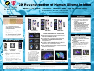
- 1. ● GBM is a highly aggressive and invasive primary brain tumor whose migration patterns are not yet well understood 1 . ● We used bioluminescent imaging (BLI) to monitor tumors in vivo. ● Goal - find a reproducible pattern of migration that can be targeted by future drug therapies. Acknowledgments We want to thank Dr. Sanjiv S. Gambhir and Google for supporting the internship program. We also want to thank our mentors, Dr. Edwin Chang and Dr. Christoph Pohling, for their guidance and support. Conclusions ● Distinct and reproducible migration pattern ○ Observed with multiple patient-derived cell lines ○ Migration into olfactory bulb and possible subventricular zone ○ Spinal metastases are more common than previously thought because even when spine metastasis was not visible in the mouse, one can see it in the spine section ○ GBM migration only surround the exterior of cerebellum and did not enter the cerebellum area. Kathryn Li*, Alex Serafini*, Tara Thakurta*, Xiaofan Wu*, Edwin Chang, and Christoph Pohling 3155 Porter Drive, Palo Alto, CA 94304, USA * A.S., T.T., K.L., and X.W. contributed equally to this project Mersereau, J., Levy, N., Staub, R., Baggett, S., Zogric, T., Chow, S., . . . Bjeldanes, L. (2008). Liquiritigenin is a plant-derived highly selective estrogen receptor β agonist. Molecular and Cellular Endocrinology, 49 1. H. Adams, J. Avendano, S.M. Raza, Z.L. Gokaslan, G.I. Jallo, A. Quinones-Hinojosa. Prognostic Factors and Survival in Primary Malignan Astrocytomas of the Spinal Cord. Spine Volume 37, Number 12: E727-E735, 2007. 2. J.S. Guillamo, F. Lisovoski, C. Christov, C. Le Guerinel, G.L. Defer, M. Peschanski and T. Lefrancois. Migration pathways of human glioblastoma cells xenografted into the immunosuppressed rat brain. Journal of Neuro-Oncology 52: 205-215, 2001. 3. J. Knierim. “Chapter 5: Cerebellum.” Neuroscience Online. 1997. <http://neuroscience.uth.tmc.edu/s3/chapter05.html>. 4. K. Onda, R. Tanaka, H. Takahashi, N. Takeda, F. Ikuta. Cerebral Glioblastoma with Cerebrospinal Fluid Dissemination: A Clinicopathological Study of 14 Cases Examined by Complete Autopsy. Neurosurgery Volume 25, Issue 4: 533-540. 5. H. Sontheimer. S. Watkins. Unique biology of gliomas: challenges and opportunities. Trends in Neurosciences Volume 35, Issue 9: 546-556, 2012. Future Directions ● Investigate the major mechanisms of migration: CSF, white matter, or vasculature tracking? ● Experiment with different sites of implantation ● FACS separation of GBM cells by forward side-scatter in order to study effects of size heterogeneity with regards to migration patterns ● Research anatomically selective drug treatments Initial BLI slice imaging of drug effects on GBM migration, with 3D reconstruction for more accurate drug targeting pending. 3D Reconstruction of Human Glioma in Mice 2015 Internship program supported by: Introduction Materials and Methods Results Conclusions Literature cited Future Directions Acknowledgments Doublet PatternSinglet Pattern Section brain and analyze w/ BLI BLI monitoring of growing tumors Tumor implantation ● pcGBM2 GFP/luc cells were implanted in nude mice. ● Mice were imaged weekly to monitor tumor growth. ● Sections were obtained from harvested mouse brains and imaged with BLI to identify migration patterns. A 3D rendering of a brain with a GBM39 doublet tumor. The olfactory bulb appears to be a niche for GBM. Correlating Migratory Pathways with Anatomical Locations ● GBM appears to travel via specific anatomical structures. In Figure 2, GBM seems to track along the white matter. ● The images also show an accumulation of tumor cells between the cerebellum and the cerebrum, suggesting migration through cerebrospinal fluid (CSF). ● The isolated BLI signals throughout the brain shown in Figure 1 are associated with GBM accumulation around vasculature. ● Future investigation is necessary to identify the cause of migration along these particular anatomical pathways. A 3D rendering of a brain implanted with pcGBM2. The model displays the migration pathway that we identified. Spinal Metastasis 8% 67% 25% 3D Reconstructions After collecting approximately 40 slices (25 microns in width) from two patient derived cell lines, ImageJ was used to render a 3D model. The BLI signal correlates to the presence of GBM xenograft, allowing for observation of both migration pathways and niches favorable for tumor formation. pcGBM2 cell line LN229 cell line U87 cell line a fe dc b Figure 2:Individual Slices Figure 1:Initial Studies Figure 3:3D Reconstructions a b
- 2. ● GBM is a highly aggressive and invasive primary brain tumor whose migration patterns are not yet well understood1 . ● We used bioluminescent imaging (BLI) to monitor tumors in vivo. ● Goal - find a reproducible pattern of migration that can be targeted by future drug therapies. We want to thank Dr. Sanjiv S. Gambhir and Google for supporting the internship program. We also want to thank our mentors, Dr. Edwin Chang and Dr. Christoph Pohling, for their guidance and support. Conclusions ● Distinct and reproducible migration pattern ○ Observed with multiple patient-derived cell lines ○ Migration into olfactory bulb and possible subventricular zone ○ Spinal metastases are more common than previously thought because even when spine metastasis was not visible in the mouse, one can see it in the spine section ○ GBM migration only surround the exterior of cerebellum and did not enter the cerebellum area. Kathryn Li*, Alex Serafini*, Tara Thakurta*, Xiaofan Wu*, Edwin Chang, and Christoph Pohling 3155 Porter Drive, Palo Alto, CA 94304, USA * A.S., T.T., K.L., and X.W. contributed equally to this project Mersereau, J., Levy, N., Staub, R., Baggett, S., Zogric, T., Chow, S., . . . Bjeldanes, L. (2008). Liquiritigenin is a plant-derived highly selective estrogen receptor β agonist. Molecular and Cellular Endocrinology, 49 1. H. Adams, J. Avendano, S.M. Raza, Z.L. Gokaslan, G.I. Jallo, A. Quinones-Hinojosa. Prognostic Factors and Survival in Primary Malignan Astrocytomas of the Spinal Cord. Spine Volume 37, Number 12: E727-E735, 2007. 2. J.S. Guillamo, F. Lisovoski, C. Christov, C. Le Guerinel, G.L. Defer, M. Peschanski and T. Lefrancois. Migration pathways of human glioblastoma cells xenografted into the immunosuppressed rat brain. Journal of Neuro-Oncology 52: 205-215, 2001. 3. J. Knierim. “Chapter 5: Cerebellum.” Neuroscience Online. 1997. <http://neuroscience.uth.tmc.edu/s3/chapter05.html>. 4. K. Onda, R. Tanaka, H. Takahashi, N. Takeda, F. Ikuta. Cerebral Glioblastoma with Cerebrospinal Fluid Dissemination: A Clinicopathological Study of 14 Cases Examined by Complete Autopsy. Neurosurgery Volume 25, Issue 4: 533-540. 5. H. Sontheimer. S. Watkins. Unique biology of gliomas: challenges and opportunities. Trends in Neurosciences Volume 35, Issue 9: 546-556, 2012. Future Directions ● Investigate the major mechanisms of migration: CSF, white matter, or vasculature tracking? ● Experiment with different sites of implantation ● FACS separation of GBM cells by forward side-scatter in order to study effects of size heterogeneity with regards to migration patterns ● Research anatomically selective drug treatments Initial BLI slice imaging of drug effects on GBM migration, with 3D reconstruction for more accurate drug targeting pending. 3D Reconstruction of Human Glioma in Mice 2015 Internship program supported by: Introduction Materials and Methods Conclusions Literature cited Future Directions Acknowledgments Doublet PatternSinglet Pattern Section brain and analyze w/ BLI BLI monitoring of growing tumors Tumor implantation ● pcGBM2 GFP/luc cells were implanted in nude mice. ● Mice were imaged weekly to monitor tumor growth. ● Sections were obtained from harvested mouse brains and imaged with BLI to identify migration patterns. A 3D rendering of a brain with a GBM39 doublet tumor. The olfactory bulb appears to be a niche for GBM. Correlating Migratory Pathways with Anatomical Locations ● GBM appears to travel via specific anatomical structures. In Figure 2, GBM seems to track along the white matter. ● The images also show an accumulation of tumor cells between the cerebellum and the cerebrum, suggesting migration through cerebrospinal fluid (CSF). ● The isolated BLI signals throughout the brain shown in Figure 1 are associated with GBM accumulation around vasculature. ● Future investigation is necessary to identify the cause of migration along these particular anatomical pathways. A 3D rendering of a brain implanted with pcGBM2. The model displays the migration pathway that we identified. Spinal Metastasis 8% 67% 25% 3D Reconstructions After collecting approximately 40 slices (25 microns in width) from two patient derived cell lines, ImageJ was used to render a 3D model. The BLI signal correlates to the presence of GBM xenograft, allowing for observation of both migration pathways and niches favorable for tumor formation. pcGBM2 cell line LN229 cell line U87 cell line a fe dc b Figure 2:Individual Slices Figure 1:Initial Studies Figure 3:3D Reconstructions a b Results
- 3. ● GBM is a highly aggressive and invasive primary brain tumor whose migration patterns are not yet well understood 1 . ● We used bioluminescent imaging (BLI) to monitor tumors in vivo. ● Goal - find a reproducible pattern of migration that can be targeted by future drug therapies. Acknowledgments We want to thank Dr. Sanjiv S. Gambhir and Google for supporting the internship program. We also want to thank our mentors, Dr. Edwin Chang and Dr. Christoph Pohling, for their guidance and support. Conclusions ● Distinct and reproducible migration pattern ○ Observed with multiple patient-derived cell lines ○ Migration into olfactory bulb and possible subventricular zone ○ Spinal metastases are more common than previously thought because even when spine metastasis was not visible in the mouse, one can see it in the spine section ○ GBM migration only surround the exterior of cerebellum and did not enter the cerebellum area. Kathryn Li*, Alex Serafini*, Tara Thakurta*, Xiaofan Wu*, Edwin Chang, and Christoph Pohling 3155 Porter Drive, Palo Alto, CA 94304, USA * A.S., T.T., K.L., and X.W. contributed equally to this project Mersereau, J., Levy, N., Staub, R., Baggett, S., Zogric, T., Chow, S., . . . Bjeldanes, L. (2008). Liquiritigenin is a plant-derived highly selective estrogen receptor β agonist. Molecular and Cellular Endocrinology, 49 1. H. Adams, J. Avendano, S.M. Raza, Z.L. Gokaslan, G.I. Jallo, A. Quinones-Hinojosa. Prognostic Factors and Survival in Primary Malignan Astrocytomas of the Spinal Cord. Spine Volume 37, Number 12: E727-E735, 2007. 2. J.S. Guillamo, F. Lisovoski, C. Christov, C. Le Guerinel, G.L. Defer, M. Peschanski and T. Lefrancois. Migration pathways of human glioblastoma cells xenografted into the immunosuppressed rat brain. Journal of Neuro-Oncology 52: 205-215, 2001. 3. J. Knierim. “Chapter 5: Cerebellum.” Neuroscience Online. 1997. <http://neuroscience.uth.tmc.edu/s3/chapter05.html>. 4. K. Onda, R. Tanaka, H. Takahashi, N. Takeda, F. Ikuta. Cerebral Glioblastoma with Cerebrospinal Fluid Dissemination: A Clinicopathological Study of 14 Cases Examined by Complete Autopsy. Neurosurgery Volume 25, Issue 4: 533-540. 5. H. Sontheimer. S. Watkins. Unique biology of gliomas: challenges and opportunities. Trends in Neurosciences Volume 35, Issue 9: 546-556, 2012. Future Directions ● Investigate the major mechanisms of migration: CSF, white matter, or vasculature tracking? ● Experiment with different sites of implantation ● FACS separation of GBM cells by forward side-scatter in order to study effects of size heterogeneity with regards to migration patterns ● Research anatomically selective drug treatments Initial BLI slice imaging of drug effects on GBM migration, with 3D reconstruction for more accurate drug targeting pending. 3D Reconstruction of Human Glioma in Mice 2015 Internship program supported by: Introduction Materials and Methods Results Conclusions Literature cited Future Directions Acknowledgments Doublet PatternSinglet Pattern Section brain and analyze w/ BLI BLI monitoring of growing tumors Tumor implantation ● pcGBM2 GFP/luc cells were implanted in nude mice. ● Mice were imaged weekly to monitor tumor growth. ● Sections were obtained from harvested mouse brains and imaged with BLI to identify migration patterns. A 3D rendering of a brain with a GBM39 doublet tumor. The olfactory bulb appears to be a niche for GBM. Correlating Migratory Pathways with Anatomical Locations ● GBM appears to travel via specific anatomical structures. In Figure 2, GBM seems to track along the white matter. ● The images also show an accumulation of tumor cells between the cerebellum and the cerebrum, suggesting migration through cerebrospinal fluid (CSF). ● The isolated BLI signals throughout the brain shown in Figure 1 are associated with GBM accumulation around vasculature. ● Future investigation is necessary to identify the cause of migration along these particular anatomical pathways. A 3D rendering of a brain implanted with pcGBM2. The model displays the migration pathway that we identified. Spinal Metastasis 8% 67% 25% 3D Reconstructions After collecting approximately 40 slices (25 microns in width) from two patient derived cell lines, ImageJ was used to render a 3D model. The BLI signal correlates to the presence of GBM xenograft, allowing for observation of both migration pathways and niches favorable for tumor formation. pcGBM2 cell line LN229 cell line U87 cell line a fe dc b Figure 2:Individual Slices Figure 1:Initial Studies Figure 3:3D Reconstructions a b