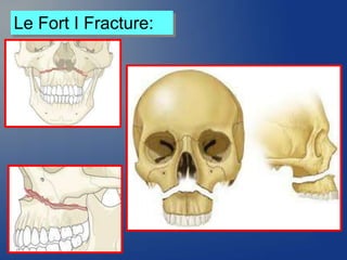
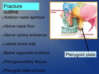
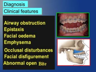
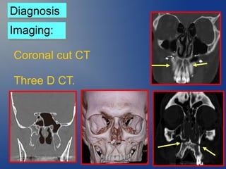
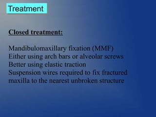
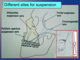
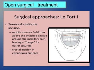

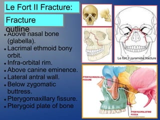
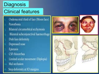
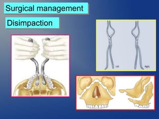
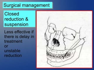
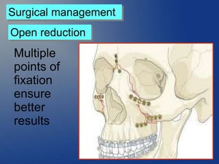
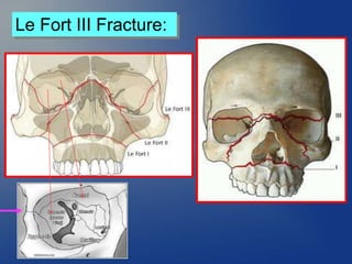
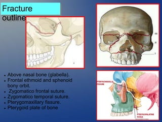
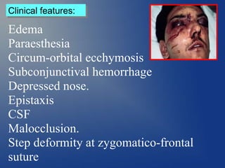
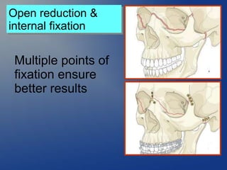


A Le Fort I fracture involves the maxilla below the nasal floor, canine eminence, lateral antral wall, pterygomaxillary fissure, and pterygoid plate. It is typically treated using closed reduction with mandibulomaxillary fixation or open reduction with plate fixation. A Le Fort II fracture extends above the nasal bone and involves the orbit. It requires surgical management like closed reduction with suspension or open reduction with multiple fixation points. A Le Fort III fracture extends through the frontal bone and involves the orbital roof. It presents with symptoms like edema and requires open reduction and internal fixation at multiple sites.


![Part [2] local anesthesia for dental students](https://cdn.slidesharecdn.com/ss_thumbnails/partiil-170222143550-thumbnail.jpg?width=640&height=640&fit=bounds)
![Part [1] local anesthesia for dental students](https://cdn.slidesharecdn.com/ss_thumbnails/partil-170222143343-thumbnail.jpg?width=640&height=640&fit=bounds)

![CTEV [ clubfoot] DR ARUN LAL ,DR MOHAMED ASHRAF travancore medical college k...](https://cdn.slidesharecdn.com/ss_thumbnails/ctevclubfootdrarunlaldrmohamedashraftravancoremedicalcollegekollamkeralaindia-260208063247-18fc466c-thumbnail.jpg?width=640&height=640&fit=bounds)


