SlideShare a Scribd company logo
J Neurosurg 53:205-221, 1980
Mierosurgical anatomy of the pineal region
ISAO YAMAMOTO,M.D., AND NAOK! KAGEYAMA,M.D.
Department of Neurosurgery, Nagoya University, Nagoya, Japan
w' Thirty cadaver brains were examined under • 6to 16magnification in order to define the microsurgical
anatomy of the pineal region, particularly the relationship of the pineal body, posterior cerebral artery,
superior cerebellar artery, vein of Galen, basal vein of Rosenthal, internal cerebral vein, straight sinus,
bridging vein, the size of the tentorial notch, and the third and the fourth cranial nerves. The infratentorial
and supratentorial approaches to the pineal region are compared from the viewpoint of microsurgical
anatomy.
KEY WoRI)S 9 anatomical study 9 pineal region 9 pineal tumor
t"I~UMORS in the pineal region are some of the
excise surgically.difficult lesions tomost
There has been a great debate about their man-
agement since the first attempt at direct removal of a
pineal tumor by Horsley in 1910)e However, 20% to
30% of tumors in this region are benign and are not
responsive to irradiation therapy, u'~8,55'ea'68.s~This has
led to renewed interest in the direct surgical removal
of pineal region tumors with microsurgical technique.
The purpose of this study was to define the
microsurgical anatomy of the pineal region.
Materials and Methods
Formalin-fixed brains complete with dura mater
from 30 adult cadavers were examined under • 6 to 16
magnification. We defined the relationship of the
pineal body, posterior cerebral artery, superior
cerebellar artery, vein of Galen, basal vein of
Rosenthal, internal cerebral vein, straight sinus,
bridging vein, the size of the tentorial notch, and the
third and the fourth cranial nerves.
Description of Anatomy
Pineal Body
The pineal body was round or oval in shape and
measured on the average 7.4 mm (5 to 10 ram) in
longitudinal length, 6.9 mm (5 to 9 mm) in transverse
width, and 2.5 mm (1.5 to 4 mm) in thickness (Fig. 1).
The distance from the anterior end of the vein of
Galen to the pineal body varied from 0 to 15 mm, with
an average measurement of 5.6 mm.
Relationship of the Pineal Body,
Artery, and Vein
The pineal artery is one of the branches of the
medial posterior choroidal artery (MPChA) and
supplies the pineal body and/or habenula trigone.
Although Wackenheim, et al./~ reported that this
artery is single, in our study an average of 1.5 (range 0
to 5) pineal arteries arose from the MPChA in one
hemisphere (Table 2). In 30% of brains (nine brains),
the pineal body was supplied from branches of the
MPChA in one hemisphere (Fig. 3 right), and in
70% (21 brains), it was supplied from those in both
hemispheres (Fig. 1 center and lower). The pineal
artery usually penetrated into the lateral portion of the
pineal body.
The pineal vein,88,8~as defined in our study, drains
from the pineal body or habenula trigone. The pineal
vein has also been called the "epithalamic vein,''72 the
"medial posterior thalamic vein,''4~ and the "latero-
epiphyseal vein.''2~ Rosenbaum and Stein65 and
Giudicelli and Salamon ~7made a distinction between
the pineal vein and the posterior thalamic vein, but
from the view point of microsurgical anatomy, it was
hard to differentiate between these two veins. An
average of 1.9 pineal veins, ranging from one to five,
emptied in the Galenic system. We classified the
pineal veins into five types, according to the mode of
draining into these major veins. In Type 1, the pineal
vein emptied into the terminal portion of one of the in-
ternal cerebral veins (13 cases) (Fig. 1 upper left). In
Type 2, pineal veins emptied into the terminal portion
of both internal cerebral veins (two cases) (Fig. 1 up-
J. Neurosurg. / Volume 53 / August, 1980 205
I. Yamamoto and N. Kageyama
FIG. 1. Posterosuperior views of the pineal region after
the removal of the corpus callosum, illustrating the rela-
tionship of the pineal body to its surrounding struc-
tures. Upper Left: Type 1: the pineal vein (PV) empties
into the terminal portion of one internal cerebral vein
(ICV). Upper Right: Type 2: the PV's empty into the ter-
minal portion of both ICV's. Center Left: Type 3: the PV's
drain into the vein of Galen (G). Center Right: Type 4:
The PV's drain into one of the ICV's and G. Lower: Type
5: the PV empties into the precentral cerebellar vein (PrCV).
The PrCV is seen flowing into either side of the ICV, and the
posterior ventricular vein (PVV) drains into the ICV (upper
left and lower). The internal occipital vein (IOV) ends in G
(upper right and center left). The pineal artery (PA) in both
hemispheres supplies the pineal body (P), basal vein of
Rosenthal (R), cerebellum (Cbl 1), massa intermedia (M),
and medial posterior choroidal artery (MPchA) (center and
lower).
206 J. Neurosurg. / Volume 53 / August, 1980
Microsurgieal anatomy of the pineal region
TABLE 1
Branches of theposterior cerebralartery (PCA) in 60 hemispheres
Branches of PCA
Branches Site of Origin
Present (% of hemispheres)
(% of
hemispheres) P-I P-2A P-2P P-3
No. Branches Present
per Hemisphere
Cortical Average Range
Branches
circumflex artery
short circumflex artery 65.0
long circumflex artery 96.7
thalamogeniculate artery 100.0
posterior choroidal artery
medial posterior choroidal artery 100.0
lateral posterior choroidal artery 100.0
cerebral branches
hippocampal artery 82.8
anterior temporal artery 80.0
middle temporal artery 80.0
posterior temporal artery 77.1
common temporal artery 20.0
parieto-occipital artery 80.0
calcarine artery 100.0
splenial artery 62.8
56.7 8.3
63.3 33.4
46.0 53.3 0.7
13.5 57.5 13.5 7.7
19.5 44.8 9.2
48.6 20.0 8.6
40.0 37.2 2.8
25.7 45.7 8.6
2.8 17.2 54.3
14.3 5.7
8.6 8.6 62.8
8.6 17.2 74.2
2.8
1.2 0-4
2.1 0-4
3.7 1-7
7.7 1.4 1~
26.5 2.6 13
2.8
2.8
51.4
per right). In Type 3, pineal veins drained into the vein
of Galen (11 cases) (Fig. 1 center left). In Type 4,
pineal veins drained into one of the internal cerebral
veins and the vein of Galen (three cases) (Fig. 1 center
right). The pineal vein that emptied into the precentral
cerebellar vein was defined as Type 5 (one case) (Fig. 1
lower). However, Salamon and Huang e7 reported that
the medial posterior thalamic vein drained not only
into the internal cerebral vein or the vein of Galen, but
also into the basal vein of Rosenthal. Tamaki, et al.,~'~
divided pineal veins into three types, but, in our study,
20 brains did not come under their classification. We
found that 40.9% of pineal veins traversed both
superior and inferior surfaces of the pineal body (Fig.
1 center left). In other specimens, the pineal veins
traversed only one surface of the pineal body: the
superior surface in 31.8%, and the inferior surface
in 27.3%.
Posterior Cerebral Artery
Several different classifications have been used for
the posterior cerebral artery (PCA). 2~'48'5~ We
followed the classification of the PCA by Zeal and
Rhotonfl 4 which divided this artery into three
segments from P-1 to P-3. The P-2 segment, which
began at the posterior communicating artery and ter-
minated at the posterior aspect of the midbrain, was
further subdivided into an anterior and posterior half,
and designated as P-2A and P-2P segments. In our
study, we mainly examined the branches originating
from P-2 and P-3 segments.
Circumflex Artery. The circumflex arteries are
divided into short and long circumflex segments (Fig.
2 upper left, center and lower). 2~
The short circumflex artery was present in 65.0% of
the specimens and the number in one hemisphere
ranged from zero to four, the average being 1.2. Most
of the short circumflex arteries arose directly from the
P-I segment (56.7%), the remaining 8.3% arose from
P-2A (Table 1). They ran medial to the PCA around
the midbrain into the thalamogeniculate sulcus and
supplied small perforating branches to the cerebral
peduncle and substantia nigra. As previously
describedfl4 some branches of this artery penetrated
the mesencephalon near the geniculate bodies.
The long circumflex arteries were present in 96.7%
of the specimens and consisted of zero to four, with an
average of 2.1 branches, which arose from the P- 1 seg-
ment in 63.3%, and from the P-2A segment in 33.4%
(Table 1, Fig. 2). They encircled the midbrain between
the PCA and the short circumflex artery, and then
sent rami to the cerebral peduncle, geniculate bodies,
and tegmentum, and the terminal branches reached
the quadrigeminal plate, more on the superior
colliculus)' Some of these terminal branches
anastomosed with the branches of the superior
cerebellar arteries in the region of the quadrigeminal
plate. 26,5',9'As these terminal branches supply the por-
tion of the dorsolateral midbrain containing the
pathways and pretectal neurons serving vertical eye
movement)' occlusion of them may give rise to
Parinaud's syndrome)TM
Thalamogeniculate Artery. In our series, 99.3% of
thalamogeniculate arteries arose from the P-2 seg-
ment in the ambient cistern near the medial and
lateral geniculate bodies. Only 0.7% of them arose in
the P-3 segment. The average incidence of the
thalamogeniculate artery was 3.7, ranging from one to
J. Neurosurg. / Volume 53 / August, 1980 207
I. Yamamoto and N. Kageyama
FIG. 2. Upper Left: Interior view of the course of the
posterior cerebral artery, basilar artery (BA), short cir-
cumflex artery (SCiA), long circumflex artery (LCiA),
medial posterior choroidal artery (MPchA), lateral
posterior choroidal artery (LPchA), anterior temporal
artery (ATA), middle temporal artery (MTA), posterior
temporal artery (PTA), parieto-occipital artery (POA),
calcarine artery (CaA), vein of Galen (G), internal cerebral
vein (ICV), basal vein of Rosenthal (R), and precentral
cerebellar vein (PrCV). Upper Right." The medial part of
the temporal lobe has been removed to expose the LPchA (1,
2, 3, 4), and the hippocampal artery (HiA). Center Left."
The ATA and MTA have been removed to expose the
thalamogeniculate arteries (ThGA), and the pineal body
(P). Center Right." Anterolateral view of the mesen-
cephalon, showing the main arterial trunks, the anterior
choroidal artery (AchA), and the posterior communicating
artery (PCoA). Lower." Inferolateral view of the mes-
encephalon, illustrating absence of the HiA branch of the
PCA. The AchA supplies the hippocampal formation (Hi).
The internal carotid artery (ICA), optic chiasm (OC), and
mamillary body (MB) are seen.
208 J. Neurosurg. / Volume 53 / August, 1980
Microsurgical anatomy of the pineal region
FIG. 3. The corpus caUosum has been removed to expose the terminal portion of the medial posterior
choroidal artery (MPchA). Right internal cerebral vein (ICV) has been retracted (left) to expose the pineal
artery (PA). The vein of Galen (G), basal veinof Rosenthal (R), internal occipital vein (IOV), posterior ven-
tricular vein (PVV), pineal vein (PV), posterior pericallosal vein (PPV), pineal body (P), and massa in-
termedia (M) are visualized.
seven per hemisphere (Table 1, Fig. 2 center left).
Although Duvernoy~1 stated that some of their
branches anastomosed with the branches of the
anterior choroidal artery (AChA) on the surface of the
lateral geniculate body, in our study no anastomosis
could be found under the operating microscope
between the thalamogeniculate arteries and the
anterior choroidal arteries. However, some branches
of the AChA, MPChA, and circumflex arteries
penetrated the mesencephalon in the thalamo-
geniculate sulcus? ~ Thalamogeniculate arteries
penetrated the thalamus and supplied the medial
geniculate, the medial half of the lateral geniculate,
the posterior part of the lateral posterior nucleus, the
ventral lateral pulvinar, the ventral posterior and ven-
tral lateral nuclei, and the nucleus centrum medianum
and intralaminar nuclei/' Damage to this artery
results in the thalamic syndrome of Dejerine-
Roussy.5~
Medial Posterior Choroidal Artery. The MPChA
arose from the P-2A segment in 57.5% of the
specimens, but occasionally arose from P-I (13.5%),
P-2P (13.5%), P-3 (7.7%), or the cortical branches
(7.7%) (Table 1). Berland and Haughton6 dem-
onstrated angiographically an anomalous origin of the
MPChA from the basilar artery. The number of
MPChA's in one hemisphere ranged from one to
three, with an average of 1.4 (Table 1). In our study,
the MPChA was single in 62% and multiple in 38%, as
compared to single in 54% and multiple in 46% as
reported by Zeal and Rhotonfl4 or single in 60% and
multiple in 40% as reported by Margolis, et al. 53 The
MPChA has been reported by others to be a single
artery. 2sa9 It ran parallel and usually medial to the
PCA, and coursed to the quadrigeminal cistern (Fig.
2). Then it lay lateral to the pineal body, and coursed
in the roof of the third ventricle parallel and medial to
the internal cerebral vein in the midline, and finally
supplied the choroid plexus of the third ventricle at the
foramen of Monro (Fig. 3). Along its course, the
MPChA sent an average of 3.6 branches to the
tegmentum (zero to eight), 1.5 branches to the lateral
geniculate body (zero to six), 3.9 branches to the
medial geniculate body (zero to 12), 5.3 branches to
the quadrigeminal plate (zero to 11), 1.6 branches to
the pulvinar (zero to four), 1.5 branches to the pineal
body (zero to five), and 2.7 branches to the medial
thalamus (zero to five). The MPChA arising from the
P-1 segment also sent zero to two branches to the
peduncle with an average number of 0.5 (Table 2). The
number of branches to the quadrigeminal plate was
usually inversely proportional to the number of
branches of the long circumflex arteries to the
quadrigeminal plate. As previously described,sa,5*,a~
when the MPChA arose from the distal cortical
branches of the PCA, this MPChA coursed behind the
pulvinar in a retrograde fashion to supply the tela
choroidea of the third ventricle, pineal body, or the
medial thalamus.
J. Neurosurg. / Volume 53 / August, 1980 209
TABLE 2
Branches of the medial posterior choroidal artery in
60 hemispheres
Site of Termination
No. Present per Hemisphere
Average Range
peduncle 0.5 0-2
tegmentum 3.6 0-8
lateral geniculate body 1.5 0--6
medial geniculate body 3.9 0-12
quadrigeminal plate 5.3 0-11
pulvinar 1.6 0-4
pineal body and/or habenula
trigone 1.5 0-5
medial thalamus 2.7 0-5
Lateral Posterior Choroidal Artery. The lateral
posterior choroidal arteries (LPChA's) arose from the
P-2P segment in 44.8% samples, the P-2A in 19.5%,
the P-3 in 9.2%, and the cortical branches in 26.5%.
The PCA gave origin to one to five LPChA's in one
hemisphere (average 2.6) (Table 1, Fig. 2 upper and
center). There was no difference in number between
right and left LPChA's, that is, the average incidence
was 2.6 on the left and 2.7 on the right. Zeal and
Rhoton94 reported that the average number of
LPChA's in one hemisphere was four, ranging from
one to nine. There was a single LPChA in 9% of
hemispheres, two in 38%, three in 35%, four in 15%,
and five in 3%, as compared to a single trunk in 60%
and multiple in 40%, as reported by Margolis, et al.) 4
or single in 12% and multiple in 88%, as reported by
Zeal and Rhoton)*When more than one LPChA were
present, they were divided into two branches, that is,
anterior and posterior (Fig. 4 upper.left). 2s,~8,53,~,sT,~9
The anterior branches usually arose from P:2A or its
cortical branches and coursed laterally to enter the
choroid fissure and supplied the choroid plexus of the
temporal horn. In our study, 31% of the anterior
branches anastomosed with the AChA, as determined
under the operating microscope (Fig. 4 upper right).
According to Carpenter, et al., TM anastomoses were
found between these two arteries in 93%. Although it
has been reported that the size of the LPChA was
usually inversely proportional to the size of the
AChA,2s'48,Ss'54in our anatomical studies there was no
obvious relationship between the size of these two
arteries except in 6% of the LPChA's which were
definitely smaller than the AChA (Fig. 4 lower). The
posterior branches arose from P-2P, P-3, or their cor-
tical branches, and coursed posteriorly around the
pulvinar, supplying the choroid plexus of the trigone.
Along its course, the LPChA supplied the crus, com-
missure, body and part of the anterior columns of the
fornix, the dorsomedial thalamic nucleus, pulvinar,
and the lateral geniculate body)3 Margolis, et al.)TM
reported that branches arising from the distal portion
of the LPChA extended medially and anastomosed
I. Yamamoto and N. Kageyama
with the MPChA, but in this study no definite anas-
tomosis was found between these two arteries under
the operating microscope.
Cortical Branches of the Posterior Cerebral Artery.
The cortical branches of the PCA are divided into four
segments, that is, the inferior temporal artery,
parieto-occipital artery, calcarine artery, and splenial
artery. The inferior temporal arteries include hip-
pocampal and anterior, middle, posterior and com-
mon temporal arteries.9'
Hippocampal Artery. The hippocampal artery,
which supplies the uncus, hippocampal gyrus, hip-
pocampal formation, and the dentate gyrus, was pres-
ent in 82.8% of specimens examined, as compared to
64% as reported by Zeal and Rhoton)4 This artery
arose from the P-1 segment in 2.8% of specimens, in
48.6% from P-2A, in 20% from P-2P, in 8.6% from
P-3 and in 2.8% from the common temporal artery
(Table 1, Fig. 2 upper right and center). The AChA
also gave off branches to the hippocampus. However,
the distribution of the hippocampal artery varied with
that of the AChA. In the absence of a hippocampal
arterial branch of the PCA, the AChA supplied the
major portion of the hippocampal formation (Fig. 2
lower).
Anterior Temporal Artery. The anterior temporal
artery was present in 80% of hemispheres, as com-
pared to 84% as reported by Zeal and Rhoton.9' It
arose from P-2A in 40.0% of specimens, P-2P in
37.2%, and P-3 in 2.8% (Table 1, Fig. 2 upper left and
center right). It supplies the inferior aspect of the
anterior portion of the temporal lobe.5~
Middle Temporal Artery. The middle temporal
artery was present in 80.0% of hemispheres, as com-
pared to 38% as reported by Zeal and Rhoton)~ It
arose from P-2A in 25.7%, from P-2P in 45.7%, and
from P-3 in 8.6% (Table 1, Fig. 2 upper left and center
right). It supplies the inferior surface of the temporal
lobe)4
Posterior Temporal A rtery. The posterior temporal
artery, which supplies the inferior temporal and oc-
cipital surface, including the occipital pole and lingual
gyrus, was present in 77.1% of hemispheres, as com-
pared to 96% as reported by Zeal and Rhoton.9~ It
originated from the P-2A segment in 2.8% of
specimens, from P-2P in 17.2%, from P-3 in 54.3%,
and, from the calcarine artery in 2.8% (Table 1, Fig. 2
upper left and center). Branches of the posterior tem-
poral artery also helped to supply the visual area, par-
ticularly a part of the "macular" area of the cortex,
and could therefore be a factor in the preservation of
central vision when the calcarine artery was
occluded.7~
Common Temporal Artery. The common temporal
artery was present in 20.0% of specimens, as com-
pared to 16% as reported by Zeal and Rhoton.9~ It
arose from the P-2P segment in 14.3% of specimens,
210 J. Neurosurg. / Volume53 / August, 1980
Microsurgieal anatomy of the pineal region
FIG. 4. Basal views of the mesencephalon illustrating
variations of lateral posterior choroidal artery (LPchA). The
medial part ofthe temporal lobehas been removedto expose
the choroid plexus (ch.pl.) of the temporal horn. Upper
Left: Anterior (l) and posterior branches (2) of the
LPchA. Upper Right: Anastomosis is shownbetween the
LPchA and the anterior choroidal artery (AchA) (curved
arrow). Lower: The AchA is larger than the LPchA. Other
structures seen are: the internal carotid artery (ICA),
posterior cerebral artery (PCA), basilar artery (BA),
posterior communicating artery (PCoA), medial posterior
choroidal artery (MPchA), posterior temporal artery
(PTA), calcarine artery (CaA), basal vein of Rosenthal (R),
tentorium cerebelli (T), and the mamillary body (MB).
and from P-3 in 5.7%. It supplies the majority of the
inferior surface of the temporo-occipital lobes?4
Parieto-Occipital Artery. The parieto-occipital
artery was seen in 80.0% of our specimens, as com-
pared to 96% noted by Zeal and Rhoton?4 It arose as
a single branch from P-2A in 8.6%, from P-2P in
8.6%, and from P-3 in 62.8% (Table 1, Fig. 2 upper
and center). It supplies the superior part of the cuneus,
the posterior fifth of the precuneus, part of the
superior parietal lobule, and the superior occipital
gyrus/*
Calcarine Artery. The calcarine artery was present
in all specimens examined and arose from P-2A in
8.6%, P-2P in 17.2%, and P-3 in 74.2% (Table 1, Fig. 2
upper and center right). It ran in the calcarine sulcus
and supplied the inferior cuneus and the superior and
posterior part of the lingual gyrus/a Smith and
RichardsonTM reported that the visual area was
supplied by the calcarine artery in only 25% of cases;
by the calcarine and the posterior temporal arteries in
34.4%; by the calcarine and the parieto-occipital
arteries in 18.8%; by the calcarine and both parieto-
occipital and posterior temporal arteries in 15.5%; by
the calcarine plus the middle cerebral and the parieto-
occipital arteries in 3.1%, and by all the above arteries
in 3.1%. Infarction of the calcarine cortex causes a
homonymous visual defect with macular sparing.37,87
J. Neurosurg. / Volume 53 / August, 1980 21 ]
I. Yamamoto and N. Kageyama
FI~. 5. Left: Anterolateral view of the left mesencephalon, showing the arrangement of the main
arterial trunks. Also visibleare: the basilar artery (BA),superior cerebellar artery (SCA), posterior cerebral
artery (PCA), anterior inferior cerebellar artery (AICA), posterior communicating artery (PCoA), medial
posterior choroidal artery (MPchA), anterior temporal artery (ATA), middle temporal artery (MTA),
posterior temporal artery (PTA), parieto-occipital artery (POA), and the calcarine artery (CaA). Right:
Superolateral view of the left cerebellum, showingthe course of the SCA. 1: lateral marginal branch, 2:
hemispheric branch and 3: superior vermis branch. The cerebellum (Cbll) is also indicated.
Splenial Artery. The splenial artery, also called the
"posterior pericallosal artery,''5''~~or "dorsal callosal
artery,'''8 was seen in 62.8% of specimens. It has
been reported to be present in 35%5. to 100% of
specimens?' It originated from the parieto-occipital
artery in 31.4%, calcarine artery in 17.2%,the LPChA
in 8.6%, the MPChA in 2.8%, and the common tem-
poral artery in 2.8% (Table 1). It coursed around the
splenium close to the midline and anastomosed with
the distal branches of the pericallosal artery of the
anterior cerebral artery.
Superior Cerebellar Artery
The superior cerebellar artery originated from the
basilar artery an average of 1.8 mm proximal to the
basilar bifurcation, as compared to 2.5 mm reported
by Saeki and Rhoton86(Fig. 5 left). Although duplica-
tion or triplication of the superior cerebellar arteries
has been reported in 28% or 2%, respectively,82'5~in
our anatomical studies 4.2% (two of 48) were dupli-
cated. The superior cerebellar artery circled around
the upper pons or lower mesencephalon parallel
to the course of the basal vein of Rosenthal, the
PCA, and the free edge of the tentorium, and coursed
toward the superior surface of the cerebellar
hemisphere and the superior vermis. The proximal
trunk of this artery divides into anterior pontine, am-
bient, and quadrigeminal segment,32 but this division
is not anatomically precise. There are three main cor-
tical branches and perforating branches of the
superior cerebellar arterya2,52 (Fig. 5 right). An
average of 1.3 lateral marginal branches (range zero
to two) usually originated close to the junction
between the pontine and ambient segments, which
supply the superior cerebellar peduncle, the dentate
nucleus, the roof nuclei, and the middle cerebellar
peduncles2"79(Table 3). The lateral marginal branches
were usually inversely proportional in size to the cor-
tical branches of the anterior inferior cerebellar
artery?2 An average of 3.8 hemispheric branches
(range, two to five) on each side arose distal to the
origin of the lateral marginal branches and coursed
over the superior surface of the cerebellum (Table 3).
The superior vermis branches originated from
the quadrigeminal segment and numbered 2.3 on the
average (range, zero to three) on each side. The right
and left superior vermis branches usually
anastomosed with each other in the quadrigeminal
cistern and then coursed posteriorly over the vermis
close to the midline. Within the quadrigeminal cistern,
small arterial branches from the proximal portion of
the superior vermis branches ran into the precentral
cerebellar fissure, and are called the precentral
cerebellar arteries?2 These arteries were identified in
all our specimens examined. From the medial wall of
the superior cerebellar artery around the brain stem,
an average of 7.1 (range, three to 13) perforating
branches arose and supplied the superior cerebellar
peduncle and the quadrigeminal plate (Table 3). Some
of the branches that reached the_quad_rigeminal plate
anastomosed with the terminal branches of the long
circumflex arteries.28'"3'94
Galenic Venous System
Vein of Galen. The vein of Galen was formed by the
union of the paired internal cerebral veins. Its average
length was 12 mm, ranging from 8 to 25 mm.
Tributaries that joined the vein of Galen were the in-
212 J. Neurosurg. / Volume53/August, 1980
Mierosurgieal anatomy of the pineal region
TABLE 3
Branches of the superior cerebellar artery (SCA) in
60 hemispheres
Branchof SCA
No. Presentper Hemisphere
Average Range
lateralmarginalbranch 1.3 0-2
hemisphericbranch 3.8 2-5
superiorvermisbranch 2.3 0-3
perforatingbranch 7.1 3-13
TABLE 4
The incidence of tributaries emptying into the vein of Galen
Percentof
Tributary Specimens
internalcerebralvein 100.0
precentralcerebellarvein 86.4
internaloccipitalvein 77.0
basalveinof Rosenthal 51.7
posteriorpericallosalvein 41.5
pinealvein 40.9
posteriormesencephalicvein 38.4
posteriorventricularvein 6.7
FIG. 6. Posteroinferior view of the pineal region of the
left occipital lobe (Occ L), showing the relationship of the
vein of Galen (G) and its tributaries. Precentral cerebellar
vein (PrCV) draining into G. The internal occipital vein
(IOV) flows into the basal vein of Rosenthal (R). Ab-
breviations as in previous figures.
ternal cerebral vein in 100% of specimens, precentral
cerebellar vein (86.4%), internal occipital vein (77%),
basal vein of Rosenthal (51.7%), posterior pericallosal
vein (41.5%), pineal vein (40.9%), posterior
mesencephalic vein (38.4%), and the posterior ven-
tricular vein (6.7%) (Table 4, Fig. 6). This vein then
coursed superoposteriorly under the splenium of the
corpus callosum and joined the inferior sagittal sinus
to form the straight sinus. Although internal
hydrocephalus resulting from the occlusion of the vein
of Galen, particularly near its origin, was reported by
Dandy,15,17,79experimental occlusion by others did not
result in hydrocephalus.2,3a8,~2,79
Basal Vein of Rosenthal: Salamon and Huang67
divided the basal vein of Rosenthal into three
segments: the first, anterior or striate segment, the
second, middle or peduncular segment, and the third,
posterior or posterior mesencephalic segment. The
second segment is further subdivided into anterior and
posterior portions by the most lateral point of the vein
as it turns around the peduncle. In this study, the
posterior portions of the second and the third
segments were examined.
Inferior Ventricular Vein. The inferior ventricular
vein drains the subependymal and choroidal veins of
the temporal horn as well as the veins of hippocampal
formation.38,57'~3As a common trunk, it flowed into
the second segment of the basal vein of Rosenthal at
the junction between the anterior and posterior por-
tions in 88.1% of the specimens examined (Fig. 7 left).
Only 11.9% of them were double. The inferior ventric-
ular vein rarely joins the lateral or straight sinus via a
tentorial sinus)s
Lateral Mesencephalic Vein. The lateral mesen-
cephalic vein runs along the lateral mesencephalic sul-
cus and drains upward to the third segment of the
basal vein of Rosenthal or downward to the brachial
tributary of the petrosal vein.3~176 It was present in
69% of the specimens examined (29 of 42) in our ana-
tomical studies. Angiographically it is seen in 30% to
35% of normal vertebral angiograms.TM Wacken-
heim, et al.fl2 reported six types of variations in the
drainage of the lateral mesencephalic vein.
Posterior Mesencephalic Vein. The posterior mes-
encephalic vein was present in 21.7% of the hemi-
spheres (13 of 60) and emptied into the vein of Galen
in 8.3% (five), the basal vein of Rosenthal in 8.3%
(five), and the internal cerebral vein in 5% (three).
The terminal portion of the basal vein of Rosenthal
drained into the vein of Galen or the posterior portion
of the internal cerebral vein. The mode of drainage
into these major veins was classified into three types in
our study; 1) both basal veins of Rosenthal emptied
into the vein of Galen in 11 specimens (Fig. 8 upper
left), 2) into the internal cerebral veins in 10 (Fig. 8 up-
per right), and 3) into the internal cerebral vein in one
J. Neurosurg. / Volume53 / August, 1980 213
I. Yarnamoto and N. Kageyama
FIG. 7. Left: The medial part of the temporal lobe has been removed to expose the inferior ventricular
vein (IVV), basilar artery (BA) posterior cerebral artery (PCA), posterior"communicating artery (PCoA),
anterior choroidal artery (AchA), medial posterior choroidal artery (MPchA), lateral posterior choroidal
artery (LPchA), and the basal veinof Rosenthal (R). Right: Posterolateral viewof the left mesencephaion,
illustrating the arrangement of the main arterial and venous trunks, and showing the superior cerebellar
artery (SCA), long circumflex artery (LCiA), precentral cerebellar vein (PrCV), superior vermian vein
(SVV), lateral mesencephalic vein (LMV), pineal vein (PV), cerebellum (Cbll), occipital lobe (Occ L), and
the pineal body (P).
side and into the vein of Galen in the other side in nine
(Fig. 8 lower left). Three basal veins of Rosenthal ter-
minated at the posterior portion of the vein of Galen
(Fig. 8 lower right). Many anatomical variations in
draining of the basal vein of Rosenthal have been
reported, including draining into the straight sinus,
lateral sinus, superior petrosal sinus via the
anastomotic lateral mesencephalic vein, or the
sphenoparietal sinus.67
Internal Occipital Vein. The internal occipital vein
originated on the inferior and medial surface of the oc-
cipital lobe and then coursed anteromedially to end in
the vein of Galen in 77.0% of specimens (Figs. 1 upper
right and lower left, 8 upper left and lower, and 10
right). More rarely, it also joined the internal cerebral
vein in 11.5% of specimens (Fig. 9), and the basal vein
of Rosenthal in 11.5% (Fig. 6). Left homonymous
hemianopsia was reported as a result of a right
supratentorial approach for pineal tumor by Harris,
et al.a~They ascribed the hemianopsia to division of
the internal occipital vein.
Posterior Pericallosal Vein. The posterior perical-
losal vein has also been called the "splenial vein,''67the
"posterior cerebral vein,''5 the "posterior marginal
vein, ''86 the "posterior vein of corpus callosum, ''5 and
the "dorsal callosal vein.''78 It originated on the dorsal
surface of the corpus callosum and traversed around
the splenium parallel to the posterior pericallosal
artery to enter the internal cerebral vein in 48.8% of
specimens (Fig. 11 right), the vein of Galen in 41.5%
(Figs. 8 upper left and 10 left), the basal vein of Rosen-
thai in 7.3% (Fig. 8 lower right), and the internal occipi-
tal vein in 2.4% (Figs. 8 left, and 10 right). Salamon and
Huang67reported that the splenial vein might also join
the medial atrial vein. The posterior pericallosal vein
was usually double (62.5%), each lying in the im-
mediate paramidline; it was single in 33.3%, and
rarely triple (4.2%).
Precentral Cerebellar Vein. The precentral
cerebellar vein originated in the precentral cerebellar
fissure, usually as two brachial veins uniting into a
single common trunk. This vessel coursed upward to
join the vein of Galen or the posterior portion of the
internal cerebral vein. In 86.4% (19 of 22) of the
specimens, the precentral cerebellar veins drained into
the vein of Galen (Fig. 6), and in 13.6% (three of 22)
they flowed into either side of the internal cerebral
vein (Fig. 1 upper left and lower). Occasionally, the
brachial veins did not unite but drained independently
into the vein of Galen. Small veins arising from the
anterior surface of the vermis and the adjacent
cerebellar hemisphere, the superior vermian, or the
superior hemispheric veins (Figs. 6 and 7 right) usually
joined the precentral cerebellar vein at the junction
2]4 J. Neurosurg. / Volume 53 / August, 1980
Mierosurgieal anatomy of the pineal region
FIG. 8. Variations of the terminal portion of the basal vein of Rosenthal (R). Upper Left: Both R's
emptying into the vein of Galen (G). The left posterior pericallosal vein (PPV) ends in G and the right PPV
ends in the internal occipital vein (IOV). Upper Right: Both R's empty into the internal cerebral veins
(ICV). Lower Left: Inferior view of the course of the terminal portion of R, one side of which empties into
the ICV (left) and the other side into G (right). Lower Right: Both R's terminate at the posterior portion of
G. Structures visualized are: the medial posterior choroidal artery (MPchA), long circumflex artery (LC!A),
calcarine artery (CaA), posterior ventricular vein (PVV), pineal vein (PV), pineal body (P), tentorxum
cerebelli (T), cerebellum (Cbll), and the straight sinus (SS).
between the inferior colliculi and the anterior superior
margin of the cerebellum?9'4~
Internal Cerebral Vein. The internal cerebral vein
originates just behind the foramen of Monro by the
union of the septal, thalamostriate, and choroidal
veins.48 Paired internal cerebral veins run posteriorly
within the tela choroidea, adjacent to the midline, and
unite with the subependymal veins, the basal vein of
Rosenthal, and/or the internal occipital vein to form
the vein of Galen. Caron, et al., TM reported that the
surgical ligature of both internal cerebral veins was
well tolerated in two pineal tumors.
Various terms have been used for the subependymal
veins of the lateral ventricles, including the "subepen-
dymal veins,''78,g3 the "vein of the posterior horn, ''43
and the "posterior ventricular vein.''8,9 Billewicz and
Bel-Amor8.9 defined four veins: the medial atrial vein,
the direct medial vein, the direct lateral vein, and the
lateral atrial vein, which were described by Wolf and
FIG. 9. Posterosuperior view of the pineal region. The
left internal occipital vein (IOV) joins the internal cerebral
vein (ICV). The medial posterior choroidal artery (MPchA),
basal vein of Rosenthal (R), pineal vein (PV), pineal body
(P), cerebellum (Cbll), massa intermedia (M) are seen.
J. Neurosurg. / Volume 53 / August, 1980 2 ]5
I. Yamamoto and N. Kageyama
FIG. 10. Medial views of a midsagittal section, showing the relationship of the posterior pericaltosal vein
(PPV) to its tributaries. Left: The PPV enters the vein of Galen (G). Right: The PPV joins the internal
occipital vein (IOV) and then flows into G. The medial posterior choroidal artery (MPchA), internal
cerebral vein (ICV), posterior ventricular vein(PVV),pineal body (P), and spleniumof the corpus callosum
(S) can be seen.
Huang93 as the posterior ventricular vein. From the
viewpoint of microsurgical anatomy, we could not
differentiate between these four veins, so in our study
we used the designation of the posterior ventricular
vein. The posterior ventricular vein, which drains the
posterior part of the body, the atrium, and the
posterior horn of the lateral ventricle,' flowed in
almost all specimens (55 of 60 hemispheres) into the
internal cerebral vein (Figs. 1 upper left and lower, 3,
8 rightpair, 10, and 11 right). It flowed into the vein of
Galen in four cases, and the basal vein of Rosenthal in
one. In 11 brains, we dissected the posterior ven-
tricular vein distally to identify the relationship
between the medial and lateral atrial veins (Fig. 11
FIG. 11. Left: Medial view of a midsagittal section, illustrating the medial and lateral atrial veins
(MAV, LAV) of the posterior ventricular vein (PVV). Right: The PVV drains into the internal cerebral
vein (ICV) as a common trunk. Other structures visualized are the medial posterior choroidal artery
(MPchA), posterior cerebral artery (PCA), internal occipital vein (IOV), posterior pericallosal vein (PPV),
pineal body (P), vein of Galen (G), and the splenium of the corpus callosum (S).
216 J. Neurosurg. Volume 53 / August, 1980
Mierosurgieal anatomy of the pineal region
FIG. 12. Left: Posterolateral view of the inferior occipital lobe (Occ L), illustrating the bridging veins
between the lateral occipital pole and the transverse sinus (TS). Right: Posterior view of the superior
cerebellum (Cbll), illustrating finebridging veins between the hemispheric cerebellar veins and TS.
left). In seven of 22 hemispheres, the medial and
lateral atrial veins united into a common trunk to
form the common atrial vein,TM or the posterior
paraventricular vein/ and drained into the internal
cerebral vein (Fig. 11 right). Bekov' found the
posterior paraventricular vein in 24% of instances. In
this situation, the posterior paraventricular vein was
almost the same size as the basal vein of Rosenthal, so
it was sometimes difficult to differentiate between
these two veins; however, the diverging point of the
basal vein of Rosenthal was closer to the vein of Galen
(Figs. 1 upper left and 8 right pair). In other
situations, the basal vein of Rosenthal was usually
larger than the posterior ventricular vein (Fig. 3 right).
Although the lateral atrial vein has been reported in
139 of 184 hemispheres,~in our study the medial and
lateral atrial veins were found in 68.2% (15 of 22) and
95.5% (21 of 22), respectively.
Straight Sinus. The straight sinus was formed by
the union of the inferior sagittal sinus and the vein of
Galen at the posterior end of the splenium and then
continued posteroinferiorly, following the line ofjunc-
tion of the falx cerebri with the tentorium cerebelli, to
the torcular Herophili. Saxena, et al./9'7~ reported a
double straight sinus in 13.95% of cadavers, but we
could not find a double straight sinus in our review of
30 specimens. Knott~e reported the absence of the
straight sinus in one of 44 specimens. In our study, the
average length of the straight sinus was 52.3 mm, with
a minimum of 45 mm and maximum of 58 mm. Sax-
ena, et al./~ reported that the straight sinus was 50
mm in average length.
Bridging Vein. No bridging veins could be found
between the medial occipital pole and the transverse
sinus. However, two to seven bridging veins per
hemisphere were present, lying between the lateral oc-
cipital pole and the transverse sinus (Fig. 12 left).
Between the tentorium cerebelli and the cerebeUar sur-
face, numerous fine bridging veins could be found
medially; there were three to five bridging veins per
hemisphere laterally (Fig. 12 right).
Tentorial Notch. The distance from the posterior
boundaries of the tentorial notch to the pineal body
was 18.6 mm on the average, ranging from 10 to 30
mm (Fig. 13).
The Third and Fourth Cranial Nerves. The third
nerve left the midbrain on the medial side of the
cerebral peduncle, and passed under the PCA and
above the superior cerebellar artery (Fig. 5 left). In
four of 60 specimens, the circumflex artery penetrated
into the substance of the third nerve (Fig. 14). The
fourth nerve originated from the dorsal surface of the
midbrain, then curved anteriorly between the PCA
and the superior cerebellar artery along the tentorial
edge and lay lateral to the third nerve (Figs. 5 right
and 7 right).
This relationship between the third and the fourth
cranial nerves and the posterior cerebral and superior
cerebellar arteries was constant in all specimens ex-
amined, even in the presence of duplication of the
superior cerebellar artery.
Discussion
Various surgical approaches to the pineal region
have been reported since 1910, when Horsleya8 first
attempted direct removal of a pineal tumor. The sur-
gical approach to the pineal region has been roughly
classified into three techniques: infratentorial su-
pracerebellar,sS'41,47,Ss'59'84,75-~'89 supratentorial, and
transventricular approaches?8 The supratentorial
J. Neurosurg. / Volume 53/August, 1980 2] 7
I. Yamamoto and N. Kageyama
FIG. 13. Three inferior views of the tentorium, illustrating variations in the interval separating the boun-
daries of the tentorial notch from the mesencephalon.
FIG. 14. Anterolateral view of the right mesencephalon,
illustrating that the short circumflex artery (SCiA) pene-
trates into the bundle of the third cranial nerve (curved
arrow). The internal carotid artery (ICA), basilar artery
(BA), posterior cerebral artery (PCA), superior cerebellar
artery (SCA), posterior communicating artery (PCoA), and
mamillary body (MB) can be seen.
technique has been further subdivided into two ap-
proaches, namely, the unilateral transtentorial ap-
proachl~176 and the interhemispheric
posterior parietal approach?,x8,33a5,49,85,82
There have been few reports comparing the two
basic surgical approaches to the pineal region, that is,
the supratentorial and the infratentorial ap-
proaches.",~6,8s Reid and Clark63compared these two
different approaches and preferred the occipital
transtentorial technique. Stein,77who did not confine
his report to tumors of the pineal body, claimed that
the infratentorial supracerebellar approach was still
preferable to the supratentorial approach. Obrador, et
al.? ~stated that the infratentorial approach was useful
for the tumors projecting downward to the posterior
fossa, and, in more anteriorly located tumors en-
croaching on the posterior third ventricle, the
supratentorial approach was preferable.
From the view point of microsurgical anatomy of
the pineal region, we have to consider the following
four important anatomical points in the determination
of the surgical approach.
First, we must consider the location of the bridging
veins. When using an infratentorial approach, all
bridging veins over the cerebellar surface must be
sacrificed to reach the quadrigeminal region. Stein77
never encountered cerebellar edema from the transec-
tion of these bridging veins, but Page~8 reported at
least one case with cerebellar swelling after they were
218 J. Neurosurg. / Volume 53 / August, 1980
Mierosurgieal anatomy of the pineal region
divided. In contrast, as no bridging veins lie between
the medial occipital pole and the transverse sinus when
a supratentorial approach is used, the occipital lobe
can be retracted superolaterally without sacrificing
bridging veins. But this retraction is at times related to
postoperative visual complications, such as transient
or permanent hemianopsia.
Second, the size of the tentorial notch is variable. 81
In our anatomical studies, the distance between the
posterior boundaries of the tentorial notch and the
pineal body ranged from 10 to 30 mm. The shorter
this distance becomes, the more difficult is the ex-
posure above the tentorium when viewing from an
infratentorial approach. In contrast, when using a
transtentorial approach, the edge of the tentorium is
transected 1 cm lateral to the straight sinus to afford
excellent visualization of the vein of Galen and its
branches. But this approach is hampered by these ma-
jor veins and does not provide a wide enough view of
the quadrigeminal plate, particularly over the con-
tralateral side.
Third, it goes without saying that a supratentorial
approach is hampered by the vein of Galen and its
major tributaries. Injury to these deep veins is one of
the main causes of surgical morbidity. At times, we
have transected the internal occipital vein or posterior
pericallosal vein. Harris, et al., s~ reported a case of
hemianopsia as a result of the section of the internal
occipital vein. Using an infratentorial approach,
Page58 reported the precentral cerebellar vein was
preserved in five of nine cases. When necessary, this
vein must be transected in order to provide a wide ex-
posure by either approach; however, this does not
result in postoperative neurological deficits.
Fourth, several authors reported that the splenium
could be spared routinely in a supratentorial ap-
proach; 42,51,6~ however, when the tumor is located
above the hiatus tentorii, bulges anteriorly into the
posterior third ventricle, and compresses the superior
colliculus (Type A of Poppen6~ the splenium is
sometimes incised even in a transtentorial approach.
In that event, a resultant disorder of the higher cor-
tical function, an alexia without agraphia, has been
reported. ~~ In Dandy's parasagittal approach,
an incision of the posterior half of the corpus callosum
exposes the tumor? 6 Dimond, et al., TM reported that
the surgical section of the posterior portion of the
body of the corpus callosum produced some memory
defect. In an infratentorial approach, even after the
transection of the precentral cerebellar vein, the lower
portion of the quadrigeminal plate is still concealed by
the superior vermis, particularly the culmen. So a
splitting and retraction of the upper vermis is oc-
casionally necessary to give enough exposure.
Based upon these anatomical considerations, the
selection of the appropriate surgical approach to the
pineal region should depend mainly upon the location
and the extent of the tumor, and partly upon the sur-
geon's experience.
References
1. Araki C: [Removal of the pineal tumor.] Gekashinryo
2:517-524, 1960 (Jpn)
2. Bedford THB: The great vein of Galen and the syn-
drome of increased intracranial pressure. Brain 57:1-24,
1934
3. Bedford THB: The venous system of the velum inter-
positum of the rhesus monkey and the effect of ex-
perimental occlusion of the great vein of Galen. Brain
57:255-265, 1934
4. Bekov DB: Some structural differences among postero-
lateral tributaries of great cerebral vein. Arkh Anat
Gistoi Embrlol 46:166-170, 1964
5. Ben-Amor M, Billewicz O: The posterior cerebral vein.
Neuroradiology 1:179-182, 1970
6. Berland LL, Haughton VM: Anomalous origin of
posterior choroidal artery from basilar artery. AJR
132:674-675, 1979
7. Billewicz O: The normal and pathological radio-
anatomy of the lateral mesencephalic vein. Neuro-
radiology 8:295-299, 1975
8. Billewicz O, Ben-Amor M: The posterior ventricular
branches of the internal cerebral and basilar vein.
Neuroradiology 2:37-45, 1971
9. Billewicz O, Ben-Amor M: The posterior ventricular
branches of the internal cerebral and basilar veins in
cases of deep cerebral tumours infiltrating the basal
ganglia. Neuroradiology 1:223-227, 1970
10. Brunner C: Cited by Rorschach H: Zur Pathologie und
operabilit/it der Tumoren der Zirbeldrfise. Beitr Kiln
Chit 83:451-474, 1913
11. Camins MB, Schlesinger EB: Treatment of tumours of
the posterior part of the third ventricle and the pineal
region: a tong term follow-up. Acta Neuroehir 40:
131-143, 1978
12. Caron JP, Debrun G, Sichez JP, et al: Ligature des
veines c6r6brales internes et survie. Apropos de deux
pin~alomectomies. Neurochirurgie 20:81-90, 1974
13. Carpenter MB, Noback CR, Moss ML: The anterior
choroidal artery: its origins, course, distribution and
variations. Arch Neurol Psychiatry 71:714-722, 1954
14. Chou SN: The treatment of third ventricular tumors
with particular reference to surgical excision, in Morley
TP (ed): Current Controversies in Neurosurgery.
Philadelphia: WB Saunders, 1976, pp 367-373
15. Dandy WE: Experimental hydrocephalus. Ann Surg
70:129-142, 1919
16. Dandy WE: An operation for the removal of pineal
tumors. Surg Gynecol Obstet 33:113-119, 1921
17. Dandy WE, Blackfan KD: Internal hydrocephalus. An
experimental, clinical and pathological study. Am J Dis
Child 8:406-482, 1914
18. DeGirolami U, Schmidek H: Clinicopathological study
of 53 tumors of the pineal region. J Neurosurg
39:455-462, 1973
19. Dimond SJ, Scammell RE, Brouwers EYM, et al:
Functions of the centre section (trunk) of the corpus
callosum in man. Brain 100:543-562, 1977
20. Duvernoy HM: Human Brainstem Vessels.
Berlin/Heidelberg/New York: Springer-Verlag, 1978,
188 pp
21. Duvernoy HM: The Superficial Veins of the Human
Brain. Berlin/Heidelberg/New York: Springer-Verlag,
1975, 110 pp
J. Neurosurg. / Volume 53 / August, 1980 2"19
22. Ecker A, Riemenschneider PA: Angiographic Localiza-
tion of lntracranial Masses. Springfield, II1: Charles C
Thomas, 1955, 433 pp
23. Galloway JR, Greitz T: The medial and lateral
choroidal arteries. An anatomic and roentgenographic
study. Acta Radiol 53:353-366, 1960
24. Geschwind N: Disconnexion syndromes in animals and
man. I. Brain. Brain 88:237-294, 1965
25. Geschwind N: Disconnexion syndromes in animals and
man. II. Brain. Brain 88:585-644, 1965
26. Gillilan LA: The arterial and venous blood supplies to
the forebrain (including the internal capsule) of
primates. Neurology 18:653-670, 1968
27. Giudicelli G, Salamon G: The veins of the thalamus.
Neuroradiology 1:92-98, 1970
28. Glasauer FE: An operative approach to pineal tumors.
Acta Neurochir 22:177-180, 1970
29. Hammock MK, Milhorat TH, Earle K, et al: Vein of
Galen ligation in the primate. Angiographic, gross and
light microscopic evaluation. J Neurosurg 34:77-83,
1971
30. Harris W, Cairns H, Adelaide MB: Diagnosis and treat-
ment of the pineal tumours with report of a case. Lancet
1:3-9, 1932
31. Heppner F: Zur Operationstechnik bei pinealomen. Zbl
Neurochir 19:219-224, 1959
32. Hoffman HB, Margolis MT, Newton TH: The superior
cerebellar artery. I. Normal gross and radiographic
anatomy, in Newton TH, Putts DG (eds): Radiology of
the Skull and Brain. St. Louis: CV Mosby, 1974, Vol II,
Book 2, pp 1809-1830
33. Horrax G: The diagnosis and treatment of pineal
tumors. Radiology 52:186-192, 1949
34. Horrax G: Extirpation of a huge pinealoma from a
patient with pubertas praecox: a new operative ap-
proach. Arch Neuroi Psychiatry 37:385-397, 1937
35. Horrax G: Treatment of tumors of the pineal body: ex-
perience in a series of twenty-two cases. Arch Neurol
Psychiatry 64:227-242, 1950
36. Horsley V: Discussion. Proc R Sue Med 3:2, 1910
37. Hoyt WF, Newton TH: Angiographic changes with
occlusion of arteries that supply the visual cortex. N Z
Med J 72:310-317, 1970
38. Huang YP, Wolf BS: The basal cerebral vein and its
tributaries, in Newton TH, Putts DG (eds): Radiology
of the Skull and Brain. St Louis: CV Mosby, 1974, Vol
II, Book 3, pp 2111-2154
39. Huang YP, Wolf BS: Precentral cerebellar vein in
angiography. Acta Radiol (Diagn) 5:250-262, 1966
40. Huang YP, Wolf BS: Veins of the posterior fossa, in
Newton TH, Putts DG (eds): Radiology of the Skull and
Brain. St Louis: CV Mosby, 1974, Vol II, Book 3, pp
2155-22i6
41. Hubschmann O, Kasoff S, Doniger D, et al: Cavernous
haemangioma in the pineal region. Surg Neurol
6:349-351, 1976
42. Jamieson KG: Excision of pineal tumors. J Neurosurg
35:550-553, 1971
43. Johanson C: The central veins and deep dural sinuses of
the brain; an anatomical and angiographic study. Acta
Radiol [Snppl 107]:1-184, 1954
44. Kempe LG: Operative Neurosurgery. Berlin/
Heidelberg/New York: Springer-Verlag, 1968, Vol 1,
pp 145-155
I. Yarnamoto and N. Kageyama
45. Kirsch WM, Stears JC: Radiographic identification and
surgical excision of an epidermoid tumor of the pineal
gland. Case report. J Neurusurg 33:708-713, 1970
46. Knott JF: On the cerebral sinuses and their variations.
J Anat Physiol 16:27-42, 1881
47. Krause F: Operative Freilegung der Vierhugel Nebst
Beobachtungen uber Hirndruck und Dekompression.
Zentralbl Chir 53:2812-2819, 1926
48. Krayenb/ihl HA, Ya~argil MG: Cerebral Angiography,
ed 2. Philadelphia: JB Lippincott, 1968, pp 20-123
49. Kunicki A: Operative experiences in 8 cases of pineal
tumor. J Neurosurg 17:815-823, 1960
50. Langworthy OR, Fox HM: Thalamic syndrome. Syn-
drome of the posterior cerebral artery; a review. Arch
Intern Med 60:203-224, 1937
51. Lazar ML, Clark K: Direct surgical management of
masses in the region of the vein of Galen. Surg Neurol
2:17-21, 1974
52. Mani RL, Newton TH, Glickman MG: The superior
cerebellar artery: an anatomic-roentgenographic cor-
relation. Radiology 91:1102-1108, 1968
53. Margolis MT, Hoffman HB, Newton TH: Choroidal
arteries in the diagnosis of thalamic tumors. J
Neurosurg 36:287-298, 1972
54. Margolis MT, Newton TH, Hoyt WF: The posterior
cerebral artery. II. Gross and roentgenographic
anatomy, in Newton TH, Putts DG (eds): Radiology of
the Skull and Brain. St Louis: CV Mosby, 1974, Vol II,
Book 2, pp 1551-1576
55. McDonnell DE: Pineal epidermoid cyst: its surgical
therapy~ Surg Neurol 7:387-391, 1977
56. Obrador S, Soto M, Gutierrez-Diaz JA: Surgical
management of tumours of the pineal region. Acta
Neurochir 34:159-171, 1976
57. Padget DH: The cranial venous system in man in
reference to development, adult configuration, and rela-
tion to the arteries. Am J Anat 98:307-355, 1956
58. Page LK: The infratentorial-supracerebellar exposure
of tumors in the pineal area. Neurosurgery 1:36--40,
1977
59. Pendl G: Infratentorlal Approach to Mesencephalic
Tumors. Clinical Microneurosurgery. Stuttgart: Georg
Thieme, 1976, pp 143-150
60. Poppen JL: The right occipital approach to a
pinealoma. J Neurosurg 25:706-710, 1966
61. Poppen JL, Marino R Jr: Pinealomas and tumors of the
posterior portion of the third ventricle. J Neurosurg
28:357-364, 1968
62. Pratt DW, Brooks EF: Successful excision of a tumour
of the pineal gland. Can Med Assoc J 39:240-243, 1938
63. Reid WS, Clark WK: Comparison of the infratentorial
and transtentorial approaches to the pineal region.
Neurosurgery 3:1-8, 1978
64. Rorschach H: Zur Pathologie und operabilit~it der
Tumoren der Zirbeldriise. Beitr Klin Chit 83:451--474,
1913
65. Rosenbaum AE, stein RL: Deep supratentorial veins.
II. Abnormal supratentorial deep cerebral veins, in
Newton TH, Putts DG (eds): Radiology of the Skull and
Brain. St Louis: CV Mosby, 1974, Vol II, Book 3, pp
1999-2110
66. Saeki N, Rhoton AL Jr: Microsurgical anatomy of the
upper basilar artery and the posterior circle of Willis.
J Neurosurg 46:563-578, 1977
220 J. Neurosurg. / Volume 53 / August, 1980
Microsurgieal anatomy of the pineal region
67. Salamon G, Huang YP: Radiologic Anatomy of the
Brain. Berlin/Heidelberg/New York: Springer-Verlag,
1976, 404 pp
68. Sano K: Diagnosis and treatment of tumours in the
pineal region. Acta Neurochir 34:153-157, 1976
69. Saxena RC, Beg MAQ, Das AC: Double straight sinus.
Report of six cases. J Neurosurg 39:540-542, 1973
70. Saxena RC, Beg MAQ, Das AC: The straight sinus.
J Neurosurg 41:724-727, 1974
71. Schlesinger B: The Upper Brainstem in the Human. Its
Nuclear Configuration and Vascular Supply.
Berlin/Heidelberg/New York: Springer-Verlag, 1976,
266 pp
72. Schlesinger B: The venous drainage of the brain, with
special reference to the Galenic system. Brain 62:
274-291, 1939
73. Schmidek HH: Surgical management of pineal region
tumors, in Schmidek HH: Pineal Tumors. New
York/Paris: Masson Publishing, 1977, pp 99-113
74. Smith CG, Richardson WFG: The course and distribu-
tion of the arteries supplying the visual (striate) cortex.
Am J Ophthalmol 61:1391-1396, 1966
75. Stein BM: The infratentorial supracerebellar approach
to pineal lesions. J Neurosurg 35:197-202, 1971
76. Stein BM: Supracerebellar approach for pineal region
neoplasms, in Schmidek HH, Sweet WH (eds): Current
Techniques in Operative Neurosurgery. New York/San
Francisco/London: Grune and Stratton, 1976, pp
257-264
77. Stein BM: Supracerebellar-infratentorial approach to
pineal tumors. Sorg Neurol 11:331-337, 1979
78. Stein RL, Rosenbaum AE: Deep supratentorial veins. I.
Normal deep cerebral venous system, in Newton TH,
Potts DG (eds): Radiology of the Skull and Brain. St
Louis: CV Mosby, 1974, Vol II, Book 3, pp 1904-1998
79. Stephens RB, Stilwell DL: Arteries and Veins of the
Human Brain. Springfield, IL: Charles C Thomas,
1969, 181 pp
80. Stern WE, Batzdorf U, Rich JR: Challenges of surgical
excision of tumors in the pineal region. Bull Los Angeles
Neurol Soc 36:105-118, 1971
81. Sunderland S: The tentorial notch and complications
produced by herniations of the brain through that aper-
ture. Br J Surg 45:422--438, 1958
82. Suzuki J, Iwabuchi T: Surgical removal of pineal
tumors (pinealomas and teratomas). Experience in a
series of 19 cases. J Neurosurg 23:565-571, 1965
83. Tamaki N, Fujiwara K, Matsumoto S: [Pineal veins --
anatomy and angiography.] Neurol Surg 2:379-387,
1974 (Jpn)
84. Tamaki N, Fujiwara K, Matsumoto S, et al: Veins
draining the pineal body. An anatomical and
neuroradiological study of "pineal veins." J Neurosurg
39:448-454, 1973
85. Tani E, Morimura T, Ametani T: [Pineal teratoma --
case report.] Neurol Surg 7:211-218, 1979 (Jpn)
86. Tod PA, Porter AJ, Jamieson KG: Pineal tumors. Am J
Roentgenol 120:19-26, 1974
87. Toole JF, Cole M: Ischemic cerebrovascular disease,
in Baker AB, Baker LH (eds): Clinical Neurology.
Hagerstown, MD: Harper and Row, 1977, Vol 1, chap
10, p 15
88. Van Wagenen WP: A surgical approach for the removal
of certain pineal tumors: report of a case. Surg Gynecol
Obstet 53:216-220, 1931
89. Ventureyra ECG, Ivan LP: Venous malformation of the
pineal region. Surg Neurol 11:225-228, 1979
90. Wackenheim A, Braun JP: Angiography of the
Mesencephalon. Normal and Pathological Findings.
Berlin/Heidelberg/New York: Springer-Verlag, 1970,
154 pp
91. Wackenheim A, Braun JP: The Veins of the Posterior
Fossa. Normal and Abnormal Findings. Berlin/Heidel-
berg/New York: Springer-Verlag, 1978, 157 pp
92. Wackenheim A, Heldt N, Ben-Amor M: Variations in
the drainage of the lateral mesencephalic vein.
Neuroradiology 2:154-161, 1971
93. Wolf BS, Huang YP: The subependymal veins of the
lateral ventricles. Am J Roeotgenol 91:406-426, 1964
94. Zeal AA, Rhoton AL Jr: Microsurgical anatomy of the
posterior cerebral artery. J Neurosurg 48:534-559, 1978
Address reprint requests to: Isao Yamamoto, M.D.,
Department of Neurosurgery, Nagoya University, 65
Tsuruma-cho, Schowa-Ku, Nagoya, 466, Japan.
J. Neurosurg. / Volume 53 / August, 1980 221

More Related Content

What's hot

Applied cross sectional anatomy of spinal cord
Applied cross sectional anatomy of spinal cordApplied cross sectional anatomy of spinal cord
Applied cross sectional anatomy of spinal cord
Tanat Tabtieang
 
Spinal cord Anatomy by Dr. Naseer (RMC)
Spinal cord Anatomy by Dr. Naseer (RMC)Spinal cord Anatomy by Dr. Naseer (RMC)
Spinal cord Anatomy by Dr. Naseer (RMC)
Hassan Ahmad
 
Abnormal branching pattern of the axillary artery
Abnormal branching pattern of the axillary arteryAbnormal branching pattern of the axillary artery
Abnormal branching pattern of the axillary arteryRamendra Raman
 
Head and cervical spine imaging
Head and cervical spine imagingHead and cervical spine imaging
Head and cervical spine imaging
SCGH ED CME
 
Anatomy of Spinal cord
Anatomy of Spinal cordAnatomy of Spinal cord
Anatomy of Spinal cord
Dr Harshanand Popalwar
 
Blood supply of spinal cord
Blood supply of spinal cordBlood supply of spinal cord
Blood supply of spinal cord
Serge Eddy Mba
 
Motor tracts
Motor tractsMotor tracts
Motor tracts
Prisma Health Upstate
 
Pyramidal system
Pyramidal systemPyramidal system
Pyramidal system
Ranjeet Singha
 
Anatomy of spinal cord
Anatomy of spinal cordAnatomy of spinal cord
Anatomy of spinal cord
Kurian Joseph
 
03.09.09(b): Spinal Cord & Spinal Nerves, part I
03.09.09(b): Spinal Cord & Spinal Nerves, part I03.09.09(b): Spinal Cord & Spinal Nerves, part I
03.09.09(b): Spinal Cord & Spinal Nerves, part I
Open.Michigan
 
Brachial plexopathies
Brachial plexopathiesBrachial plexopathies
Brachial plexopathies
rzgar hamed
 
独中高中生物Chapter 12 structure of different sense organs
独中高中生物Chapter 12 structure of different sense organs独中高中生物Chapter 12 structure of different sense organs
独中高中生物Chapter 12 structure of different sense organs
Yee Sing Ong
 
Spinal cord
Spinal cordSpinal cord
Craniopharyngioma - What is the best approach
Craniopharyngioma - What is the best approach Craniopharyngioma - What is the best approach
Craniopharyngioma - What is the best approach
Murali Chand Nallamothu
 
Motor system2
Motor system2Motor system2
Motor system2
profgoodnewszion
 
Spinal cord
Spinal cordSpinal cord
Spinal cord
DAWN V TOMY
 
SPINAL CORD NEUROANATOMY BY Dr.Deepika.T
SPINAL CORD NEUROANATOMY BY Dr.Deepika.TSPINAL CORD NEUROANATOMY BY Dr.Deepika.T
SPINAL CORD NEUROANATOMY BY Dr.Deepika.T
Dr.Deepika T
 

What's hot (20)

Applied cross sectional anatomy of spinal cord
Applied cross sectional anatomy of spinal cordApplied cross sectional anatomy of spinal cord
Applied cross sectional anatomy of spinal cord
 
Spinal cord Anatomy by Dr. Naseer (RMC)
Spinal cord Anatomy by Dr. Naseer (RMC)Spinal cord Anatomy by Dr. Naseer (RMC)
Spinal cord Anatomy by Dr. Naseer (RMC)
 
Abnormal branching pattern of the axillary artery
Abnormal branching pattern of the axillary arteryAbnormal branching pattern of the axillary artery
Abnormal branching pattern of the axillary artery
 
Head and cervical spine imaging
Head and cervical spine imagingHead and cervical spine imaging
Head and cervical spine imaging
 
Anatomy of Spinal cord
Anatomy of Spinal cordAnatomy of Spinal cord
Anatomy of Spinal cord
 
Blood supply of spinal cord
Blood supply of spinal cordBlood supply of spinal cord
Blood supply of spinal cord
 
Motor tracts
Motor tractsMotor tracts
Motor tracts
 
Pyramidal system
Pyramidal systemPyramidal system
Pyramidal system
 
Anatomy of spinal cord
Anatomy of spinal cordAnatomy of spinal cord
Anatomy of spinal cord
 
03.09.09(b): Spinal Cord & Spinal Nerves, part I
03.09.09(b): Spinal Cord & Spinal Nerves, part I03.09.09(b): Spinal Cord & Spinal Nerves, part I
03.09.09(b): Spinal Cord & Spinal Nerves, part I
 
Brachial plexopathies
Brachial plexopathiesBrachial plexopathies
Brachial plexopathies
 
独中高中生物Chapter 12 structure of different sense organs
独中高中生物Chapter 12 structure of different sense organs独中高中生物Chapter 12 structure of different sense organs
独中高中生物Chapter 12 structure of different sense organs
 
Spinal cord
Spinal cordSpinal cord
Spinal cord
 
Craniopharyngioma - What is the best approach
Craniopharyngioma - What is the best approach Craniopharyngioma - What is the best approach
Craniopharyngioma - What is the best approach
 
Motor system2
Motor system2Motor system2
Motor system2
 
Cisterns 360°
Cisterns 360°Cisterns 360°
Cisterns 360°
 
Spinal cord
Spinal cordSpinal cord
Spinal cord
 
Anatomy of spinal cord
Anatomy of spinal cordAnatomy of spinal cord
Anatomy of spinal cord
 
Cavernous sinus 360°
Cavernous sinus 360°Cavernous sinus 360°
Cavernous sinus 360°
 
SPINAL CORD NEUROANATOMY BY Dr.Deepika.T
SPINAL CORD NEUROANATOMY BY Dr.Deepika.TSPINAL CORD NEUROANATOMY BY Dr.Deepika.T
SPINAL CORD NEUROANATOMY BY Dr.Deepika.T
 

Viewers also liked

Standards of medical care in diabetes—2015
Standards of medical care in diabetes—2015Standards of medical care in diabetes—2015
Standards of medical care in diabetes—2015
Sachin Shende
 
SOUTH EAST ASIA IDF DIABETES FACTSHEET
SOUTH EAST ASIA IDF DIABETES FACTSHEETSOUTH EAST ASIA IDF DIABETES FACTSHEET
SOUTH EAST ASIA IDF DIABETES FACTSHEET
Sachin Shende
 
Obesity
ObesityObesity
Obesity
Sachin Shende
 
Follow up care of high risk newborn
Follow up care of high risk newbornFollow up care of high risk newborn
Follow up care of high risk newbornSachin Shende
 
Circulation 2015-executive summary heart disease and stroke
Circulation 2015-executive summary heart disease and strokeCirculation 2015-executive summary heart disease and stroke
Circulation 2015-executive summary heart disease and stroke
Sachin Shende
 
American association of clinical endocrinologists and ace comprehensive care ...
American association of clinical endocrinologists and ace comprehensive care ...American association of clinical endocrinologists and ace comprehensive care ...
American association of clinical endocrinologists and ace comprehensive care ...
Sachin Shende
 
Management of non st elevation acs
Management of non st elevation acsManagement of non st elevation acs
Management of non st elevation acs
Sachin Shende
 
Type 1 diabetes mellitus and cardiovascular disease
Type 1 diabetes mellitus and cardiovascular diseaseType 1 diabetes mellitus and cardiovascular disease
Type 1 diabetes mellitus and cardiovascular disease
Sachin Shende
 
Prevention of stroke in patients with tia
Prevention of stroke in patients with tiaPrevention of stroke in patients with tia
Prevention of stroke in patients with tia
Sachin Shende
 
Effectiveness and safety of glimepiride and iDPP4, associated with metformin ...
Effectiveness and safety of glimepiride and iDPP4, associated with metformin ...Effectiveness and safety of glimepiride and iDPP4, associated with metformin ...
Effectiveness and safety of glimepiride and iDPP4, associated with metformin ...
Sachin Shende
 
Circulation 2014-amsterdam-e344-426
Circulation 2014-amsterdam-e344-426Circulation 2014-amsterdam-e344-426
Circulation 2014-amsterdam-e344-426
Sachin Shende
 
Aace algorithm 2015
Aace algorithm 2015Aace algorithm 2015
Aace algorithm 2015
Sachin Shende
 
Guidelines 2015-midyear-review-cardio
Guidelines 2015-midyear-review-cardioGuidelines 2015-midyear-review-cardio
Guidelines 2015-midyear-review-cardio
Sachin Shende
 
Incidence of diabetes india
Incidence of diabetes indiaIncidence of diabetes india
Incidence of diabetes india
Sachin Shende
 
Cerib catalogue formations 2015
Cerib   catalogue formations 2015Cerib   catalogue formations 2015
Cerib catalogue formations 2015
Novabuild
 
Hiscox "la maison de demain"
Hiscox "la maison de demain"Hiscox "la maison de demain"
Hiscox "la maison de demain"
Novabuild
 
Etude Adyoulike/Ifop "Les français et la publicité en ligne"
Etude Adyoulike/Ifop "Les français et la publicité en ligne"Etude Adyoulike/Ifop "Les français et la publicité en ligne"
Etude Adyoulike/Ifop "Les français et la publicité en ligne"
Cedric Buisson
 
Marketing opérationnel
Marketing opérationnel Marketing opérationnel
Marketing opérationnel
raouia mira
 
Humanisme et Renaissance, imprimerie.doc
Humanisme et Renaissance, imprimerie.docHumanisme et Renaissance, imprimerie.doc
Humanisme et Renaissance, imprimerie.docxandrey
 

Viewers also liked (20)

Standards of medical care in diabetes—2015
Standards of medical care in diabetes—2015Standards of medical care in diabetes—2015
Standards of medical care in diabetes—2015
 
SOUTH EAST ASIA IDF DIABETES FACTSHEET
SOUTH EAST ASIA IDF DIABETES FACTSHEETSOUTH EAST ASIA IDF DIABETES FACTSHEET
SOUTH EAST ASIA IDF DIABETES FACTSHEET
 
Obesity
ObesityObesity
Obesity
 
Follow up care of high risk newborn
Follow up care of high risk newbornFollow up care of high risk newborn
Follow up care of high risk newborn
 
Circulation 2015-executive summary heart disease and stroke
Circulation 2015-executive summary heart disease and strokeCirculation 2015-executive summary heart disease and stroke
Circulation 2015-executive summary heart disease and stroke
 
American association of clinical endocrinologists and ace comprehensive care ...
American association of clinical endocrinologists and ace comprehensive care ...American association of clinical endocrinologists and ace comprehensive care ...
American association of clinical endocrinologists and ace comprehensive care ...
 
Management of non st elevation acs
Management of non st elevation acsManagement of non st elevation acs
Management of non st elevation acs
 
Type 1 diabetes mellitus and cardiovascular disease
Type 1 diabetes mellitus and cardiovascular diseaseType 1 diabetes mellitus and cardiovascular disease
Type 1 diabetes mellitus and cardiovascular disease
 
Prevention of stroke in patients with tia
Prevention of stroke in patients with tiaPrevention of stroke in patients with tia
Prevention of stroke in patients with tia
 
Effectiveness and safety of glimepiride and iDPP4, associated with metformin ...
Effectiveness and safety of glimepiride and iDPP4, associated with metformin ...Effectiveness and safety of glimepiride and iDPP4, associated with metformin ...
Effectiveness and safety of glimepiride and iDPP4, associated with metformin ...
 
Circulation 2014-amsterdam-e344-426
Circulation 2014-amsterdam-e344-426Circulation 2014-amsterdam-e344-426
Circulation 2014-amsterdam-e344-426
 
Aace algorithm 2015
Aace algorithm 2015Aace algorithm 2015
Aace algorithm 2015
 
Pitavastatin
PitavastatinPitavastatin
Pitavastatin
 
Guidelines 2015-midyear-review-cardio
Guidelines 2015-midyear-review-cardioGuidelines 2015-midyear-review-cardio
Guidelines 2015-midyear-review-cardio
 
Incidence of diabetes india
Incidence of diabetes indiaIncidence of diabetes india
Incidence of diabetes india
 
Cerib catalogue formations 2015
Cerib   catalogue formations 2015Cerib   catalogue formations 2015
Cerib catalogue formations 2015
 
Hiscox "la maison de demain"
Hiscox "la maison de demain"Hiscox "la maison de demain"
Hiscox "la maison de demain"
 
Etude Adyoulike/Ifop "Les français et la publicité en ligne"
Etude Adyoulike/Ifop "Les français et la publicité en ligne"Etude Adyoulike/Ifop "Les français et la publicité en ligne"
Etude Adyoulike/Ifop "Les français et la publicité en ligne"
 
Marketing opérationnel
Marketing opérationnel Marketing opérationnel
Marketing opérationnel
 
Humanisme et Renaissance, imprimerie.doc
Humanisme et Renaissance, imprimerie.docHumanisme et Renaissance, imprimerie.doc
Humanisme et Renaissance, imprimerie.doc
 

Similar to Microsurgical anatomy of the pineal region

Coronaria derecha porcinos ijm
Coronaria derecha porcinos ijmCoronaria derecha porcinos ijm
Coronaria derecha porcinos ijmfagoto
 
Ijm 31(42)1289 1296, 2013
Ijm 31(42)1289 1296, 2013Ijm 31(42)1289 1296, 2013
Ijm 31(42)1289 1296, 2013fagoto
 
Presentation1.pptx, radiological anatomy of the brain and pituitary gland
Presentation1.pptx, radiological anatomy of the brain and pituitary glandPresentation1.pptx, radiological anatomy of the brain and pituitary gland
Presentation1.pptx, radiological anatomy of the brain and pituitary glandAbdellah Nazeer
 
Spinal cord Gross anatomy with Clinical Anatomy.pptx
Spinal cord Gross anatomy with Clinical Anatomy.pptxSpinal cord Gross anatomy with Clinical Anatomy.pptx
Spinal cord Gross anatomy with Clinical Anatomy.pptx
siddharthroy26587
 
RADIOLOGICAL ANATOMY OF THE PITUITARY GLAND AND TECHNIQUES.pptx
RADIOLOGICAL ANATOMY OF THE PITUITARY GLAND AND TECHNIQUES.pptxRADIOLOGICAL ANATOMY OF THE PITUITARY GLAND AND TECHNIQUES.pptx
RADIOLOGICAL ANATOMY OF THE PITUITARY GLAND AND TECHNIQUES.pptx
muhammadibrahim759561
 
Anatomy of the external carotid artery (ECA)
Anatomy of the external carotid artery (ECA)Anatomy of the external carotid artery (ECA)
Anatomy of the external carotid artery (ECA)
Mohamed M.A. Zaitoun
 
SPINAL CORD PRESENTATION (1) (1).pptx
SPINAL CORD PRESENTATION (1) (1).pptxSPINAL CORD PRESENTATION (1) (1).pptx
SPINAL CORD PRESENTATION (1) (1).pptx
ganta rajasekhar
 
Blood supply of brain and spinal cord.pdf
Blood supply of brain and spinal cord.pdfBlood supply of brain and spinal cord.pdf
Blood supply of brain and spinal cord.pdf
OmpriyaS
 
Transoral & Transorbital approaches of skull base
Transoral & Transorbital approaches of skull baseTransoral & Transorbital approaches of skull base
Transoral & Transorbital approaches of skull base
Murali Chand Nallamothu
 
Cerebral Vascular Anatomy and Technique
Cerebral Vascular Anatomy and TechniqueCerebral Vascular Anatomy and Technique
Cerebral Vascular Anatomy and Technique
Mohamed M.A. Zaitoun
 
Brachial plexus in the Upper Extremity. By Dr. G Kamau
Brachial plexus in the Upper Extremity. By Dr. G KamauBrachial plexus in the Upper Extremity. By Dr. G Kamau
Brachial plexus in the Upper Extremity. By Dr. G Kamau
MathewJude
 
Anatomy of mitral valve echo evaluation
Anatomy of mitral valve echo evaluationAnatomy of mitral valve echo evaluation
Anatomy of mitral valve echo evaluation
madhusiva03
 
Blood supply of spinal cord
Blood supply of spinal cordBlood supply of spinal cord
Blood supply of spinal cord
Raj Pannem
 
Blood supply of spinal cord
Blood supply of spinal cord Blood supply of spinal cord
Blood supply of spinal cord
RajkumarPannem
 
Dr bikal paraplegia
Dr bikal paraplegiaDr bikal paraplegia
Dr bikal paraplegia
Bikal Lamichhane
 
Syndromes Caused With Partial Postganglionic Paresis of N. Sympathicus
Syndromes Caused With Partial Postganglionic Paresis of N. SympathicusSyndromes Caused With Partial Postganglionic Paresis of N. Sympathicus
Syndromes Caused With Partial Postganglionic Paresis of N. Sympathicus
iosrjce
 

Similar to Microsurgical anatomy of the pineal region (20)

Coronaria derecha porcinos ijm
Coronaria derecha porcinos ijmCoronaria derecha porcinos ijm
Coronaria derecha porcinos ijm
 
Ijm 31(42)1289 1296, 2013
Ijm 31(42)1289 1296, 2013Ijm 31(42)1289 1296, 2013
Ijm 31(42)1289 1296, 2013
 
Presentation1.pptx, radiological anatomy of the brain and pituitary gland
Presentation1.pptx, radiological anatomy of the brain and pituitary glandPresentation1.pptx, radiological anatomy of the brain and pituitary gland
Presentation1.pptx, radiological anatomy of the brain and pituitary gland
 
SPINAL CORD.pptx
SPINAL CORD.pptxSPINAL CORD.pptx
SPINAL CORD.pptx
 
Spinal cord Gross anatomy with Clinical Anatomy.pptx
Spinal cord Gross anatomy with Clinical Anatomy.pptxSpinal cord Gross anatomy with Clinical Anatomy.pptx
Spinal cord Gross anatomy with Clinical Anatomy.pptx
 
RADIOLOGICAL ANATOMY OF THE PITUITARY GLAND AND TECHNIQUES.pptx
RADIOLOGICAL ANATOMY OF THE PITUITARY GLAND AND TECHNIQUES.pptxRADIOLOGICAL ANATOMY OF THE PITUITARY GLAND AND TECHNIQUES.pptx
RADIOLOGICAL ANATOMY OF THE PITUITARY GLAND AND TECHNIQUES.pptx
 
Anatomy of the external carotid artery (ECA)
Anatomy of the external carotid artery (ECA)Anatomy of the external carotid artery (ECA)
Anatomy of the external carotid artery (ECA)
 
Pituitary 360°
Pituitary 360°Pituitary 360°
Pituitary 360°
 
SPINAL CORD PRESENTATION (1) (1).pptx
SPINAL CORD PRESENTATION (1) (1).pptxSPINAL CORD PRESENTATION (1) (1).pptx
SPINAL CORD PRESENTATION (1) (1).pptx
 
Blood supply of brain and spinal cord.pdf
Blood supply of brain and spinal cord.pdfBlood supply of brain and spinal cord.pdf
Blood supply of brain and spinal cord.pdf
 
Transoral & Transorbital approaches of skull base
Transoral & Transorbital approaches of skull baseTransoral & Transorbital approaches of skull base
Transoral & Transorbital approaches of skull base
 
Anatomy of Cardiac Conduction System
Anatomy of Cardiac Conduction SystemAnatomy of Cardiac Conduction System
Anatomy of Cardiac Conduction System
 
Cerebral Vascular Anatomy and Technique
Cerebral Vascular Anatomy and TechniqueCerebral Vascular Anatomy and Technique
Cerebral Vascular Anatomy and Technique
 
Trigeminal nerve
Trigeminal nerveTrigeminal nerve
Trigeminal nerve
 
Brachial plexus in the Upper Extremity. By Dr. G Kamau
Brachial plexus in the Upper Extremity. By Dr. G KamauBrachial plexus in the Upper Extremity. By Dr. G Kamau
Brachial plexus in the Upper Extremity. By Dr. G Kamau
 
Anatomy of mitral valve echo evaluation
Anatomy of mitral valve echo evaluationAnatomy of mitral valve echo evaluation
Anatomy of mitral valve echo evaluation
 
Blood supply of spinal cord
Blood supply of spinal cordBlood supply of spinal cord
Blood supply of spinal cord
 
Blood supply of spinal cord
Blood supply of spinal cord Blood supply of spinal cord
Blood supply of spinal cord
 
Dr bikal paraplegia
Dr bikal paraplegiaDr bikal paraplegia
Dr bikal paraplegia
 
Syndromes Caused With Partial Postganglionic Paresis of N. Sympathicus
Syndromes Caused With Partial Postganglionic Paresis of N. SympathicusSyndromes Caused With Partial Postganglionic Paresis of N. Sympathicus
Syndromes Caused With Partial Postganglionic Paresis of N. Sympathicus
 

Recently uploaded

ANATOMY AND PHYSIOLOGY OF URINARY SYSTEM.pptx
ANATOMY AND PHYSIOLOGY OF URINARY SYSTEM.pptxANATOMY AND PHYSIOLOGY OF URINARY SYSTEM.pptx
ANATOMY AND PHYSIOLOGY OF URINARY SYSTEM.pptx
Swetaba Besh
 
basicmodesofventilation2022-220313203758.pdf
basicmodesofventilation2022-220313203758.pdfbasicmodesofventilation2022-220313203758.pdf
basicmodesofventilation2022-220313203758.pdf
aljamhori teaching hospital
 
HOT NEW PRODUCT! BIG SALES FAST SHIPPING NOW FROM CHINA!! EU KU DB BK substit...
HOT NEW PRODUCT! BIG SALES FAST SHIPPING NOW FROM CHINA!! EU KU DB BK substit...HOT NEW PRODUCT! BIG SALES FAST SHIPPING NOW FROM CHINA!! EU KU DB BK substit...
HOT NEW PRODUCT! BIG SALES FAST SHIPPING NOW FROM CHINA!! EU KU DB BK substit...
GL Anaacs
 
ACUTE SCROTUM.....pdf. ACUTE SCROTAL CONDITIOND
ACUTE SCROTUM.....pdf. ACUTE SCROTAL CONDITIONDACUTE SCROTUM.....pdf. ACUTE SCROTAL CONDITIOND
ACUTE SCROTUM.....pdf. ACUTE SCROTAL CONDITIOND
DR SETH JOTHAM
 
Are There Any Natural Remedies To Treat Syphilis.pdf
Are There Any Natural Remedies To Treat Syphilis.pdfAre There Any Natural Remedies To Treat Syphilis.pdf
Are There Any Natural Remedies To Treat Syphilis.pdf
Little Cross Family Clinic
 
ARTIFICIAL INTELLIGENCE IN HEALTHCARE.pdf
ARTIFICIAL INTELLIGENCE IN  HEALTHCARE.pdfARTIFICIAL INTELLIGENCE IN  HEALTHCARE.pdf
ARTIFICIAL INTELLIGENCE IN HEALTHCARE.pdf
Anujkumaranit
 
How to Give Better Lectures: Some Tips for Doctors
How to Give Better Lectures: Some Tips for DoctorsHow to Give Better Lectures: Some Tips for Doctors
How to Give Better Lectures: Some Tips for Doctors
LanceCatedral
 
Lung Cancer: Artificial Intelligence, Synergetics, Complex System Analysis, S...
Lung Cancer: Artificial Intelligence, Synergetics, Complex System Analysis, S...Lung Cancer: Artificial Intelligence, Synergetics, Complex System Analysis, S...
Lung Cancer: Artificial Intelligence, Synergetics, Complex System Analysis, S...
Oleg Kshivets
 
Pulmonary Thromboembolism - etilogy, types, medical- Surgical and nursing man...
Pulmonary Thromboembolism - etilogy, types, medical- Surgical and nursing man...Pulmonary Thromboembolism - etilogy, types, medical- Surgical and nursing man...
Pulmonary Thromboembolism - etilogy, types, medical- Surgical and nursing man...
VarunMahajani
 
Couples presenting to the infertility clinic- Do they really have infertility...
Couples presenting to the infertility clinic- Do they really have infertility...Couples presenting to the infertility clinic- Do they really have infertility...
Couples presenting to the infertility clinic- Do they really have infertility...
Sujoy Dasgupta
 
Prix Galien International 2024 Forum Program
Prix Galien International 2024 Forum ProgramPrix Galien International 2024 Forum Program
Prix Galien International 2024 Forum Program
Levi Shapiro
 
BENIGN PROSTATIC HYPERPLASIA.BPH. BPHpdf
BENIGN PROSTATIC HYPERPLASIA.BPH. BPHpdfBENIGN PROSTATIC HYPERPLASIA.BPH. BPHpdf
BENIGN PROSTATIC HYPERPLASIA.BPH. BPHpdf
DR SETH JOTHAM
 
BRACHYTHERAPY OVERVIEW AND APPLICATORS
BRACHYTHERAPY OVERVIEW  AND  APPLICATORSBRACHYTHERAPY OVERVIEW  AND  APPLICATORS
BRACHYTHERAPY OVERVIEW AND APPLICATORS
Krishan Murari
 
Triangles of Neck and Clinical Correlation by Dr. RIG.pptx
Triangles of Neck and Clinical Correlation by Dr. RIG.pptxTriangles of Neck and Clinical Correlation by Dr. RIG.pptx
Triangles of Neck and Clinical Correlation by Dr. RIG.pptx
Dr. Rabia Inam Gandapore
 
Pharynx and Clinical Correlations BY Dr.Rabia Inam Gandapore.pptx
Pharynx and Clinical Correlations BY Dr.Rabia Inam Gandapore.pptxPharynx and Clinical Correlations BY Dr.Rabia Inam Gandapore.pptx
Pharynx and Clinical Correlations BY Dr.Rabia Inam Gandapore.pptx
Dr. Rabia Inam Gandapore
 
NVBDCP.pptx Nation vector borne disease control program
NVBDCP.pptx Nation vector borne disease control programNVBDCP.pptx Nation vector borne disease control program
NVBDCP.pptx Nation vector borne disease control program
Sapna Thakur
 
Flu Vaccine Alert in Bangalore Karnataka
Flu Vaccine Alert in Bangalore KarnatakaFlu Vaccine Alert in Bangalore Karnataka
Flu Vaccine Alert in Bangalore Karnataka
addon Scans
 
Alcohol_Dr. Jeenal Mistry MD Pharmacology.pdf
Alcohol_Dr. Jeenal Mistry MD Pharmacology.pdfAlcohol_Dr. Jeenal Mistry MD Pharmacology.pdf
Alcohol_Dr. Jeenal Mistry MD Pharmacology.pdf
Dr Jeenal Mistry
 
24 Upakrama.pptx class ppt useful in all
24 Upakrama.pptx class ppt useful in all24 Upakrama.pptx class ppt useful in all
24 Upakrama.pptx class ppt useful in all
DrSathishMS1
 
Charaka Samhita Sutra sthana Chapter 15 Upakalpaniyaadhyaya
Charaka Samhita Sutra sthana Chapter 15 UpakalpaniyaadhyayaCharaka Samhita Sutra sthana Chapter 15 Upakalpaniyaadhyaya
Charaka Samhita Sutra sthana Chapter 15 Upakalpaniyaadhyaya
Dr KHALID B.M
 

Recently uploaded (20)

ANATOMY AND PHYSIOLOGY OF URINARY SYSTEM.pptx
ANATOMY AND PHYSIOLOGY OF URINARY SYSTEM.pptxANATOMY AND PHYSIOLOGY OF URINARY SYSTEM.pptx
ANATOMY AND PHYSIOLOGY OF URINARY SYSTEM.pptx
 
basicmodesofventilation2022-220313203758.pdf
basicmodesofventilation2022-220313203758.pdfbasicmodesofventilation2022-220313203758.pdf
basicmodesofventilation2022-220313203758.pdf
 
HOT NEW PRODUCT! BIG SALES FAST SHIPPING NOW FROM CHINA!! EU KU DB BK substit...
HOT NEW PRODUCT! BIG SALES FAST SHIPPING NOW FROM CHINA!! EU KU DB BK substit...HOT NEW PRODUCT! BIG SALES FAST SHIPPING NOW FROM CHINA!! EU KU DB BK substit...
HOT NEW PRODUCT! BIG SALES FAST SHIPPING NOW FROM CHINA!! EU KU DB BK substit...
 
ACUTE SCROTUM.....pdf. ACUTE SCROTAL CONDITIOND
ACUTE SCROTUM.....pdf. ACUTE SCROTAL CONDITIONDACUTE SCROTUM.....pdf. ACUTE SCROTAL CONDITIOND
ACUTE SCROTUM.....pdf. ACUTE SCROTAL CONDITIOND
 
Are There Any Natural Remedies To Treat Syphilis.pdf
Are There Any Natural Remedies To Treat Syphilis.pdfAre There Any Natural Remedies To Treat Syphilis.pdf
Are There Any Natural Remedies To Treat Syphilis.pdf
 
ARTIFICIAL INTELLIGENCE IN HEALTHCARE.pdf
ARTIFICIAL INTELLIGENCE IN  HEALTHCARE.pdfARTIFICIAL INTELLIGENCE IN  HEALTHCARE.pdf
ARTIFICIAL INTELLIGENCE IN HEALTHCARE.pdf
 
How to Give Better Lectures: Some Tips for Doctors
How to Give Better Lectures: Some Tips for DoctorsHow to Give Better Lectures: Some Tips for Doctors
How to Give Better Lectures: Some Tips for Doctors
 
Lung Cancer: Artificial Intelligence, Synergetics, Complex System Analysis, S...
Lung Cancer: Artificial Intelligence, Synergetics, Complex System Analysis, S...Lung Cancer: Artificial Intelligence, Synergetics, Complex System Analysis, S...
Lung Cancer: Artificial Intelligence, Synergetics, Complex System Analysis, S...
 
Pulmonary Thromboembolism - etilogy, types, medical- Surgical and nursing man...
Pulmonary Thromboembolism - etilogy, types, medical- Surgical and nursing man...Pulmonary Thromboembolism - etilogy, types, medical- Surgical and nursing man...
Pulmonary Thromboembolism - etilogy, types, medical- Surgical and nursing man...
 
Couples presenting to the infertility clinic- Do they really have infertility...
Couples presenting to the infertility clinic- Do they really have infertility...Couples presenting to the infertility clinic- Do they really have infertility...
Couples presenting to the infertility clinic- Do they really have infertility...
 
Prix Galien International 2024 Forum Program
Prix Galien International 2024 Forum ProgramPrix Galien International 2024 Forum Program
Prix Galien International 2024 Forum Program
 
BENIGN PROSTATIC HYPERPLASIA.BPH. BPHpdf
BENIGN PROSTATIC HYPERPLASIA.BPH. BPHpdfBENIGN PROSTATIC HYPERPLASIA.BPH. BPHpdf
BENIGN PROSTATIC HYPERPLASIA.BPH. BPHpdf
 
BRACHYTHERAPY OVERVIEW AND APPLICATORS
BRACHYTHERAPY OVERVIEW  AND  APPLICATORSBRACHYTHERAPY OVERVIEW  AND  APPLICATORS
BRACHYTHERAPY OVERVIEW AND APPLICATORS
 
Triangles of Neck and Clinical Correlation by Dr. RIG.pptx
Triangles of Neck and Clinical Correlation by Dr. RIG.pptxTriangles of Neck and Clinical Correlation by Dr. RIG.pptx
Triangles of Neck and Clinical Correlation by Dr. RIG.pptx
 
Pharynx and Clinical Correlations BY Dr.Rabia Inam Gandapore.pptx
Pharynx and Clinical Correlations BY Dr.Rabia Inam Gandapore.pptxPharynx and Clinical Correlations BY Dr.Rabia Inam Gandapore.pptx
Pharynx and Clinical Correlations BY Dr.Rabia Inam Gandapore.pptx
 
NVBDCP.pptx Nation vector borne disease control program
NVBDCP.pptx Nation vector borne disease control programNVBDCP.pptx Nation vector borne disease control program
NVBDCP.pptx Nation vector borne disease control program
 
Flu Vaccine Alert in Bangalore Karnataka
Flu Vaccine Alert in Bangalore KarnatakaFlu Vaccine Alert in Bangalore Karnataka
Flu Vaccine Alert in Bangalore Karnataka
 
Alcohol_Dr. Jeenal Mistry MD Pharmacology.pdf
Alcohol_Dr. Jeenal Mistry MD Pharmacology.pdfAlcohol_Dr. Jeenal Mistry MD Pharmacology.pdf
Alcohol_Dr. Jeenal Mistry MD Pharmacology.pdf
 
24 Upakrama.pptx class ppt useful in all
24 Upakrama.pptx class ppt useful in all24 Upakrama.pptx class ppt useful in all
24 Upakrama.pptx class ppt useful in all
 
Charaka Samhita Sutra sthana Chapter 15 Upakalpaniyaadhyaya
Charaka Samhita Sutra sthana Chapter 15 UpakalpaniyaadhyayaCharaka Samhita Sutra sthana Chapter 15 Upakalpaniyaadhyaya
Charaka Samhita Sutra sthana Chapter 15 Upakalpaniyaadhyaya
 

Microsurgical anatomy of the pineal region

  • 1. J Neurosurg 53:205-221, 1980 Mierosurgical anatomy of the pineal region ISAO YAMAMOTO,M.D., AND NAOK! KAGEYAMA,M.D. Department of Neurosurgery, Nagoya University, Nagoya, Japan w' Thirty cadaver brains were examined under • 6to 16magnification in order to define the microsurgical anatomy of the pineal region, particularly the relationship of the pineal body, posterior cerebral artery, superior cerebellar artery, vein of Galen, basal vein of Rosenthal, internal cerebral vein, straight sinus, bridging vein, the size of the tentorial notch, and the third and the fourth cranial nerves. The infratentorial and supratentorial approaches to the pineal region are compared from the viewpoint of microsurgical anatomy. KEY WoRI)S 9 anatomical study 9 pineal region 9 pineal tumor t"I~UMORS in the pineal region are some of the excise surgically.difficult lesions tomost There has been a great debate about their man- agement since the first attempt at direct removal of a pineal tumor by Horsley in 1910)e However, 20% to 30% of tumors in this region are benign and are not responsive to irradiation therapy, u'~8,55'ea'68.s~This has led to renewed interest in the direct surgical removal of pineal region tumors with microsurgical technique. The purpose of this study was to define the microsurgical anatomy of the pineal region. Materials and Methods Formalin-fixed brains complete with dura mater from 30 adult cadavers were examined under • 6 to 16 magnification. We defined the relationship of the pineal body, posterior cerebral artery, superior cerebellar artery, vein of Galen, basal vein of Rosenthal, internal cerebral vein, straight sinus, bridging vein, the size of the tentorial notch, and the third and the fourth cranial nerves. Description of Anatomy Pineal Body The pineal body was round or oval in shape and measured on the average 7.4 mm (5 to 10 ram) in longitudinal length, 6.9 mm (5 to 9 mm) in transverse width, and 2.5 mm (1.5 to 4 mm) in thickness (Fig. 1). The distance from the anterior end of the vein of Galen to the pineal body varied from 0 to 15 mm, with an average measurement of 5.6 mm. Relationship of the Pineal Body, Artery, and Vein The pineal artery is one of the branches of the medial posterior choroidal artery (MPChA) and supplies the pineal body and/or habenula trigone. Although Wackenheim, et al./~ reported that this artery is single, in our study an average of 1.5 (range 0 to 5) pineal arteries arose from the MPChA in one hemisphere (Table 2). In 30% of brains (nine brains), the pineal body was supplied from branches of the MPChA in one hemisphere (Fig. 3 right), and in 70% (21 brains), it was supplied from those in both hemispheres (Fig. 1 center and lower). The pineal artery usually penetrated into the lateral portion of the pineal body. The pineal vein,88,8~as defined in our study, drains from the pineal body or habenula trigone. The pineal vein has also been called the "epithalamic vein,''72 the "medial posterior thalamic vein,''4~ and the "latero- epiphyseal vein.''2~ Rosenbaum and Stein65 and Giudicelli and Salamon ~7made a distinction between the pineal vein and the posterior thalamic vein, but from the view point of microsurgical anatomy, it was hard to differentiate between these two veins. An average of 1.9 pineal veins, ranging from one to five, emptied in the Galenic system. We classified the pineal veins into five types, according to the mode of draining into these major veins. In Type 1, the pineal vein emptied into the terminal portion of one of the in- ternal cerebral veins (13 cases) (Fig. 1 upper left). In Type 2, pineal veins emptied into the terminal portion of both internal cerebral veins (two cases) (Fig. 1 up- J. Neurosurg. / Volume 53 / August, 1980 205
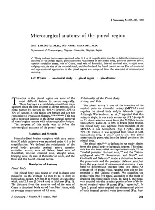
  • 2. I. Yamamoto and N. Kageyama FIG. 1. Posterosuperior views of the pineal region after the removal of the corpus callosum, illustrating the rela- tionship of the pineal body to its surrounding struc- tures. Upper Left: Type 1: the pineal vein (PV) empties into the terminal portion of one internal cerebral vein (ICV). Upper Right: Type 2: the PV's empty into the ter- minal portion of both ICV's. Center Left: Type 3: the PV's drain into the vein of Galen (G). Center Right: Type 4: The PV's drain into one of the ICV's and G. Lower: Type 5: the PV empties into the precentral cerebellar vein (PrCV). The PrCV is seen flowing into either side of the ICV, and the posterior ventricular vein (PVV) drains into the ICV (upper left and lower). The internal occipital vein (IOV) ends in G (upper right and center left). The pineal artery (PA) in both hemispheres supplies the pineal body (P), basal vein of Rosenthal (R), cerebellum (Cbl 1), massa intermedia (M), and medial posterior choroidal artery (MPchA) (center and lower). 206 J. Neurosurg. / Volume 53 / August, 1980
  • 3. Microsurgieal anatomy of the pineal region TABLE 1 Branches of theposterior cerebralartery (PCA) in 60 hemispheres Branches of PCA Branches Site of Origin Present (% of hemispheres) (% of hemispheres) P-I P-2A P-2P P-3 No. Branches Present per Hemisphere Cortical Average Range Branches circumflex artery short circumflex artery 65.0 long circumflex artery 96.7 thalamogeniculate artery 100.0 posterior choroidal artery medial posterior choroidal artery 100.0 lateral posterior choroidal artery 100.0 cerebral branches hippocampal artery 82.8 anterior temporal artery 80.0 middle temporal artery 80.0 posterior temporal artery 77.1 common temporal artery 20.0 parieto-occipital artery 80.0 calcarine artery 100.0 splenial artery 62.8 56.7 8.3 63.3 33.4 46.0 53.3 0.7 13.5 57.5 13.5 7.7 19.5 44.8 9.2 48.6 20.0 8.6 40.0 37.2 2.8 25.7 45.7 8.6 2.8 17.2 54.3 14.3 5.7 8.6 8.6 62.8 8.6 17.2 74.2 2.8 1.2 0-4 2.1 0-4 3.7 1-7 7.7 1.4 1~ 26.5 2.6 13 2.8 2.8 51.4 per right). In Type 3, pineal veins drained into the vein of Galen (11 cases) (Fig. 1 center left). In Type 4, pineal veins drained into one of the internal cerebral veins and the vein of Galen (three cases) (Fig. 1 center right). The pineal vein that emptied into the precentral cerebellar vein was defined as Type 5 (one case) (Fig. 1 lower). However, Salamon and Huang e7 reported that the medial posterior thalamic vein drained not only into the internal cerebral vein or the vein of Galen, but also into the basal vein of Rosenthal. Tamaki, et al.,~'~ divided pineal veins into three types, but, in our study, 20 brains did not come under their classification. We found that 40.9% of pineal veins traversed both superior and inferior surfaces of the pineal body (Fig. 1 center left). In other specimens, the pineal veins traversed only one surface of the pineal body: the superior surface in 31.8%, and the inferior surface in 27.3%. Posterior Cerebral Artery Several different classifications have been used for the posterior cerebral artery (PCA). 2~'48'5~ We followed the classification of the PCA by Zeal and Rhotonfl 4 which divided this artery into three segments from P-1 to P-3. The P-2 segment, which began at the posterior communicating artery and ter- minated at the posterior aspect of the midbrain, was further subdivided into an anterior and posterior half, and designated as P-2A and P-2P segments. In our study, we mainly examined the branches originating from P-2 and P-3 segments. Circumflex Artery. The circumflex arteries are divided into short and long circumflex segments (Fig. 2 upper left, center and lower). 2~ The short circumflex artery was present in 65.0% of the specimens and the number in one hemisphere ranged from zero to four, the average being 1.2. Most of the short circumflex arteries arose directly from the P-I segment (56.7%), the remaining 8.3% arose from P-2A (Table 1). They ran medial to the PCA around the midbrain into the thalamogeniculate sulcus and supplied small perforating branches to the cerebral peduncle and substantia nigra. As previously describedfl4 some branches of this artery penetrated the mesencephalon near the geniculate bodies. The long circumflex arteries were present in 96.7% of the specimens and consisted of zero to four, with an average of 2.1 branches, which arose from the P- 1 seg- ment in 63.3%, and from the P-2A segment in 33.4% (Table 1, Fig. 2). They encircled the midbrain between the PCA and the short circumflex artery, and then sent rami to the cerebral peduncle, geniculate bodies, and tegmentum, and the terminal branches reached the quadrigeminal plate, more on the superior colliculus)' Some of these terminal branches anastomosed with the branches of the superior cerebellar arteries in the region of the quadrigeminal plate. 26,5',9'As these terminal branches supply the por- tion of the dorsolateral midbrain containing the pathways and pretectal neurons serving vertical eye movement)' occlusion of them may give rise to Parinaud's syndrome)TM Thalamogeniculate Artery. In our series, 99.3% of thalamogeniculate arteries arose from the P-2 seg- ment in the ambient cistern near the medial and lateral geniculate bodies. Only 0.7% of them arose in the P-3 segment. The average incidence of the thalamogeniculate artery was 3.7, ranging from one to J. Neurosurg. / Volume 53 / August, 1980 207
  • 4. I. Yamamoto and N. Kageyama FIG. 2. Upper Left: Interior view of the course of the posterior cerebral artery, basilar artery (BA), short cir- cumflex artery (SCiA), long circumflex artery (LCiA), medial posterior choroidal artery (MPchA), lateral posterior choroidal artery (LPchA), anterior temporal artery (ATA), middle temporal artery (MTA), posterior temporal artery (PTA), parieto-occipital artery (POA), calcarine artery (CaA), vein of Galen (G), internal cerebral vein (ICV), basal vein of Rosenthal (R), and precentral cerebellar vein (PrCV). Upper Right." The medial part of the temporal lobe has been removed to expose the LPchA (1, 2, 3, 4), and the hippocampal artery (HiA). Center Left." The ATA and MTA have been removed to expose the thalamogeniculate arteries (ThGA), and the pineal body (P). Center Right." Anterolateral view of the mesen- cephalon, showing the main arterial trunks, the anterior choroidal artery (AchA), and the posterior communicating artery (PCoA). Lower." Inferolateral view of the mes- encephalon, illustrating absence of the HiA branch of the PCA. The AchA supplies the hippocampal formation (Hi). The internal carotid artery (ICA), optic chiasm (OC), and mamillary body (MB) are seen. 208 J. Neurosurg. / Volume 53 / August, 1980
  • 5. Microsurgical anatomy of the pineal region FIG. 3. The corpus caUosum has been removed to expose the terminal portion of the medial posterior choroidal artery (MPchA). Right internal cerebral vein (ICV) has been retracted (left) to expose the pineal artery (PA). The vein of Galen (G), basal veinof Rosenthal (R), internal occipital vein (IOV), posterior ven- tricular vein (PVV), pineal vein (PV), posterior pericallosal vein (PPV), pineal body (P), and massa in- termedia (M) are visualized. seven per hemisphere (Table 1, Fig. 2 center left). Although Duvernoy~1 stated that some of their branches anastomosed with the branches of the anterior choroidal artery (AChA) on the surface of the lateral geniculate body, in our study no anastomosis could be found under the operating microscope between the thalamogeniculate arteries and the anterior choroidal arteries. However, some branches of the AChA, MPChA, and circumflex arteries penetrated the mesencephalon in the thalamo- geniculate sulcus? ~ Thalamogeniculate arteries penetrated the thalamus and supplied the medial geniculate, the medial half of the lateral geniculate, the posterior part of the lateral posterior nucleus, the ventral lateral pulvinar, the ventral posterior and ven- tral lateral nuclei, and the nucleus centrum medianum and intralaminar nuclei/' Damage to this artery results in the thalamic syndrome of Dejerine- Roussy.5~ Medial Posterior Choroidal Artery. The MPChA arose from the P-2A segment in 57.5% of the specimens, but occasionally arose from P-I (13.5%), P-2P (13.5%), P-3 (7.7%), or the cortical branches (7.7%) (Table 1). Berland and Haughton6 dem- onstrated angiographically an anomalous origin of the MPChA from the basilar artery. The number of MPChA's in one hemisphere ranged from one to three, with an average of 1.4 (Table 1). In our study, the MPChA was single in 62% and multiple in 38%, as compared to single in 54% and multiple in 46% as reported by Zeal and Rhotonfl4 or single in 60% and multiple in 40% as reported by Margolis, et al. 53 The MPChA has been reported by others to be a single artery. 2sa9 It ran parallel and usually medial to the PCA, and coursed to the quadrigeminal cistern (Fig. 2). Then it lay lateral to the pineal body, and coursed in the roof of the third ventricle parallel and medial to the internal cerebral vein in the midline, and finally supplied the choroid plexus of the third ventricle at the foramen of Monro (Fig. 3). Along its course, the MPChA sent an average of 3.6 branches to the tegmentum (zero to eight), 1.5 branches to the lateral geniculate body (zero to six), 3.9 branches to the medial geniculate body (zero to 12), 5.3 branches to the quadrigeminal plate (zero to 11), 1.6 branches to the pulvinar (zero to four), 1.5 branches to the pineal body (zero to five), and 2.7 branches to the medial thalamus (zero to five). The MPChA arising from the P-1 segment also sent zero to two branches to the peduncle with an average number of 0.5 (Table 2). The number of branches to the quadrigeminal plate was usually inversely proportional to the number of branches of the long circumflex arteries to the quadrigeminal plate. As previously described,sa,5*,a~ when the MPChA arose from the distal cortical branches of the PCA, this MPChA coursed behind the pulvinar in a retrograde fashion to supply the tela choroidea of the third ventricle, pineal body, or the medial thalamus. J. Neurosurg. / Volume 53 / August, 1980 209
  • 6. TABLE 2 Branches of the medial posterior choroidal artery in 60 hemispheres Site of Termination No. Present per Hemisphere Average Range peduncle 0.5 0-2 tegmentum 3.6 0-8 lateral geniculate body 1.5 0--6 medial geniculate body 3.9 0-12 quadrigeminal plate 5.3 0-11 pulvinar 1.6 0-4 pineal body and/or habenula trigone 1.5 0-5 medial thalamus 2.7 0-5 Lateral Posterior Choroidal Artery. The lateral posterior choroidal arteries (LPChA's) arose from the P-2P segment in 44.8% samples, the P-2A in 19.5%, the P-3 in 9.2%, and the cortical branches in 26.5%. The PCA gave origin to one to five LPChA's in one hemisphere (average 2.6) (Table 1, Fig. 2 upper and center). There was no difference in number between right and left LPChA's, that is, the average incidence was 2.6 on the left and 2.7 on the right. Zeal and Rhoton94 reported that the average number of LPChA's in one hemisphere was four, ranging from one to nine. There was a single LPChA in 9% of hemispheres, two in 38%, three in 35%, four in 15%, and five in 3%, as compared to a single trunk in 60% and multiple in 40%, as reported by Margolis, et al.) 4 or single in 12% and multiple in 88%, as reported by Zeal and Rhoton)*When more than one LPChA were present, they were divided into two branches, that is, anterior and posterior (Fig. 4 upper.left). 2s,~8,53,~,sT,~9 The anterior branches usually arose from P:2A or its cortical branches and coursed laterally to enter the choroid fissure and supplied the choroid plexus of the temporal horn. In our study, 31% of the anterior branches anastomosed with the AChA, as determined under the operating microscope (Fig. 4 upper right). According to Carpenter, et al., TM anastomoses were found between these two arteries in 93%. Although it has been reported that the size of the LPChA was usually inversely proportional to the size of the AChA,2s'48,Ss'54in our anatomical studies there was no obvious relationship between the size of these two arteries except in 6% of the LPChA's which were definitely smaller than the AChA (Fig. 4 lower). The posterior branches arose from P-2P, P-3, or their cor- tical branches, and coursed posteriorly around the pulvinar, supplying the choroid plexus of the trigone. Along its course, the LPChA supplied the crus, com- missure, body and part of the anterior columns of the fornix, the dorsomedial thalamic nucleus, pulvinar, and the lateral geniculate body)3 Margolis, et al.)TM reported that branches arising from the distal portion of the LPChA extended medially and anastomosed I. Yamamoto and N. Kageyama with the MPChA, but in this study no definite anas- tomosis was found between these two arteries under the operating microscope. Cortical Branches of the Posterior Cerebral Artery. The cortical branches of the PCA are divided into four segments, that is, the inferior temporal artery, parieto-occipital artery, calcarine artery, and splenial artery. The inferior temporal arteries include hip- pocampal and anterior, middle, posterior and com- mon temporal arteries.9' Hippocampal Artery. The hippocampal artery, which supplies the uncus, hippocampal gyrus, hip- pocampal formation, and the dentate gyrus, was pres- ent in 82.8% of specimens examined, as compared to 64% as reported by Zeal and Rhoton)4 This artery arose from the P-1 segment in 2.8% of specimens, in 48.6% from P-2A, in 20% from P-2P, in 8.6% from P-3 and in 2.8% from the common temporal artery (Table 1, Fig. 2 upper right and center). The AChA also gave off branches to the hippocampus. However, the distribution of the hippocampal artery varied with that of the AChA. In the absence of a hippocampal arterial branch of the PCA, the AChA supplied the major portion of the hippocampal formation (Fig. 2 lower). Anterior Temporal Artery. The anterior temporal artery was present in 80% of hemispheres, as com- pared to 84% as reported by Zeal and Rhoton.9' It arose from P-2A in 40.0% of specimens, P-2P in 37.2%, and P-3 in 2.8% (Table 1, Fig. 2 upper left and center right). It supplies the inferior aspect of the anterior portion of the temporal lobe.5~ Middle Temporal Artery. The middle temporal artery was present in 80.0% of hemispheres, as com- pared to 38% as reported by Zeal and Rhoton)~ It arose from P-2A in 25.7%, from P-2P in 45.7%, and from P-3 in 8.6% (Table 1, Fig. 2 upper left and center right). It supplies the inferior surface of the temporal lobe)4 Posterior Temporal A rtery. The posterior temporal artery, which supplies the inferior temporal and oc- cipital surface, including the occipital pole and lingual gyrus, was present in 77.1% of hemispheres, as com- pared to 96% as reported by Zeal and Rhoton.9~ It originated from the P-2A segment in 2.8% of specimens, from P-2P in 17.2%, from P-3 in 54.3%, and, from the calcarine artery in 2.8% (Table 1, Fig. 2 upper left and center). Branches of the posterior tem- poral artery also helped to supply the visual area, par- ticularly a part of the "macular" area of the cortex, and could therefore be a factor in the preservation of central vision when the calcarine artery was occluded.7~ Common Temporal Artery. The common temporal artery was present in 20.0% of specimens, as com- pared to 16% as reported by Zeal and Rhoton.9~ It arose from the P-2P segment in 14.3% of specimens, 210 J. Neurosurg. / Volume53 / August, 1980
  • 7. Microsurgieal anatomy of the pineal region FIG. 4. Basal views of the mesencephalon illustrating variations of lateral posterior choroidal artery (LPchA). The medial part ofthe temporal lobehas been removedto expose the choroid plexus (ch.pl.) of the temporal horn. Upper Left: Anterior (l) and posterior branches (2) of the LPchA. Upper Right: Anastomosis is shownbetween the LPchA and the anterior choroidal artery (AchA) (curved arrow). Lower: The AchA is larger than the LPchA. Other structures seen are: the internal carotid artery (ICA), posterior cerebral artery (PCA), basilar artery (BA), posterior communicating artery (PCoA), medial posterior choroidal artery (MPchA), posterior temporal artery (PTA), calcarine artery (CaA), basal vein of Rosenthal (R), tentorium cerebelli (T), and the mamillary body (MB). and from P-3 in 5.7%. It supplies the majority of the inferior surface of the temporo-occipital lobes?4 Parieto-Occipital Artery. The parieto-occipital artery was seen in 80.0% of our specimens, as com- pared to 96% noted by Zeal and Rhoton?4 It arose as a single branch from P-2A in 8.6%, from P-2P in 8.6%, and from P-3 in 62.8% (Table 1, Fig. 2 upper and center). It supplies the superior part of the cuneus, the posterior fifth of the precuneus, part of the superior parietal lobule, and the superior occipital gyrus/* Calcarine Artery. The calcarine artery was present in all specimens examined and arose from P-2A in 8.6%, P-2P in 17.2%, and P-3 in 74.2% (Table 1, Fig. 2 upper and center right). It ran in the calcarine sulcus and supplied the inferior cuneus and the superior and posterior part of the lingual gyrus/a Smith and RichardsonTM reported that the visual area was supplied by the calcarine artery in only 25% of cases; by the calcarine and the posterior temporal arteries in 34.4%; by the calcarine and the parieto-occipital arteries in 18.8%; by the calcarine and both parieto- occipital and posterior temporal arteries in 15.5%; by the calcarine plus the middle cerebral and the parieto- occipital arteries in 3.1%, and by all the above arteries in 3.1%. Infarction of the calcarine cortex causes a homonymous visual defect with macular sparing.37,87 J. Neurosurg. / Volume 53 / August, 1980 21 ]
  • 8. I. Yamamoto and N. Kageyama FI~. 5. Left: Anterolateral view of the left mesencephalon, showing the arrangement of the main arterial trunks. Also visibleare: the basilar artery (BA),superior cerebellar artery (SCA), posterior cerebral artery (PCA), anterior inferior cerebellar artery (AICA), posterior communicating artery (PCoA), medial posterior choroidal artery (MPchA), anterior temporal artery (ATA), middle temporal artery (MTA), posterior temporal artery (PTA), parieto-occipital artery (POA), and the calcarine artery (CaA). Right: Superolateral view of the left cerebellum, showingthe course of the SCA. 1: lateral marginal branch, 2: hemispheric branch and 3: superior vermis branch. The cerebellum (Cbll) is also indicated. Splenial Artery. The splenial artery, also called the "posterior pericallosal artery,''5''~~or "dorsal callosal artery,'''8 was seen in 62.8% of specimens. It has been reported to be present in 35%5. to 100% of specimens?' It originated from the parieto-occipital artery in 31.4%, calcarine artery in 17.2%,the LPChA in 8.6%, the MPChA in 2.8%, and the common tem- poral artery in 2.8% (Table 1). It coursed around the splenium close to the midline and anastomosed with the distal branches of the pericallosal artery of the anterior cerebral artery. Superior Cerebellar Artery The superior cerebellar artery originated from the basilar artery an average of 1.8 mm proximal to the basilar bifurcation, as compared to 2.5 mm reported by Saeki and Rhoton86(Fig. 5 left). Although duplica- tion or triplication of the superior cerebellar arteries has been reported in 28% or 2%, respectively,82'5~in our anatomical studies 4.2% (two of 48) were dupli- cated. The superior cerebellar artery circled around the upper pons or lower mesencephalon parallel to the course of the basal vein of Rosenthal, the PCA, and the free edge of the tentorium, and coursed toward the superior surface of the cerebellar hemisphere and the superior vermis. The proximal trunk of this artery divides into anterior pontine, am- bient, and quadrigeminal segment,32 but this division is not anatomically precise. There are three main cor- tical branches and perforating branches of the superior cerebellar arterya2,52 (Fig. 5 right). An average of 1.3 lateral marginal branches (range zero to two) usually originated close to the junction between the pontine and ambient segments, which supply the superior cerebellar peduncle, the dentate nucleus, the roof nuclei, and the middle cerebellar peduncles2"79(Table 3). The lateral marginal branches were usually inversely proportional in size to the cor- tical branches of the anterior inferior cerebellar artery?2 An average of 3.8 hemispheric branches (range, two to five) on each side arose distal to the origin of the lateral marginal branches and coursed over the superior surface of the cerebellum (Table 3). The superior vermis branches originated from the quadrigeminal segment and numbered 2.3 on the average (range, zero to three) on each side. The right and left superior vermis branches usually anastomosed with each other in the quadrigeminal cistern and then coursed posteriorly over the vermis close to the midline. Within the quadrigeminal cistern, small arterial branches from the proximal portion of the superior vermis branches ran into the precentral cerebellar fissure, and are called the precentral cerebellar arteries?2 These arteries were identified in all our specimens examined. From the medial wall of the superior cerebellar artery around the brain stem, an average of 7.1 (range, three to 13) perforating branches arose and supplied the superior cerebellar peduncle and the quadrigeminal plate (Table 3). Some of the branches that reached the_quad_rigeminal plate anastomosed with the terminal branches of the long circumflex arteries.28'"3'94 Galenic Venous System Vein of Galen. The vein of Galen was formed by the union of the paired internal cerebral veins. Its average length was 12 mm, ranging from 8 to 25 mm. Tributaries that joined the vein of Galen were the in- 212 J. Neurosurg. / Volume53/August, 1980
  • 9. Mierosurgieal anatomy of the pineal region TABLE 3 Branches of the superior cerebellar artery (SCA) in 60 hemispheres Branchof SCA No. Presentper Hemisphere Average Range lateralmarginalbranch 1.3 0-2 hemisphericbranch 3.8 2-5 superiorvermisbranch 2.3 0-3 perforatingbranch 7.1 3-13 TABLE 4 The incidence of tributaries emptying into the vein of Galen Percentof Tributary Specimens internalcerebralvein 100.0 precentralcerebellarvein 86.4 internaloccipitalvein 77.0 basalveinof Rosenthal 51.7 posteriorpericallosalvein 41.5 pinealvein 40.9 posteriormesencephalicvein 38.4 posteriorventricularvein 6.7 FIG. 6. Posteroinferior view of the pineal region of the left occipital lobe (Occ L), showing the relationship of the vein of Galen (G) and its tributaries. Precentral cerebellar vein (PrCV) draining into G. The internal occipital vein (IOV) flows into the basal vein of Rosenthal (R). Ab- breviations as in previous figures. ternal cerebral vein in 100% of specimens, precentral cerebellar vein (86.4%), internal occipital vein (77%), basal vein of Rosenthal (51.7%), posterior pericallosal vein (41.5%), pineal vein (40.9%), posterior mesencephalic vein (38.4%), and the posterior ven- tricular vein (6.7%) (Table 4, Fig. 6). This vein then coursed superoposteriorly under the splenium of the corpus callosum and joined the inferior sagittal sinus to form the straight sinus. Although internal hydrocephalus resulting from the occlusion of the vein of Galen, particularly near its origin, was reported by Dandy,15,17,79experimental occlusion by others did not result in hydrocephalus.2,3a8,~2,79 Basal Vein of Rosenthal: Salamon and Huang67 divided the basal vein of Rosenthal into three segments: the first, anterior or striate segment, the second, middle or peduncular segment, and the third, posterior or posterior mesencephalic segment. The second segment is further subdivided into anterior and posterior portions by the most lateral point of the vein as it turns around the peduncle. In this study, the posterior portions of the second and the third segments were examined. Inferior Ventricular Vein. The inferior ventricular vein drains the subependymal and choroidal veins of the temporal horn as well as the veins of hippocampal formation.38,57'~3As a common trunk, it flowed into the second segment of the basal vein of Rosenthal at the junction between the anterior and posterior por- tions in 88.1% of the specimens examined (Fig. 7 left). Only 11.9% of them were double. The inferior ventric- ular vein rarely joins the lateral or straight sinus via a tentorial sinus)s Lateral Mesencephalic Vein. The lateral mesen- cephalic vein runs along the lateral mesencephalic sul- cus and drains upward to the third segment of the basal vein of Rosenthal or downward to the brachial tributary of the petrosal vein.3~176 It was present in 69% of the specimens examined (29 of 42) in our ana- tomical studies. Angiographically it is seen in 30% to 35% of normal vertebral angiograms.TM Wacken- heim, et al.fl2 reported six types of variations in the drainage of the lateral mesencephalic vein. Posterior Mesencephalic Vein. The posterior mes- encephalic vein was present in 21.7% of the hemi- spheres (13 of 60) and emptied into the vein of Galen in 8.3% (five), the basal vein of Rosenthal in 8.3% (five), and the internal cerebral vein in 5% (three). The terminal portion of the basal vein of Rosenthal drained into the vein of Galen or the posterior portion of the internal cerebral vein. The mode of drainage into these major veins was classified into three types in our study; 1) both basal veins of Rosenthal emptied into the vein of Galen in 11 specimens (Fig. 8 upper left), 2) into the internal cerebral veins in 10 (Fig. 8 up- per right), and 3) into the internal cerebral vein in one J. Neurosurg. / Volume53 / August, 1980 213
  • 10. I. Yarnamoto and N. Kageyama FIG. 7. Left: The medial part of the temporal lobe has been removed to expose the inferior ventricular vein (IVV), basilar artery (BA) posterior cerebral artery (PCA), posterior"communicating artery (PCoA), anterior choroidal artery (AchA), medial posterior choroidal artery (MPchA), lateral posterior choroidal artery (LPchA), and the basal veinof Rosenthal (R). Right: Posterolateral viewof the left mesencephaion, illustrating the arrangement of the main arterial and venous trunks, and showing the superior cerebellar artery (SCA), long circumflex artery (LCiA), precentral cerebellar vein (PrCV), superior vermian vein (SVV), lateral mesencephalic vein (LMV), pineal vein (PV), cerebellum (Cbll), occipital lobe (Occ L), and the pineal body (P). side and into the vein of Galen in the other side in nine (Fig. 8 lower left). Three basal veins of Rosenthal ter- minated at the posterior portion of the vein of Galen (Fig. 8 lower right). Many anatomical variations in draining of the basal vein of Rosenthal have been reported, including draining into the straight sinus, lateral sinus, superior petrosal sinus via the anastomotic lateral mesencephalic vein, or the sphenoparietal sinus.67 Internal Occipital Vein. The internal occipital vein originated on the inferior and medial surface of the oc- cipital lobe and then coursed anteromedially to end in the vein of Galen in 77.0% of specimens (Figs. 1 upper right and lower left, 8 upper left and lower, and 10 right). More rarely, it also joined the internal cerebral vein in 11.5% of specimens (Fig. 9), and the basal vein of Rosenthal in 11.5% (Fig. 6). Left homonymous hemianopsia was reported as a result of a right supratentorial approach for pineal tumor by Harris, et al.a~They ascribed the hemianopsia to division of the internal occipital vein. Posterior Pericallosal Vein. The posterior perical- losal vein has also been called the "splenial vein,''67the "posterior cerebral vein,''5 the "posterior marginal vein, ''86 the "posterior vein of corpus callosum, ''5 and the "dorsal callosal vein.''78 It originated on the dorsal surface of the corpus callosum and traversed around the splenium parallel to the posterior pericallosal artery to enter the internal cerebral vein in 48.8% of specimens (Fig. 11 right), the vein of Galen in 41.5% (Figs. 8 upper left and 10 left), the basal vein of Rosen- thai in 7.3% (Fig. 8 lower right), and the internal occipi- tal vein in 2.4% (Figs. 8 left, and 10 right). Salamon and Huang67reported that the splenial vein might also join the medial atrial vein. The posterior pericallosal vein was usually double (62.5%), each lying in the im- mediate paramidline; it was single in 33.3%, and rarely triple (4.2%). Precentral Cerebellar Vein. The precentral cerebellar vein originated in the precentral cerebellar fissure, usually as two brachial veins uniting into a single common trunk. This vessel coursed upward to join the vein of Galen or the posterior portion of the internal cerebral vein. In 86.4% (19 of 22) of the specimens, the precentral cerebellar veins drained into the vein of Galen (Fig. 6), and in 13.6% (three of 22) they flowed into either side of the internal cerebral vein (Fig. 1 upper left and lower). Occasionally, the brachial veins did not unite but drained independently into the vein of Galen. Small veins arising from the anterior surface of the vermis and the adjacent cerebellar hemisphere, the superior vermian, or the superior hemispheric veins (Figs. 6 and 7 right) usually joined the precentral cerebellar vein at the junction 2]4 J. Neurosurg. / Volume 53 / August, 1980
  • 11. Mierosurgieal anatomy of the pineal region FIG. 8. Variations of the terminal portion of the basal vein of Rosenthal (R). Upper Left: Both R's emptying into the vein of Galen (G). The left posterior pericallosal vein (PPV) ends in G and the right PPV ends in the internal occipital vein (IOV). Upper Right: Both R's empty into the internal cerebral veins (ICV). Lower Left: Inferior view of the course of the terminal portion of R, one side of which empties into the ICV (left) and the other side into G (right). Lower Right: Both R's terminate at the posterior portion of G. Structures visualized are: the medial posterior choroidal artery (MPchA), long circumflex artery (LC!A), calcarine artery (CaA), posterior ventricular vein (PVV), pineal vein (PV), pineal body (P), tentorxum cerebelli (T), cerebellum (Cbll), and the straight sinus (SS). between the inferior colliculi and the anterior superior margin of the cerebellum?9'4~ Internal Cerebral Vein. The internal cerebral vein originates just behind the foramen of Monro by the union of the septal, thalamostriate, and choroidal veins.48 Paired internal cerebral veins run posteriorly within the tela choroidea, adjacent to the midline, and unite with the subependymal veins, the basal vein of Rosenthal, and/or the internal occipital vein to form the vein of Galen. Caron, et al., TM reported that the surgical ligature of both internal cerebral veins was well tolerated in two pineal tumors. Various terms have been used for the subependymal veins of the lateral ventricles, including the "subepen- dymal veins,''78,g3 the "vein of the posterior horn, ''43 and the "posterior ventricular vein.''8,9 Billewicz and Bel-Amor8.9 defined four veins: the medial atrial vein, the direct medial vein, the direct lateral vein, and the lateral atrial vein, which were described by Wolf and FIG. 9. Posterosuperior view of the pineal region. The left internal occipital vein (IOV) joins the internal cerebral vein (ICV). The medial posterior choroidal artery (MPchA), basal vein of Rosenthal (R), pineal vein (PV), pineal body (P), cerebellum (Cbll), massa intermedia (M) are seen. J. Neurosurg. / Volume 53 / August, 1980 2 ]5
  • 12. I. Yamamoto and N. Kageyama FIG. 10. Medial views of a midsagittal section, showing the relationship of the posterior pericaltosal vein (PPV) to its tributaries. Left: The PPV enters the vein of Galen (G). Right: The PPV joins the internal occipital vein (IOV) and then flows into G. The medial posterior choroidal artery (MPchA), internal cerebral vein (ICV), posterior ventricular vein(PVV),pineal body (P), and spleniumof the corpus callosum (S) can be seen. Huang93 as the posterior ventricular vein. From the viewpoint of microsurgical anatomy, we could not differentiate between these four veins, so in our study we used the designation of the posterior ventricular vein. The posterior ventricular vein, which drains the posterior part of the body, the atrium, and the posterior horn of the lateral ventricle,' flowed in almost all specimens (55 of 60 hemispheres) into the internal cerebral vein (Figs. 1 upper left and lower, 3, 8 rightpair, 10, and 11 right). It flowed into the vein of Galen in four cases, and the basal vein of Rosenthal in one. In 11 brains, we dissected the posterior ven- tricular vein distally to identify the relationship between the medial and lateral atrial veins (Fig. 11 FIG. 11. Left: Medial view of a midsagittal section, illustrating the medial and lateral atrial veins (MAV, LAV) of the posterior ventricular vein (PVV). Right: The PVV drains into the internal cerebral vein (ICV) as a common trunk. Other structures visualized are the medial posterior choroidal artery (MPchA), posterior cerebral artery (PCA), internal occipital vein (IOV), posterior pericallosal vein (PPV), pineal body (P), vein of Galen (G), and the splenium of the corpus callosum (S). 216 J. Neurosurg. Volume 53 / August, 1980
  • 13. Mierosurgieal anatomy of the pineal region FIG. 12. Left: Posterolateral view of the inferior occipital lobe (Occ L), illustrating the bridging veins between the lateral occipital pole and the transverse sinus (TS). Right: Posterior view of the superior cerebellum (Cbll), illustrating finebridging veins between the hemispheric cerebellar veins and TS. left). In seven of 22 hemispheres, the medial and lateral atrial veins united into a common trunk to form the common atrial vein,TM or the posterior paraventricular vein/ and drained into the internal cerebral vein (Fig. 11 right). Bekov' found the posterior paraventricular vein in 24% of instances. In this situation, the posterior paraventricular vein was almost the same size as the basal vein of Rosenthal, so it was sometimes difficult to differentiate between these two veins; however, the diverging point of the basal vein of Rosenthal was closer to the vein of Galen (Figs. 1 upper left and 8 right pair). In other situations, the basal vein of Rosenthal was usually larger than the posterior ventricular vein (Fig. 3 right). Although the lateral atrial vein has been reported in 139 of 184 hemispheres,~in our study the medial and lateral atrial veins were found in 68.2% (15 of 22) and 95.5% (21 of 22), respectively. Straight Sinus. The straight sinus was formed by the union of the inferior sagittal sinus and the vein of Galen at the posterior end of the splenium and then continued posteroinferiorly, following the line ofjunc- tion of the falx cerebri with the tentorium cerebelli, to the torcular Herophili. Saxena, et al./9'7~ reported a double straight sinus in 13.95% of cadavers, but we could not find a double straight sinus in our review of 30 specimens. Knott~e reported the absence of the straight sinus in one of 44 specimens. In our study, the average length of the straight sinus was 52.3 mm, with a minimum of 45 mm and maximum of 58 mm. Sax- ena, et al./~ reported that the straight sinus was 50 mm in average length. Bridging Vein. No bridging veins could be found between the medial occipital pole and the transverse sinus. However, two to seven bridging veins per hemisphere were present, lying between the lateral oc- cipital pole and the transverse sinus (Fig. 12 left). Between the tentorium cerebelli and the cerebeUar sur- face, numerous fine bridging veins could be found medially; there were three to five bridging veins per hemisphere laterally (Fig. 12 right). Tentorial Notch. The distance from the posterior boundaries of the tentorial notch to the pineal body was 18.6 mm on the average, ranging from 10 to 30 mm (Fig. 13). The Third and Fourth Cranial Nerves. The third nerve left the midbrain on the medial side of the cerebral peduncle, and passed under the PCA and above the superior cerebellar artery (Fig. 5 left). In four of 60 specimens, the circumflex artery penetrated into the substance of the third nerve (Fig. 14). The fourth nerve originated from the dorsal surface of the midbrain, then curved anteriorly between the PCA and the superior cerebellar artery along the tentorial edge and lay lateral to the third nerve (Figs. 5 right and 7 right). This relationship between the third and the fourth cranial nerves and the posterior cerebral and superior cerebellar arteries was constant in all specimens ex- amined, even in the presence of duplication of the superior cerebellar artery. Discussion Various surgical approaches to the pineal region have been reported since 1910, when Horsleya8 first attempted direct removal of a pineal tumor. The sur- gical approach to the pineal region has been roughly classified into three techniques: infratentorial su- pracerebellar,sS'41,47,Ss'59'84,75-~'89 supratentorial, and transventricular approaches?8 The supratentorial J. Neurosurg. / Volume 53/August, 1980 2] 7
  • 14. I. Yamamoto and N. Kageyama FIG. 13. Three inferior views of the tentorium, illustrating variations in the interval separating the boun- daries of the tentorial notch from the mesencephalon. FIG. 14. Anterolateral view of the right mesencephalon, illustrating that the short circumflex artery (SCiA) pene- trates into the bundle of the third cranial nerve (curved arrow). The internal carotid artery (ICA), basilar artery (BA), posterior cerebral artery (PCA), superior cerebellar artery (SCA), posterior communicating artery (PCoA), and mamillary body (MB) can be seen. technique has been further subdivided into two ap- proaches, namely, the unilateral transtentorial ap- proachl~176 and the interhemispheric posterior parietal approach?,x8,33a5,49,85,82 There have been few reports comparing the two basic surgical approaches to the pineal region, that is, the supratentorial and the infratentorial ap- proaches.",~6,8s Reid and Clark63compared these two different approaches and preferred the occipital transtentorial technique. Stein,77who did not confine his report to tumors of the pineal body, claimed that the infratentorial supracerebellar approach was still preferable to the supratentorial approach. Obrador, et al.? ~stated that the infratentorial approach was useful for the tumors projecting downward to the posterior fossa, and, in more anteriorly located tumors en- croaching on the posterior third ventricle, the supratentorial approach was preferable. From the view point of microsurgical anatomy of the pineal region, we have to consider the following four important anatomical points in the determination of the surgical approach. First, we must consider the location of the bridging veins. When using an infratentorial approach, all bridging veins over the cerebellar surface must be sacrificed to reach the quadrigeminal region. Stein77 never encountered cerebellar edema from the transec- tion of these bridging veins, but Page~8 reported at least one case with cerebellar swelling after they were 218 J. Neurosurg. / Volume 53 / August, 1980
  • 15. Mierosurgieal anatomy of the pineal region divided. In contrast, as no bridging veins lie between the medial occipital pole and the transverse sinus when a supratentorial approach is used, the occipital lobe can be retracted superolaterally without sacrificing bridging veins. But this retraction is at times related to postoperative visual complications, such as transient or permanent hemianopsia. Second, the size of the tentorial notch is variable. 81 In our anatomical studies, the distance between the posterior boundaries of the tentorial notch and the pineal body ranged from 10 to 30 mm. The shorter this distance becomes, the more difficult is the ex- posure above the tentorium when viewing from an infratentorial approach. In contrast, when using a transtentorial approach, the edge of the tentorium is transected 1 cm lateral to the straight sinus to afford excellent visualization of the vein of Galen and its branches. But this approach is hampered by these ma- jor veins and does not provide a wide enough view of the quadrigeminal plate, particularly over the con- tralateral side. Third, it goes without saying that a supratentorial approach is hampered by the vein of Galen and its major tributaries. Injury to these deep veins is one of the main causes of surgical morbidity. At times, we have transected the internal occipital vein or posterior pericallosal vein. Harris, et al., s~ reported a case of hemianopsia as a result of the section of the internal occipital vein. Using an infratentorial approach, Page58 reported the precentral cerebellar vein was preserved in five of nine cases. When necessary, this vein must be transected in order to provide a wide ex- posure by either approach; however, this does not result in postoperative neurological deficits. Fourth, several authors reported that the splenium could be spared routinely in a supratentorial ap- proach; 42,51,6~ however, when the tumor is located above the hiatus tentorii, bulges anteriorly into the posterior third ventricle, and compresses the superior colliculus (Type A of Poppen6~ the splenium is sometimes incised even in a transtentorial approach. In that event, a resultant disorder of the higher cor- tical function, an alexia without agraphia, has been reported. ~~ In Dandy's parasagittal approach, an incision of the posterior half of the corpus callosum exposes the tumor? 6 Dimond, et al., TM reported that the surgical section of the posterior portion of the body of the corpus callosum produced some memory defect. In an infratentorial approach, even after the transection of the precentral cerebellar vein, the lower portion of the quadrigeminal plate is still concealed by the superior vermis, particularly the culmen. So a splitting and retraction of the upper vermis is oc- casionally necessary to give enough exposure. Based upon these anatomical considerations, the selection of the appropriate surgical approach to the pineal region should depend mainly upon the location and the extent of the tumor, and partly upon the sur- geon's experience. References 1. Araki C: [Removal of the pineal tumor.] Gekashinryo 2:517-524, 1960 (Jpn) 2. Bedford THB: The great vein of Galen and the syn- drome of increased intracranial pressure. Brain 57:1-24, 1934 3. Bedford THB: The venous system of the velum inter- positum of the rhesus monkey and the effect of ex- perimental occlusion of the great vein of Galen. Brain 57:255-265, 1934 4. Bekov DB: Some structural differences among postero- lateral tributaries of great cerebral vein. Arkh Anat Gistoi Embrlol 46:166-170, 1964 5. Ben-Amor M, Billewicz O: The posterior cerebral vein. Neuroradiology 1:179-182, 1970 6. Berland LL, Haughton VM: Anomalous origin of posterior choroidal artery from basilar artery. AJR 132:674-675, 1979 7. Billewicz O: The normal and pathological radio- anatomy of the lateral mesencephalic vein. Neuro- radiology 8:295-299, 1975 8. Billewicz O, Ben-Amor M: The posterior ventricular branches of the internal cerebral and basilar vein. Neuroradiology 2:37-45, 1971 9. Billewicz O, Ben-Amor M: The posterior ventricular branches of the internal cerebral and basilar veins in cases of deep cerebral tumours infiltrating the basal ganglia. Neuroradiology 1:223-227, 1970 10. Brunner C: Cited by Rorschach H: Zur Pathologie und operabilit/it der Tumoren der Zirbeldrfise. Beitr Kiln Chit 83:451-474, 1913 11. Camins MB, Schlesinger EB: Treatment of tumours of the posterior part of the third ventricle and the pineal region: a tong term follow-up. Acta Neuroehir 40: 131-143, 1978 12. Caron JP, Debrun G, Sichez JP, et al: Ligature des veines c6r6brales internes et survie. Apropos de deux pin~alomectomies. Neurochirurgie 20:81-90, 1974 13. Carpenter MB, Noback CR, Moss ML: The anterior choroidal artery: its origins, course, distribution and variations. Arch Neurol Psychiatry 71:714-722, 1954 14. Chou SN: The treatment of third ventricular tumors with particular reference to surgical excision, in Morley TP (ed): Current Controversies in Neurosurgery. Philadelphia: WB Saunders, 1976, pp 367-373 15. Dandy WE: Experimental hydrocephalus. Ann Surg 70:129-142, 1919 16. Dandy WE: An operation for the removal of pineal tumors. Surg Gynecol Obstet 33:113-119, 1921 17. Dandy WE, Blackfan KD: Internal hydrocephalus. An experimental, clinical and pathological study. Am J Dis Child 8:406-482, 1914 18. DeGirolami U, Schmidek H: Clinicopathological study of 53 tumors of the pineal region. J Neurosurg 39:455-462, 1973 19. Dimond SJ, Scammell RE, Brouwers EYM, et al: Functions of the centre section (trunk) of the corpus callosum in man. Brain 100:543-562, 1977 20. Duvernoy HM: Human Brainstem Vessels. Berlin/Heidelberg/New York: Springer-Verlag, 1978, 188 pp 21. Duvernoy HM: The Superficial Veins of the Human Brain. Berlin/Heidelberg/New York: Springer-Verlag, 1975, 110 pp J. Neurosurg. / Volume 53 / August, 1980 2"19
  • 16. 22. Ecker A, Riemenschneider PA: Angiographic Localiza- tion of lntracranial Masses. Springfield, II1: Charles C Thomas, 1955, 433 pp 23. Galloway JR, Greitz T: The medial and lateral choroidal arteries. An anatomic and roentgenographic study. Acta Radiol 53:353-366, 1960 24. Geschwind N: Disconnexion syndromes in animals and man. I. Brain. Brain 88:237-294, 1965 25. Geschwind N: Disconnexion syndromes in animals and man. II. Brain. Brain 88:585-644, 1965 26. Gillilan LA: The arterial and venous blood supplies to the forebrain (including the internal capsule) of primates. Neurology 18:653-670, 1968 27. Giudicelli G, Salamon G: The veins of the thalamus. Neuroradiology 1:92-98, 1970 28. Glasauer FE: An operative approach to pineal tumors. Acta Neurochir 22:177-180, 1970 29. Hammock MK, Milhorat TH, Earle K, et al: Vein of Galen ligation in the primate. Angiographic, gross and light microscopic evaluation. J Neurosurg 34:77-83, 1971 30. Harris W, Cairns H, Adelaide MB: Diagnosis and treat- ment of the pineal tumours with report of a case. Lancet 1:3-9, 1932 31. Heppner F: Zur Operationstechnik bei pinealomen. Zbl Neurochir 19:219-224, 1959 32. Hoffman HB, Margolis MT, Newton TH: The superior cerebellar artery. I. Normal gross and radiographic anatomy, in Newton TH, Putts DG (eds): Radiology of the Skull and Brain. St. Louis: CV Mosby, 1974, Vol II, Book 2, pp 1809-1830 33. Horrax G: The diagnosis and treatment of pineal tumors. Radiology 52:186-192, 1949 34. Horrax G: Extirpation of a huge pinealoma from a patient with pubertas praecox: a new operative ap- proach. Arch Neuroi Psychiatry 37:385-397, 1937 35. Horrax G: Treatment of tumors of the pineal body: ex- perience in a series of twenty-two cases. Arch Neurol Psychiatry 64:227-242, 1950 36. Horsley V: Discussion. Proc R Sue Med 3:2, 1910 37. Hoyt WF, Newton TH: Angiographic changes with occlusion of arteries that supply the visual cortex. N Z Med J 72:310-317, 1970 38. Huang YP, Wolf BS: The basal cerebral vein and its tributaries, in Newton TH, Putts DG (eds): Radiology of the Skull and Brain. St Louis: CV Mosby, 1974, Vol II, Book 3, pp 2111-2154 39. Huang YP, Wolf BS: Precentral cerebellar vein in angiography. Acta Radiol (Diagn) 5:250-262, 1966 40. Huang YP, Wolf BS: Veins of the posterior fossa, in Newton TH, Putts DG (eds): Radiology of the Skull and Brain. St Louis: CV Mosby, 1974, Vol II, Book 3, pp 2155-22i6 41. Hubschmann O, Kasoff S, Doniger D, et al: Cavernous haemangioma in the pineal region. Surg Neurol 6:349-351, 1976 42. Jamieson KG: Excision of pineal tumors. J Neurosurg 35:550-553, 1971 43. Johanson C: The central veins and deep dural sinuses of the brain; an anatomical and angiographic study. Acta Radiol [Snppl 107]:1-184, 1954 44. Kempe LG: Operative Neurosurgery. Berlin/ Heidelberg/New York: Springer-Verlag, 1968, Vol 1, pp 145-155 I. Yarnamoto and N. Kageyama 45. Kirsch WM, Stears JC: Radiographic identification and surgical excision of an epidermoid tumor of the pineal gland. Case report. J Neurusurg 33:708-713, 1970 46. Knott JF: On the cerebral sinuses and their variations. J Anat Physiol 16:27-42, 1881 47. Krause F: Operative Freilegung der Vierhugel Nebst Beobachtungen uber Hirndruck und Dekompression. Zentralbl Chir 53:2812-2819, 1926 48. Krayenb/ihl HA, Ya~argil MG: Cerebral Angiography, ed 2. Philadelphia: JB Lippincott, 1968, pp 20-123 49. Kunicki A: Operative experiences in 8 cases of pineal tumor. J Neurosurg 17:815-823, 1960 50. Langworthy OR, Fox HM: Thalamic syndrome. Syn- drome of the posterior cerebral artery; a review. Arch Intern Med 60:203-224, 1937 51. Lazar ML, Clark K: Direct surgical management of masses in the region of the vein of Galen. Surg Neurol 2:17-21, 1974 52. Mani RL, Newton TH, Glickman MG: The superior cerebellar artery: an anatomic-roentgenographic cor- relation. Radiology 91:1102-1108, 1968 53. Margolis MT, Hoffman HB, Newton TH: Choroidal arteries in the diagnosis of thalamic tumors. J Neurosurg 36:287-298, 1972 54. Margolis MT, Newton TH, Hoyt WF: The posterior cerebral artery. II. Gross and roentgenographic anatomy, in Newton TH, Putts DG (eds): Radiology of the Skull and Brain. St Louis: CV Mosby, 1974, Vol II, Book 2, pp 1551-1576 55. McDonnell DE: Pineal epidermoid cyst: its surgical therapy~ Surg Neurol 7:387-391, 1977 56. Obrador S, Soto M, Gutierrez-Diaz JA: Surgical management of tumours of the pineal region. Acta Neurochir 34:159-171, 1976 57. Padget DH: The cranial venous system in man in reference to development, adult configuration, and rela- tion to the arteries. Am J Anat 98:307-355, 1956 58. Page LK: The infratentorial-supracerebellar exposure of tumors in the pineal area. Neurosurgery 1:36--40, 1977 59. Pendl G: Infratentorlal Approach to Mesencephalic Tumors. Clinical Microneurosurgery. Stuttgart: Georg Thieme, 1976, pp 143-150 60. Poppen JL: The right occipital approach to a pinealoma. J Neurosurg 25:706-710, 1966 61. Poppen JL, Marino R Jr: Pinealomas and tumors of the posterior portion of the third ventricle. J Neurosurg 28:357-364, 1968 62. Pratt DW, Brooks EF: Successful excision of a tumour of the pineal gland. Can Med Assoc J 39:240-243, 1938 63. Reid WS, Clark WK: Comparison of the infratentorial and transtentorial approaches to the pineal region. Neurosurgery 3:1-8, 1978 64. Rorschach H: Zur Pathologie und operabilit~it der Tumoren der Zirbeldriise. Beitr Klin Chit 83:451--474, 1913 65. Rosenbaum AE, stein RL: Deep supratentorial veins. II. Abnormal supratentorial deep cerebral veins, in Newton TH, Putts DG (eds): Radiology of the Skull and Brain. St Louis: CV Mosby, 1974, Vol II, Book 3, pp 1999-2110 66. Saeki N, Rhoton AL Jr: Microsurgical anatomy of the upper basilar artery and the posterior circle of Willis. J Neurosurg 46:563-578, 1977 220 J. Neurosurg. / Volume 53 / August, 1980
  • 17. Microsurgieal anatomy of the pineal region 67. Salamon G, Huang YP: Radiologic Anatomy of the Brain. Berlin/Heidelberg/New York: Springer-Verlag, 1976, 404 pp 68. Sano K: Diagnosis and treatment of tumours in the pineal region. Acta Neurochir 34:153-157, 1976 69. Saxena RC, Beg MAQ, Das AC: Double straight sinus. Report of six cases. J Neurosurg 39:540-542, 1973 70. Saxena RC, Beg MAQ, Das AC: The straight sinus. J Neurosurg 41:724-727, 1974 71. Schlesinger B: The Upper Brainstem in the Human. Its Nuclear Configuration and Vascular Supply. Berlin/Heidelberg/New York: Springer-Verlag, 1976, 266 pp 72. Schlesinger B: The venous drainage of the brain, with special reference to the Galenic system. Brain 62: 274-291, 1939 73. Schmidek HH: Surgical management of pineal region tumors, in Schmidek HH: Pineal Tumors. New York/Paris: Masson Publishing, 1977, pp 99-113 74. Smith CG, Richardson WFG: The course and distribu- tion of the arteries supplying the visual (striate) cortex. Am J Ophthalmol 61:1391-1396, 1966 75. Stein BM: The infratentorial supracerebellar approach to pineal lesions. J Neurosurg 35:197-202, 1971 76. Stein BM: Supracerebellar approach for pineal region neoplasms, in Schmidek HH, Sweet WH (eds): Current Techniques in Operative Neurosurgery. New York/San Francisco/London: Grune and Stratton, 1976, pp 257-264 77. Stein BM: Supracerebellar-infratentorial approach to pineal tumors. Sorg Neurol 11:331-337, 1979 78. Stein RL, Rosenbaum AE: Deep supratentorial veins. I. Normal deep cerebral venous system, in Newton TH, Potts DG (eds): Radiology of the Skull and Brain. St Louis: CV Mosby, 1974, Vol II, Book 3, pp 1904-1998 79. Stephens RB, Stilwell DL: Arteries and Veins of the Human Brain. Springfield, IL: Charles C Thomas, 1969, 181 pp 80. Stern WE, Batzdorf U, Rich JR: Challenges of surgical excision of tumors in the pineal region. Bull Los Angeles Neurol Soc 36:105-118, 1971 81. Sunderland S: The tentorial notch and complications produced by herniations of the brain through that aper- ture. Br J Surg 45:422--438, 1958 82. Suzuki J, Iwabuchi T: Surgical removal of pineal tumors (pinealomas and teratomas). Experience in a series of 19 cases. J Neurosurg 23:565-571, 1965 83. Tamaki N, Fujiwara K, Matsumoto S: [Pineal veins -- anatomy and angiography.] Neurol Surg 2:379-387, 1974 (Jpn) 84. Tamaki N, Fujiwara K, Matsumoto S, et al: Veins draining the pineal body. An anatomical and neuroradiological study of "pineal veins." J Neurosurg 39:448-454, 1973 85. Tani E, Morimura T, Ametani T: [Pineal teratoma -- case report.] Neurol Surg 7:211-218, 1979 (Jpn) 86. Tod PA, Porter AJ, Jamieson KG: Pineal tumors. Am J Roentgenol 120:19-26, 1974 87. Toole JF, Cole M: Ischemic cerebrovascular disease, in Baker AB, Baker LH (eds): Clinical Neurology. Hagerstown, MD: Harper and Row, 1977, Vol 1, chap 10, p 15 88. Van Wagenen WP: A surgical approach for the removal of certain pineal tumors: report of a case. Surg Gynecol Obstet 53:216-220, 1931 89. Ventureyra ECG, Ivan LP: Venous malformation of the pineal region. Surg Neurol 11:225-228, 1979 90. Wackenheim A, Braun JP: Angiography of the Mesencephalon. Normal and Pathological Findings. Berlin/Heidelberg/New York: Springer-Verlag, 1970, 154 pp 91. Wackenheim A, Braun JP: The Veins of the Posterior Fossa. Normal and Abnormal Findings. Berlin/Heidel- berg/New York: Springer-Verlag, 1978, 157 pp 92. Wackenheim A, Heldt N, Ben-Amor M: Variations in the drainage of the lateral mesencephalic vein. Neuroradiology 2:154-161, 1971 93. Wolf BS, Huang YP: The subependymal veins of the lateral ventricles. Am J Roeotgenol 91:406-426, 1964 94. Zeal AA, Rhoton AL Jr: Microsurgical anatomy of the posterior cerebral artery. J Neurosurg 48:534-559, 1978 Address reprint requests to: Isao Yamamoto, M.D., Department of Neurosurgery, Nagoya University, 65 Tsuruma-cho, Schowa-Ku, Nagoya, 466, Japan. J. Neurosurg. / Volume 53 / August, 1980 221