MICROBIAL GROWTH
Growth ofbacterial cultures is defined as an increase in the number of bacteria in a population
rather than in the size of individual cells. The growth of a bacterial population occurs
geometrically or exponentially. Bacterial cell division occurs primarily through binary fission, a
simple and efficient method of asexual reproduction. This process allows a single bacterial cell
to divide into two genetically identical daughter cells.
Generation time:
The time difference between one generation of bacteria to double into two daughter cells is the
definition of generation time, also known as the doubling time.
Examples of Bacterial Generation Times:
Escherichia coli (E. coli): ~20 minutes under optimal conditions
Staphylococcus aureus: ~30 minutes
Mycobacterium tuberculosis: ~12-24 hours
Clostridium perfringens: ~10 minutes (one of the fastest)
Treponema pallidum (syphilis bacterium): ~30 hours.
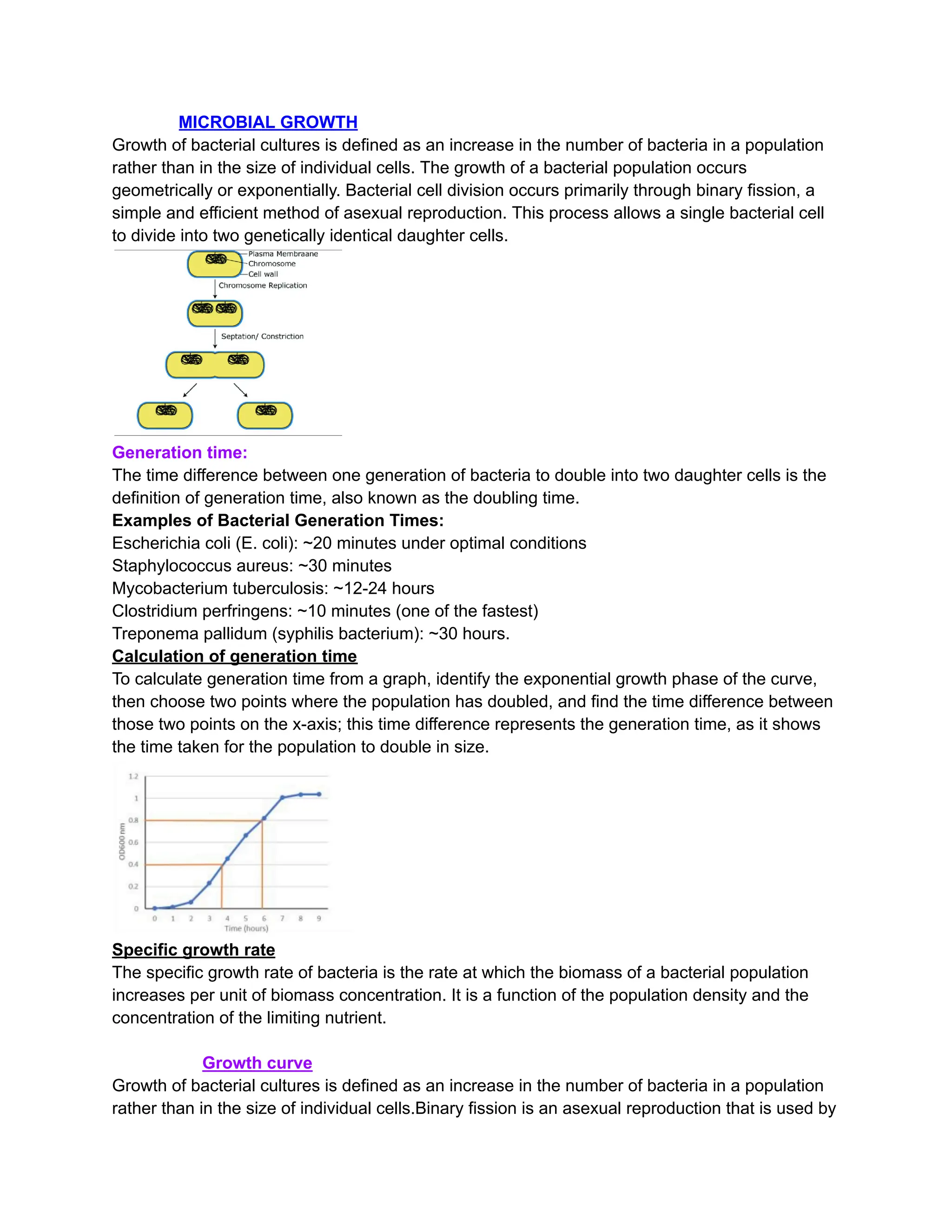
Calculation of generation time
To calculate generation time from a graph, identify the exponential growth phase of the curve,
then choose two points where the population has doubled, and find the time difference between
those two points on the x-axis; this time difference represents the generation time, as it shows
the time taken for the population to double in size.
Specific growth rate
The specific growth rate of bacteria is the rate at which the biomass of a bacterial population
increases per unit of biomass concentration. It is a function of the population density and the
concentration of the limiting nutrient.
Growth curve
Growth of bacterial cultures is defined as an increase in the number of bacteria in a population
rather than in the size of individual cells.Binary fission is an asexual reproduction that is used by
2.
prokaryotes such asbacteria to proliferate. When a broth culture is inoculated with a small
bacterial inoculum, the population size of the bacteria increases showing a classical pattern.
When plotted on a graph, a distinct curve is obtained referred to as the bacterial growth curve.In
batch culture, there is an initial phase of no growth (lag phase), which is followed by rapid
growth (exponential growth), then there is leveling off (stationary phase) and finally a decline in
the viable cell count (death or decline phase).
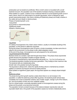
Phases of bacterial growth curve
1)Lag phase
2)Log phase or exponential phase
3)Stationary phase
4)Death or decline phase
Lag phase
On adding microorganisms into a fresh culture medium, usually no immediate doubling of the
population, so this period is called the Lag phase.
During this phase microorganisms does not grow or divide immediately, but take some time to
adjust to the environment, so this phase is also called adaptation phase.
At the end of the phase, microorganisms are well prepared for the cell division.
Log phase(exponential phase)
Bacteria divide at a constant, rapid rate through binary fission. This phase shows exponential
growth because the population doubles at regular intervals.
This phase is characterized by rapid exponential cell growth (i.e., 1 to 2 to 4 to 8 and so on).
The bacterial population doubles during every generation. They multiply at their maximum rate.
The bacterial cells are metabolically active
Growth rate is the greatest during the log phase.
The log phase is always brief, unless the rapidly dividing culture is maintained by constant
addition of nutrients and frequent removal of waste products.
When plotted on logarithmic graph paper, the log phase appears as a steeply sloped straight
line.
Stationary phase
The phase during which growth reaches a state where there is no net increase in the
population, is called the stationary phase.After log phase, the bacterial growth almost stops
completely due to lack of essential nutrients, lack of water oxygen, change in pH of the medium,
etc. and accumulation of their own toxic metabolic wastes.
Due to a closed system, eventually population growth of microorganisms ceases during this
phase and the growth curve becomes horizontal.
The population remains constant because of the balance between reproduction rate and
equivalent death rate or the growth of the population ceases but remains metabolically active.
3.
Endospores start formingduring this stage.
Decline phase:
During this phase, the bacterial population declines due to death of cells.
The decline phase starts due to
(a) accumulation of toxic products and autolytic enzymes and
(b) exhaustion of nutrients.
__________________________________
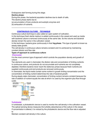
CONTINUOUS CULTURE TECHNIQUE
Continuous culture technique is also called an open system of cultivation.
In this technique fresh sterile medium is added continuously in the vessel and used up media
with bacterial culture is removed continuously at the same rate. So the volume and bacterial
density remain the same in the cultivation vessel.
In this technique, bacteria grow continuously in their log phase. This type of growth is known as
steady state growth.
The cell density in continuous culture remains constant and it is achieved by maintaining
constant dilution and flow rate
Types of approach to continuous culture
Chemostat:
It is the most common type of approach which controls the population density and growth of
culture.
Two elements are used in chemostat, the dilution rate and concentration of limiting nutrients.
In continuous culture, end products do not accumulate and nutrients are not completely
depleted, therefore bacteria never reach the stationary phase because fresh nutrients are
supplied continuously and end products are removed continuously.
In chemostat, the liquid media contain some nutrient in growth limiting concentration and the
concentration of limiting nutrient determines the rate of bacterial growth.
During steady state chemostat, concentration of limiting nutrient remains constant because the
rate of addition of nutrient equals the rate at which it is used by the organism plus flow through
outlet.
Turbidostat
In turbidostat, a photoelectric device is used to monitor the cell density in the cultivation vessel.
The optical sensing device measures the turbidity (absorbance) of the culture in the vessel.
If concentration is altered, it is noticed by the photoelectric device and the flow rate is adjusted
to
Maintain constant cell density in the culture.
4.
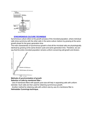
SYNCHRONOUS CULTURE TECHNIQUE
Synchronousculture refers to the growth process of the microbial population, where individual
cells show synchrony with the other cells in the same culture medium by growing at the same
growth phase for the given generation time.
The main characteristic of synchronous growth is that all the microbial cells are physiologically
identical by growing at the same division cycle and same generation time. Therefore, we can
say that the entire microbial population remains uniform concerning cell growth and division.
Methods of synchronization of growth
Selection of cells by membrane filter -
Use of membrane filters with specific pore size will help in separating cells with uniform
diameter. Such cells are then used for obtaining synchronous growth.
Another method for obtaining cells with uniform size by use of a membrane filter is
Helmstetter Cummings technique.
5.
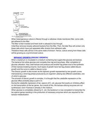
When heterogeneous cultureis filtered through a cellulose nitrate membrane filter, some cells
get adhered to the filter.
The filter is then inverted and fresh broth is allowed to low through.
Initial flow removes loosely adhered bacteria from the filter. Then, the later flow will contain only
these cells which have just separated after division from adherent cells.
Therefore these cells will be in the same state of division. Hence, culture arising from these cells
will be growing as a synchronized culture.
BIPHASIC GROWTH (DIAUXIC GROWTH)
When a bacterium is incubated in a medium containing two sugars like glucose and lactose.
The bacteria first utilize glucose and complete their lag and log phase. After completion of
glucose utilization, they used lactose and produce and another lag phase due to the synthesis
of their lactose breaking enzyme. Such types of growth have two lag phases called diauxic
growth and the curve is called a diauxic curve.
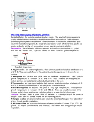
The Diauxic growth is also known as the diphasic growth represented by two growth curves
intervened by a short lag phase produced by an organism utilizing two different substrates, one
of which is glucose.
The process of diauxic growth is complex, it is thought that the catabolite repression or the
glucose effect probably plays a part in it.
During the catabolite repression of lac- operon of E. coli, glucose first exerts an inhibitory effect
on the transcription of the lac genes. As a result of this, the lactose-utilizing enzymes are not
synthesized, even if lactose is already in the medium.
When glucose is completely utilized by E. coli, the bacterium is now competent to transcribe the
lac-operon genes resulting in the production of necessary enzymes which are required for the
lactose metabolization.
6.
FACTORS INFLUENCING BACTERIALGROWTH
Controlled factors for bacterial growth and culture media The growth of microorganisms is
greatly affected by the chemical and physical nature of their surroundings. Prokaryotes are
present or grow anywhere life can exist. The environments in which some prokaryotes grow
would kill most other organisms, the major physical factors which affect microbial growth are
solutes and water activity, pH, temperature, oxygen level, pressure and radiation.
Temperature : Bacteria have a minimum, optimum, and maximum temperature for growth
and can be divided into 3 groups based on their optimum growth temperature
1. Psychrophiles are cold-loving bacteria. Their optimum growth temperature is between -5 ہC
and 15 ہC. They are usually found in the Arctic and Antarctic regions and in streams fed by
glaciers
2. Mesophiles are bacteria that grow best at moderate temperatures. Their Optimum
growth temperature is between 25 ہC and 45 ہC. Most bacteria are mesophilic and
include common soil bacteria and bacteria that live in and on the body.
3.Thermophiles are heat-loving bacteria , their optimum growth temperature is between 45 ہC
and 70 ہC and are commonly found in hot springs and in compost heaps.
4 Hyperthermophiles are bacteria that grow at very high temperatures. Their Optimum
growth temperature is between 70 ہC and 110 ہC. They are usually members of the
Archaea and are found growing near hydrothermal vents at great depths in the ocean.
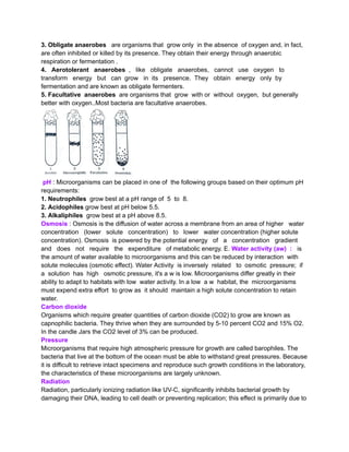
Oxygen : Bacteria show a great deal of variation in their requirements for gaseous
oxygen. Most can be placed in one of the following groups:
1.Obligate aerobes are organisms that grow only in the presence of oxygen. They Obtain their
energy through aerobic respiration .
2. Microaerophiles are organisms that require a low concentration of oxygen (2%to 10%) for
growth, but higher concentrations are inhibitory. They obtain their energy through aerobic
respiration .
7.
3. Obligate anaerobesare organisms that grow only in the absence of oxygen and, in fact,
are often inhibited or killed by its presence. They obtain their energy through anaerobic
respiration or fermentation .
4. Aerotolerant anaerobes , like obligate anaerobes, cannot use oxygen to
transform energy but can grow in its presence. They obtain energy only by
fermentation and are known as obligate fermenters.
5. Facultative anaerobes are organisms that grow with or without oxygen, but generally
better with oxygen..Most bacteria are facultative anaerobes.
pH : Microorganisms can be placed in one of the following groups based on their optimum pH
requirements:
1. Neutrophiles grow best at a pH range of 5 to 8.
2. Acidophiles grow best at pH below 5.5.
3. Alkaliphiles grow best at a pH above 8.5.
Osmosis : Osmosis is the diffusion of water across a membrane from an area of higher water
concentration (lower solute concentration) to lower water concentration (higher solute
concentration). Osmosis is powered by the potential energy of a concentration gradient
and does not require the expenditure of metabolic energy. E. Water activity (aw) : is
the amount of water available to microorganisms and this can be reduced by interaction with
solute molecules (osmotic effect). Water Activity is inversely related to osmotic pressure; if
a solution has high osmotic pressure, it's a w is low. Microorganisms differ greatly in their
ability to adapt to habitats with low water activity. In a low a w habitat, the microorganisms
must expend extra effort to grow as it should maintain a high solute concentration to retain
water.
Carbon dioxide
Organisms which require greater quantities of carbon dioxide (CO2) to grow are known as
capnophilic bacteria. They thrive when they are surrounded by 5-10 percent CO2 and 15% O2.
In the candle Jars the CO2 level of 3% can be produced.
Pressure
Microorganisms that require high atmospheric pressure for growth are called barophiles. The
bacteria that live at the bottom of the ocean must be able to withstand great pressures. Because
it is difficult to retrieve intact specimens and reproduce such growth conditions in the laboratory,
the characteristics of these microorganisms are largely unknown.
Radiation
Radiation, particularly ionizing radiation like UV-C, significantly inhibits bacterial growth by
damaging their DNA, leading to cell death or preventing replication; this effect is primarily due to
8.
the formation ofpyrimidine dimers in the DNA, rendering the bacteria unable to reproduce,
effectively killing or inactivating them.
_________________________________
MEASUREMENT OF MICROBIAL GROWTH
Estimating the growth of bacteria is extremely important. Environmental health officers regularly
inspect food premises and take samples for analysis. Water boards check water supplies daily.
Many products are produced using bacteria grown in fermenters. Measuring their growth is an
important part of the process.
There are various ways to measure microbial growth for the determination of growth rates and
generation times..
Growth can be measured by one of the following types of measurements:
1) Direct methods
2) Indirect methods
Direct methods:
1. Breed Method:
A known volume of microbial cell suspension (0.01 ml) is spread uniformly over a glass slide
covering a specific area (1 sq. cm). The smear is then fixed by heating, stained, examined
under oil immersion lens, and the cells are counted.
Customarily, cells in a few microscopic fields are counted because it is not possible to scan the
entire area of the smear. The counting of the total number of cells is determined by calculating
the total number of microscopic fields per square cm. area of the smear.
The total number of cells can be counted with the help of following calculations:
(a) Area of microscopic field = πr2
r (oil immersion lens) = 0.08 mm.
Area of the microscopic field under the oil-immersion lens
= πr2 = 3.14 x (0.08 mm)2 = 0.02 sq. mm.
(b) Area of the smear one sq. cm. = 100sq. mm.
Then, the no. of microscopic fields = 100/0.02=5000
(c) No. of cells 1 sq. cm. (or per 0.01 ml microbial cell suspension)
= Average no. of microbes per microscopic field x 5000
2. Proportional count
The proportional count method is a way to measure the number of cells in a suspension by
mixing it with a known number of particles and counting them under a microscope.
Mix an equal amount of a cell suspension with a standard suspension of particles.
Spread the mixture on a slide and fix and stain it.
Count the number of particles and cells in each microscopic field.
9.
Calculate the averagenumber of cells per field.
Use the average number of cells per field to calculate the number of cells per unit volume of the
suspension.
Electronic count
A Coulter counter is an electronic device used to count and size particles suspended in a fluid,
including bacteria. It operates based on the principle of electrical impedance, detecting changes
in electrical resistance as particles pass through a small aperture.
The bacterial sample is suspended in an electrolyte solution to ensure electrical conductivity.
The sample is drawn through a small aperture, with electrodes on either side.
As each bacterial cell passes through the aperture, it momentarily displaces the conducting
fluid, causing a measurable increase in electrical resistance.
The Coulter counter registers these impedance changes as pulses. The number of pulses
corresponds to the number of bacteria, while pulse magnitude correlates with cell size.
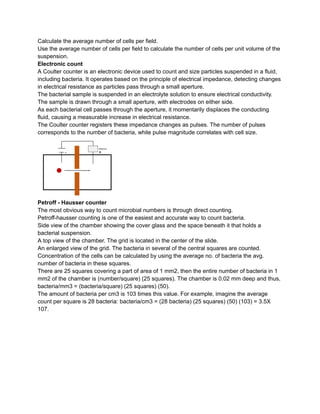
Petroff - Hausser counter
The most obvious way to count microbial numbers is through direct counting.
Petroff-hausser counting is one of the easiest and accurate way to count bacteria.
Side view of the chamber showing the cover glass and the space beneath it that holds a
bacterial suspension.
A top view of the chamber. The grid is located in the center of the slide.
An enlarged view of the grid. The bacteria in several of the central squares are counted.
Concentration of the cells can be calculated by using the average no. of bacteria the avg.
number of bacteria in these squares.
There are 25 squares covering a part of area of 1 mm2, then the entire number of bacteria in 1
mm2 of the chamber is (number/square) (25 squares). The chamber is 0.02 mm deep and thus,
bacteria/mm3 = (bacteria/square) (25 squares) (50).
The amount of bacteria per cm3 is 103 times this value. For example, imagine the average
count per square is 28 bacteria: bacteria/cm3 = (28 bacteria) (25 squares) (50) (103) = 3.5X
107.
10.
Indirect methods:
Viable count( Standard plate count)
The estimation of the number of living bacterial cells is called a viable count. Standard Plate
Count (SPC) method is the most commonly used laboratory technique for viable count of
bacterial cells in milk, food, water, and many other materials
After serial dilution, the aliquots of the diluted sample are plated on an appropriate culture
media. Then, the plates are incubated, after which the number of colonies formed is counted.
This technique is also known as plate count or colony counts.
In this method, accurate determination of total cell numbers is only possible if each colony is
formed of a single cell. However, it’s difficult for one to ensure such a case; that’s why the total
numbers of cells reported using this method are termed Colony Forming Units (CFUs) rather
than cell numbers. It’s calculated as:[3]
The number of CFUs per ml of sample = The number of colonies (30-300 plate) X The dilution
factor of the plate counted.
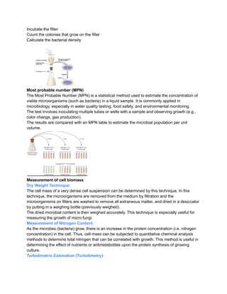
Membrane filtration method
The membrane filter count method is a technique used to count bacteria and other
microorganisms in water samples. It involves filtering water through a porous membrane, then
counting and culturing the trapped microorganisms.
Filter a known volume of water through a membrane filter
Place the filter on a culture medium
11.
Incubate the filter
Countthe colonies that grow on the filter
Calculate the bacterial density
Most probable number (MPN)
The Most Probable Number (MPN) is a statistical method used to estimate the concentration of
viable microorganisms (such as bacteria) in a liquid sample. It is commonly applied in
microbiology, especially in water quality testing, food safety, and environmental monitoring.
The test involves inoculating multiple tubes or wells with a sample and observing growth (e.g.,
color change, gas production).
The results are compared with an MPN table to estimate the microbial population per unit
volume.
Measurement of cell biomass
Dry Weight Technique:
The cell mass of a very dense cell suspension can be determined by this technique. In this
technique, the microorganisms are removed from the medium by filtration and the
microorganisms on filters are washed to remove all extraneous matter, and dried in a desiccator
by putting in a weighing bottle (previously weighed).
The dried microbial content is then weighed accurately. This technique is especially useful for
measuring the growth of micro fungi.
Measurement of Nitrogen Content:
As the microbes (bacteria) grow, there is an increase in the protein concentration (i.e. nitrogen
concentration) in the cell. Thus, cell mass can be subjected to quantitative chemical analysis
methods to determine total nitrogen that can be correlated with growth. This method is useful in
determining the effect of nutrients or antimetabolites upon the protein synthesis of growing
culture.
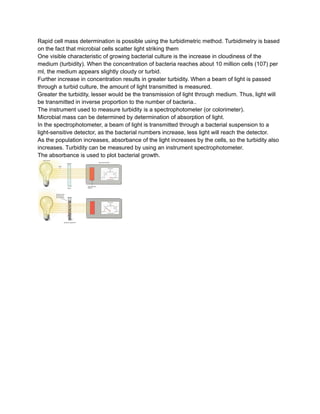
Turbidimetric Estimation (Turbidimetry):
12.
Rapid cell massdetermination is possible using the turbidimetric method. Turbidimetry is based
on the fact that microbial cells scatter light striking them
One visible characteristic of growing bacterial culture is the increase in cloudiness of the
medium (turbidity). When the concentration of bacteria reaches about 10 million cells (107) per
ml, the medium appears slightly cloudy or turbid.
Further increase in concentration results in greater turbidity. When a beam of light is passed
through a turbid culture, the amount of light transmitted is measured.
Greater the turbidity, lesser would be the transmission of light through medium. Thus, light will
be transmitted in inverse proportion to the number of bacteria..
The instrument used to measure turbidity is a spectrophotometer (or colorimeter).
Microbial mass can be determined by determination of absorption of light.
In the spectrophotometer, a beam of light is transmitted through a bacterial suspension to a
light-sensitive detector, as the bacterial numbers increase, less light will reach the detector.
As the population increases, absorbance of the light increases by the cells, so the turbidity also
increases. Turbidity can be measured by using an instrument spectrophotometer.
The absorbance is used to plot bacterial growth.

![Indirect methods:
Viable count ( Standard plate count)
The estimation of the number of living bacterial cells is called a viable count. Standard Plate
Count (SPC) method is the most commonly used laboratory technique for viable count of
bacterial cells in milk, food, water, and many other materials
After serial dilution, the aliquots of the diluted sample are plated on an appropriate culture
media. Then, the plates are incubated, after which the number of colonies formed is counted.
This technique is also known as plate count or colony counts.
In this method, accurate determination of total cell numbers is only possible if each colony is
formed of a single cell. However, it’s difficult for one to ensure such a case; that’s why the total
numbers of cells reported using this method are termed Colony Forming Units (CFUs) rather
than cell numbers. It’s calculated as:[3]
The number of CFUs per ml of sample = The number of colonies (30-300 plate) X The dilution
factor of the plate counted.
Membrane filtration method
The membrane filter count method is a technique used to count bacteria and other
microorganisms in water samples. It involves filtering water through a porous membrane, then
counting and culturing the trapped microorganisms.
Filter a known volume of water through a membrane filter
Place the filter on a culture medium](https://image.slidesharecdn.com/2bw8qkpqqaso9jhkh12k-microbial-growth-250409160026-74d5cbe7/85/Microbial_growth-growth-curve-notes_-pdf-10-320.jpg)