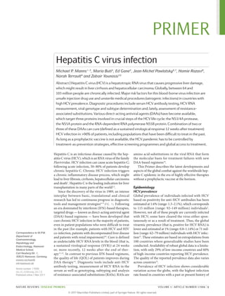
See discussions, stats, and author profiles for this publication at: https://www.researchgate.net/publication/314192710
Hepatitis C virus infection
Article in Nature Reviews Disease Primers · March 2017
DOI: 10.1038/nrdp.2017.6
CITATIONS
457
READS
12,560
7 authors, including:
Maria Buti
University Hospital Vall d'Hebron
733 PUBLICATIONS 29,630 CITATIONS
SEE PROFILE
Homie A Razavi
274 PUBLICATIONS 59,921 CITATIONS
SEE PROFILE
Norah Terrault
University of Southern California
605 PUBLICATIONS 39,493 CITATIONS
SEE PROFILE
All content following this page was uploaded by Homie A Razavi on 03 January 2018.
The user has requested enhancement of the downloaded file.
Hepatitis C is an infectious disease caused by the hep­
atitis C virus (HCV), which is an RNA virus of the family
Flaviviridae. HCV infection can cause acute hepatitis C;
following acute infection, 50–80% of patients develop
chronic hepatitis C. Chronic HCV infection triggers
a chronic inflammatory disease process, which might
lead to liver fibrosis, cirrhosis, hepatocellular ­carcinoma
and death1
. Hepatitis C is the leading indication for liver
transplantation in many parts of the world2,3
.
Since the discovery of the virus in 1989, an intense
interplay between basic, translational and clinical
research has led to continuous progress in diagnostic
tools and management strategies4–9
(FIG. 1). Following
an era dominated by interferon (IFN)-based therapies,
targeted drugs — known as direct-acting antiviral agent
(DAA)-based regimens — have been developed that
cure chronic HCV infection in the majority of patients,
even in patient populations who were difficult to treat
in the past (for example, patients with HCV and HIV
co-infection, patients with decompensated liver disease
and patients with renal impairment)10
. Cure is defined
as undetectable HCV RNA levels in the blood (that is,
a sustained virological response (SVR)) at 24 weeks
or, more recently, 12 weeks after the end of ther­
apy11,12
. In contrast to previous IFN-based regimens,
the quality of life (QOL) of patients improves during
DAA therapy13
. Diagnostic tools include anti-HCV
antibody testing, measurement of HCV RNA in the
serum as well as genotyping, subtyping and analysis
of resistance-associated substitutions (RASs). RASs are
amino acid substitutions in the viral RNA that form
the molecular basis for ­
treatment failures with new
DAA-based regimens14
.
This Primer describes the latest developments and
aspects of the global combat against the worldwide hep­
atitis C epidemic in the era of highly effective therapies
without a prophylactic vaccine at the horizon.
Epidemiology
HCV prevalence
Global prevalence of individuals infected with HCV
based on positivity for anti-HCV antibodies has been
estimated at 1.6% (range: 1.3–2.1%), which corresponds
to 115 million (range: 92–149 million) individuals2
.
However, not all of these people are currently infected
with HCV; some have cleared the virus either spon­
taneously or as a result of treatment. Thus, the global
­
viraemic prevalence (that is, positive for HCV RNA) is
lower and estimated at 1% (range: 0.8–1.14%) or 71 mil­
lion (range: 62–79 million) individuals with HCV infec­
tion15
. These estimates are based on extrapolations from
100 countries where generalizable studies have been
conducted. Availability of robust global data is a limita­
tion, with only 29% of low-income countries and 60%
of high-income countries reporting HCV prevalence.
The quality of the reported prevalence data also varies
across countries15
.
Prevalence of HCV infection shows considerable
variation across the globe, with the highest infection
rate found in countries with a past or present history of
Correspondence to M.P.M.
Department of
Gastroenterology,
Hepatology and
Endocrinology, Hannover
Medical School,
Carl-Neuberg-Str. 1,
30625 Hannover, Germany.
manns.michael@
mh‑hannover.de
Article number: 17006
doi:10.1038/nrdp.2017.6
Published online 2 Mar 2017
Hepatitis C virus infection
Michael P. Manns1–3
, Maria Buti4
, Ed Gane5
, Jean-Michel Pawlotsky6,7
, Homie Razavi8
,
Norah Terrault9
and Zobair Younossi10
Abstract | Hepatitis C virus (HCV) is a hepatotropic RNA virus that causes progressive liver damage,
which might result in liver cirrhosis and hepatocellular carcinoma. Globally, between 64 and
103 million people are chronically infected. Major risk factors for this blood-borne virus infection are
unsafe injection drug use and unsterile medical procedures (iatrogenic infections) in countries with
high HCV prevalence. Diagnostic procedures include serum HCV antibody testing, HCV RNA
measurement, viral genotype and subtype determination and, lately, assessment of resistance-
associated substitutions. Various direct-acting antiviral agents (DAAs) have become available,
which target three proteins involved in crucial steps of the HCV life cycle: the NS3/4A protease,
the NS5A protein and the RNA-dependent RNA polymerase NS5B protein. Combination of two or
three of these DAAs can cure (defined as a sustained virological response 12 weeks after treatment)
HCV infection in >90% of patients, including populations that have been difficult to treat in the past.
As long as a prophylactic vaccine is not available, the HCV pandemic has to be controlled by
treatment‑as‑prevention strategies, effective screening programmes and global access to treatment.
NATURE REVIEWS | DISEASE PRIMERS VOLUME 3 | ARTICLE NUMBER 17006 | 1
PRIMER
©
2
0
1
7
M
a
c
m
i
l
l
a
n
P
u
b
l
i
s
h
e
r
s
L
i
m
i
t
e
d
,
p
a
r
t
o
f
S
p
r
i
n
g
e
r
N
a
t
u
r
e
.
A
l
l
r
i
g
h
t
s
r
e
s
e
r
v
e
d
.
iatrogenic infections (that is, infections due to the activ­
ity of a physician or medical therapy) (FIG. 2). Cameroon,
Egypt, Gabon, Georgia, Mongolia, Nigeria and
Uzbekistan2,16–21
all have an anti-HCV antibody preva­
lence of >5% in the adult population; iatrogenic infection
is a key risk factor in these countries. The source of HCV
infection in Egypt is well documented and attributed to
intravenous treatment for schistosomiasis (flat worm)
in the 1960–1970s22,23
. Western countries account only
for a small percentage of global HCV infections, with
China, Pakistan, India, Egypt and Russia accounting for
approximately half of the total viraemic HCV infections2
.
The age distribution of the HCV-infected population
correlates to the primary source of infection in specific
countries. Countries where injection drug use is an ongo­
ing important risk factor (for example, Australia, Czech
Republic, Finland, Luxembourg, Portugal, Russia and
the United Kingdom), the HCV-infected population
has a peak age of mid‑30s years, whereas the peak age
is usually older (50–60 years) in countries where iatro­
genic infections dominate as the cause24,25
. This difference
is explained by the fact that active injection drug users
are typically young, whereas most iatrogenic infections
occurred before the 1990s when diagnostics for HCV
became available. In some countries, the age profile is
mixed owing to the presence of several risk factors.
HCV genotype distribution varies by region (FIG. 3).
This genotypic distribution has implications on clinical
courseandtherequirementsfortreatmentanddrugdevel­
opment. Pan-genotypic drugs are, in particular, needed in
lower-middle-income and low-income ­
countries where
genotype 1 accounts for less than half of all infections2
.
Risk factors
HCV is primarily transmitted through percutaneous
exposure to blood, owing to medical procedures or
sharing contaminated devices for injection drug use.
Mother-to‑infant transmission and sexual transmission
also occur, but are less common, except in the case of
HIV-infected men having unprotected sex with men26,27
.
Causes of iatrogenic infections include blood transfusion
or administration of clotting factors (with contaminated
blood before the start of blood screening in the 1990s),
long-term haemodialysis, injection of multiple individ­
uals with the same syringe and reuse of glass syringes for
medical injections. The importance of these risk factors
differs between countries. The reuse of glass syringes for
medical injections continues to this day in some regions
and remains, for example, the key risk factor in Pakistan.
In Europe and the United States, the greatest risk for HCV
infection is unsafe injection drug use, which accounts
for 50–60% of acute HCV infections24,28–31
. In the past,
healthcareexposurewasanimportantsourceoftransmis­
sion. Currently, needle-stick injuries among health care
workers and patient-to‑patient transmission are still risk
factors for HCV transmission, as is receiving a tattoo in
an unregulated setting32,33
. However, no risk factors can be
identified in a considerable proportion (up to 40% in the
western world) of patients with HCV infection.
Comorbidities and mortality
Chronic HCV infection can result in hepatic fibrosis,
cirrhosis and hepatocellular carcinoma. The progres­
sion through these stages is a function of time since
infection and age of initial infection. Japan, which has
the oldest HCV-infected population, is already seeing
a drop in hepatocellular carcinoma cases34
, as infected
individuals are dying of other causes before progress­
ing to hepato­
cellular carcinoma. However, in nearly
every other country, the projected number of hepato­
cellular carcinoma and decompensated cirrhosis cases
as a result of HCV infection has been increasing and
will continue to increase in the absence of treatment and
universal screening programmes and interventions35–37
.
A recent study showed that, worldwide, the number of
hepatitis C-related deaths as a result of hepatocellular
carcinoma and cirrhosis increased from 895,000 deaths
in 1990 to 1,454,000 deaths in 2013; the proportion of
deaths that were attributed to hepatitis C without hepato­
cellular carcinoma also increased in the same period
from 33.8% in 1990 to 48.4% in 2015 (REF. 1).
In addition to liver-related complications, HCV infec­
tion is associated with numerous extrahepatic manifes­
tations38
. Individuals with chronic HCV infection are
more likely to develop cryoglobulinaemia and non-­
Hodgkin lymphoma38
. In addition, these individ­uals are
at increased risk of developing insulin resistance and dia­
betes mellitus, which may lead to the increase in cardio­
vascular mortality as a result of stroke and myo­
cardial
perfusion defects39
. Finally, fatigue is more common
among those with chronic HCV infection and in patients
with lower health-related QOL (HRQOL); fatigue and
HRQOL scores improve after achieving a SVR.
Mechanisms/pathophysiology
HCV is a member of the Flaviviridae family and the
genus Hepacivirus, which also includes GB virus B and
the recently identified non-primate, rodent and bat
hepaci­viruses40–44
. HCV virions are 45–65nm in diameter
Author addresses
1
Department of Gastroenterology, Hepatology and
Endocrinology, Hannover Medical School,
Carl-Neuberg-Str. 1, 30625 Hannover, Germany.
2
German Center for Infection Research (DZIF), Partner Site
Hannover-Braunschweig, Germany.
3
Helmholtz Centre for Infection Research (HZI),
Braunschweig, Germany.
4
Liver Unit, Hospital Universitari Vall d’Hebron and
CIBEREHD del Instituto Carlos III, Barcelona, Spain.
5
New Zealand Liver Transplant Unit, Auckland City
Hospital, Auckland, New Zealand.
6
National Reference Center for Viral Hepatitis B, C and D,
Department of Virology, Hôpital Henri Mondor, Université
Paris-Est, Créteil, France.
7
INSERM U955, Créteil, France.
8
Center for Disease Analysis, Lafayette, Colorado, USA.
9
Viral Hepatitis Center, Division of Gastroenterology,
University of California at San Francisco, San Francisco,
California, USA.
10
Beatty Center for Integrated Research, Falls Church,
Virginia, USA.
PRIMER
2 | ARTICLE NUMBER 17006 | VOLUME 3 www.nature.com/nrdp
©
2
0
1
7
M
a
c
m
i
l
l
a
n
P
u
b
l
i
s
h
e
r
s
L
i
m
i
t
e
d
,
p
a
r
t
o
f
S
p
r
i
n
g
e
r
N
a
t
u
r
e
.
A
l
l
r
i
g
h
t
s
r
e
s
e
r
v
e
d
.
and are enveloped in a lipid bilayer in which two envel­
ope glyco­
proteins (E1 and E2) are anchored. The
­
envelope surrounds the non-­
icosahedral nucleo­
capsid,
which is composed of multiple copies of the small basic
HCV core protein and contains the positive-strand RNA
genome of approximately 9.6kb, with an open reading
frame encoding a single polyprotein of approximately
3,000 amino acids. The structural proteins (core, E1
and E2) are encoded by the amino‑­
terminal part of the
open reading frame, whereas the remaining portion
codes for the non-structural proteins (p7, NS2, NS3,
NS4A, NS4B, NS5A and NS5B)45
. HCV virions are
associ­ated with host low-density lipoproteins (LDLs) and
very-low-­density lipoproteins (VLDLs), forming what are
known as lipoviroparticles. The lipoviroparticles also
contain apolipoprotein B (APOB) and other exchange­
able apolipo­proteins, such as APOC and APOE46
. Poorly
infectious non-enveloped viral capsids may also be
­
present in the blood of infected patients.
HCV is a very heterogeneous virus. Phylogenetic
analyses of HCV strains isolated in various regions of
the world have led to the identification of seven main
HCV genotypes, designated 1–7. The HCV genotypes
comprise a large number of subtypes, identified by
lower-­
case letters (1a, 1b, and so on)44
. The genotype
influences the­disease course and the response to ­antiviral
therapy (FIG. 3).
HCV life cycle
Viral attachment involves the two envelope glyco­proteins,
E1 and E2, apolipoproteins present at the surface of
the lipoviroparticles and several cell surface mol­
ecules
(FIG. 4). Glycosaminoglycans and the LDL receptor seem
to be involved in low-affinity initial cell binding. Then,
E1–E2interactswithCD81andscavengerreceptorclassB
member 1, whereas claudin 1, occludin and possibly
other molecules, such as claudin 6 or claudin 9, epider­
mal growth factor receptor or ephrin receptor type A2,
are required for cell entry47
. This multi-receptor complex
mediates uptake and defines organ and species specifi­
city. The E2 envelope glycoprotein contains hypervariable
regions that play the part of immunodominant neutral­
ization epitopes. Antibodies against these hypervariable
regions in patient sera are protective. However, the high
variability of HCV with different viral quasi-­
species in
the same patient has so far prevented the develop­
ment a
successful prophylactic vaccine based on these viral pro­
teins. HCV–receptor complexes are seemingly associated
with tight junctions, which enable direct cell–cell trans­
mission48
. After attachment, HCV entry into cells results
in clathrin-mediated endocytosis, followed by fusion
between viral and endosomal membranes, which leads
to the release of the nucleocapsid into the cytoplasm. The
E1 envelope glycoprotein is believed to be the fusogen
(the glycoprotein that facilitates cell fusion).
Nature Reviews | Disease Primers
1986 1989 1993 1996 1998 1999 2001 2003 2005 2014 2015 2016
Advances in HCV biology
IFN treatment era
All oral, IFN-free treatment era
IFN used for the
treatment of
non-A, non-B
hepatitis
IFN plus
ribavirin
Evidence that early treatment
of acute HCV infection with IFN
prevents chronic infection
PEG-IFN plus ribavirin as standard
of care for the next decade
December
Approval of ombitasvir/paritaprevir/r
plus dasabuvir
Proof of
concept
for the
first HCV
protease
inhibitor
Nucleic acid test for HCV In vitro
system
for HCV
infection
December
Approval of sofosbuvir
November
Approval of simeprevir
July
Approval of
daclatasvir
October
Approval of sofosbuvir/ledipasvir
July
Approval of elbasvir/grazoprevir
in Europe
January
Approval of elbasvir/grazoprevir
in the United States
June
Approval of sofosbuvir/velpatasvir
Identification of HCV Crystal structure
of NS3/4A
Replicon
system
2013
Figure 1 | Milestones in HCV research and management. Recombinant
interferon‑α (IFN) injected three times a week was given to patients with
non‑A,non‑Bhepatitis—atermusedforhepatitisCbeforethehepatitisC
virus(HCV)wasdiscoveredin1989(REF. 179).Subsequently,thedoseofIFN
and duration of treatment were increased; then, IFN was empirically
combined with ribavirin, which is a non-specific antiviral agent180,181
.
Between 2001 and 2011 (REF. 182), the combination of long-acting
pegylated (PEG)-IFNs, PEG‑IFNα2a or PEG‑IFNα2b, in combination with
ribavirinbecamethestandardofcare183,184
.TreatmentofchronichepatitisC
between 12 and 72 weeks resulted in sustained virological response (SVR)
rates(thatis,undetectableHCVRNAlevelsintheblood12or24weeksafter
the end of treatment) of 40% for patients with HCV genotype 1 infection
and 80% for patients with HCV genotype 2 infection, but considerable
adverse effects were the price to pay. Unravelling of the HCV life cycle,
crystallography of HCV viral enzymes and the development of the HCV
replicon system4
paved the way towards direct-acting antiviral agents
(DAAs), which specifically interfere with the HCV life cycle55
. BILN 2061,
a NS3/4A protease inhibitor, became the first DAA to show significant
inhibition of viral replication in humans, but further clinical development
was terminated owing to safety concerns. Subsequently, DAAs against
three different targets of the HCV life cycle were approved55
. In 2011, two
HCV protease inhibitors, boceprevir and telaprevir, were approved, but
treatment had to be combined with PEG-IFN and ribavirin at the cost of
adverseeffects.SeveralNS3/4AandNS5Ainhibitorsandonenucleotideas
well as one non-nucleotide NS5B polymerase inhibitor were approved
between 2013 and 2016. When two or three DAAs are combined, cure
rates between 90% and 100% are achieved. These new all-oral, IFN-free
DAA regimens are administered for durations of 8–24 weeks and are
associatedwithexcellenttolerabilityandsafety.Paritaprevir/r,paritaprevir
ritonavir-boosted.
PRIMER
NATURE REVIEWS | DISEASE PRIMERS VOLUME 3 | ARTICLE NUMBER 17006 | 3
©
2
0
1
7
M
a
c
m
i
l
l
a
n
P
u
b
l
i
s
h
e
r
s
L
i
m
i
t
e
d
,
p
a
r
t
o
f
S
p
r
i
n
g
e
r
N
a
t
u
r
e
.
A
l
l
r
i
g
h
t
s
r
e
s
e
r
v
e
d
.
Uncoating of the viral nucleocapsid liberates the
positive-strand genomic RNA into the cytosol, where it
serves as mRNA for synthesis of the HCV polyprotein.
The HCV 5ʹ untranslated region contains an internal
ribosome entry site, which controls HCV open reading
frame translation49
. The large precursor polyprotein
gener­
ated is translated at the endoplasmic reticulum
membrane where the processing events take place, result­
ing in the generation of the three structural proteins and
the seven non-structural proteins50
. At least two host
cellular peptidases (signalase and signal peptide pepti­
dase) are required for the processing of the HCV struc­
tural proteins, whereas two viral peptidases (NS2 and
NS3/4A) are involved in the processing of the HCV non-­
structural proteins. The viral proteins remain ­associated
with ­
intracellular membranes after processing51
.
Replication is catalysed by the NS5B protein. The
NS5A protein and the helicase-NTPase domain of NS3
play an important regulatory part in virus replication52,53
.
NS5A acts as a dimer with a basic channel involved in
RNA binding. Domain I and domain II of the NS5A pro­
tein are required for HCV replication in the replication
complex. The phosphorylation state of NS5A modu­
lates the balance between replication and later stages of
the HCV life cycle. The NS3 helicase has an important
role in separating nascent and template RNA strands,
unwinding RNA secondary structures and displacing
RNA-binding proteins53
. NS4B is an integral membrane
protein with a role in membrane rearrangements that
are induced by HCV proteins, leading to the formation
of the ‘membranous web’ or replication complex that
supports and compartmentalizes HCV replication. The
positive-strand genome RNA serves as a template for the
synthesis of a negative-strand intermediate of replication.
Then, negative-­
strand RNAs serve as templates to prod­
uce numerous strands of positive polarity that are subse­
quently used for polyprotein translation, the synthesis of
new intermediates of replication or packaging into new
virusparticles52
.Varioushostfactorshavealsobeenshown
to have important functional roles in the HCV life cycle.
Cyclophilin A (also known as peptidylprolyl isomerase A)
binds to both NS5A and NS5B, thereby inducing the con­
formational changes that are required for efficient HCV
replication. MicroRNA‑122 (miR‑122), an abundant
liver-­
specific miRNA, binds to two conserved sites in the
internal ribosome entry site, which is required for effi­
cient HCV replication and RNA stabilization. Other host
factors and pathways are involved in HCV replication,
such as phosphatidylinositol 4‑kinase IIIα or the choles­
terol and fatty acid biosynthesis pathways. Viral particle
formation is initiated by the interaction of the core and
NS5A proteins with genomic RNA in cytoplasmic lipid
droplets. HCV uses the VLDL production pathway at the
later stages of assembly and for release54
.
DAAs
Targets. The multiple steps in the HCV life cycle prov­ide
the targets for DAAs14,53,55
(FIG. 4). The NS3/4A protease
was identified as a major target for antiviral intervention,
as its blockade shuts down the intra­
cellular life cycle by
inhibitingmaturation oftheviralpoly­protein.Replication
has been identified as a major target for antiviral drugs.
Replication can be directly inhibited by NS5B inhibitors.
These include nucleotide analogues, which function
Nature Reviews | Disease Primers
Viraemic prevalence (%)
<0.6
0.6–0.74
0.75–1.29
1.3–2.8
>2.9
Infected individuals
>5 million
1 million–5 million
<1 million
Figure 2 | HCV prevalence. Schematic representation of the actual viraemic hepatitis C virus (HCV) prevalence and the
extrapolated total HCV infections per country. Figure based on data obtained from REF. 15.
PRIMER
4 | ARTICLE NUMBER 17006 | VOLUME 3 www.nature.com/nrdp
©
2
0
1
7
M
a
c
m
i
l
l
a
n
P
u
b
l
i
s
h
e
r
s
L
i
m
i
t
e
d
,
p
a
r
t
o
f
S
p
r
i
n
g
e
r
N
a
t
u
r
e
.
A
l
l
r
i
g
h
t
s
r
e
s
e
r
v
e
d
.
as RNA chain terminators after being activated intra­
cellularly by two rounds of phosphoryl­
ation, and non-­
nucleoside inhibitors of NS5B that target allosteric
sites of the enzyme and make it non-­
functional. NS5A
inhibitors alter the regulatory role of NS5A and seem to
disorganize the replication complex, thereby inhibiting
HCV replication in a potent manner, enhanced by their
ability to also inhibit viral assembly and release. Besides
well-known DAA targets, host-­targeted replication inhib­
itory approaches are attractive, because of their potential
for high barrier to resistance. Cyclophilin inhibition by
non-­
immunosuppressive analo­
gues of cyclosporine A is
credible, but no drugs have reached the market thus far,
owing to adverse effects that are ­
unrelated to cyclophilin
inhibition. miR‑122 antagonists have reached clinical
development in an injectable form56
, but safety issues
caused clinical hold.
Resistance to DAAs. HCV has a viral quasi-species
distrib­ution57
. The viral populations that constitute the
quasi-­
species differ by amino acid polymorphisms that
were generated during replication and subsequently
selected based on their effect on the corresponding virus
replication capacity. Natural polymorphisms that are
presentinaregiontargetedbyaDAAmayconferreduced
susceptibility to the DAA class or the speci­fic DAA. Upon
DAA administration, selection of viral vari­
ants with
reduced susceptibility to the drug (or drugs) defines ‘viral
resistance’. The term ‘resistance-­associated variant’, which
has been often used to indiffer­ently character­ize substitu­
tions that confer reduced susceptibil­ity to a drug or drug
class and the viral variants that carry these substitutions,
is incorrect. Substitutions (that is, amino acid changes)
that confer resistance are now called RASs, whereas
the viral variants that carry these RASs and thus have
reduced susceptibility to the DAA are called ‘­
resistant
variants’ (REF. 14).
In DAA-based antiviral treatments, each viral popu­
lation that is present in the quasi-species at baseline
evolves following individual kinetics that depend on
its amount at treatment initiation, its susceptibility to
the antiviral action of the DAAs and its fitness in the
presence of the DAA combination. Sensitive viral popu­
lations are rapidly eliminated, whereas resistant variants
are selected. Failure during treatment is called break­
through. However, most often treatment failure with
current regimens is due to relapse (that is, the recurrence
of the virus in the blood after treatment). The major­
ity of viral populations are resistant to one or several of
the drugs administered. They are characterized by the
presence of class-specific or drug-specific RASs on their
genomes. After treatment failure, variants that are resist­
ant to NS3/4A protease inhibitors disappear within a few
weeks to months after termination of therapy, whereas
variants that are resistant to NS5A inhibitors persist for
years, probably for ever in most cases14
.
Immune response
Both innate and adaptive immune responses are impor­
tant for HCV viral clearance. For the innate immune
response, natural killer (NK) cells seem to be involved
in resolving HCV infection; it has been shown that
certain NK cell receptor genes (those encoding killer
cell immunoglobulin-like receptor 2DL3 (KIR2DL3)
and HLA‑C1) are associated with viral clearance58
. For
adaptive immune responses, humoral antibody and
T cell responses are usually involved in controlling viral
infections. For HCV infection, most antibodies seem to
have no relevant activity against HCV owing to the high
variability of the virus and the quasi-species populations
in a single patient. Nevertheless, neutralizing antibodies
against certain epitopes may be protective59
, and rapid
induction of neutralizing antibodies has been associated
with the control of infection60
. Most data are available
on T cell responses. Broad and multi-specific CD4+
and
CD8+
T cell responses are associated with spontaneous
viral clearance, and the persistence of infection is attrib­
uted to an insufficient or early loss of T cell responsive­
ness61
. Several studies have shown a reduction in the
frequency and proportion of subpopulations of T cells
in the circulation when acute HCV infection develops
into chronicity62,63
. Studies in chimpanzees have prov­
ided evidence that loss of T helper cell activity results in
immune evasion of CD8+
T cells and viral persistence64
.
Experimental depletion of CD8+
T cells in chimpan­
zees resulted in viral persistence until the CD8+
T cell
response recovered64
.
HCV has evolved several immune escape or evasion
strategies that are linked to the persistence of infection65
.
For example, the HCV NS3/4A protein can efficiently
cleave and inactivate two host signalling pathways that
react to HCV pathogen-associated molecular patterns to
induce the IFN pathway. Nevertheless, IFN-stimulated
genes are induced during acute HCV infection, but this
response is not very effective at clearing the virus. IFN-
responsive gene expression remains high in chronic
infection66
, which was associated with poor response to
IFN-based treatment.
Nature Reviews | Disease Primers
Distribution
of
HCV
genotypes
(%)
100
80
60
40
20
0
Upper middle
High Lower middle
Income countries
90
70
50
30
10
Low
Genotype
1a
1b
1 (other)
2
3
4
5
6
Figure 3 | HCV genotype distribution. Hepatitis C virus (HCV) is a very heterogeneous
virus; seven genotypes have been detected thus far, but this figure focuses on six.
Genotype distribution differs between countries according to the World Bank income
categories. Genotype 1 is more prevalent in the Americas, Europe, Australia,
New Zealand, Central Asia and East Asia. Genotype 3 is most common in India185
and Pakistan186
, whereas genotype 4 dominates in Egypt187–189
and Central sub-Saharan
Africa15
. Genotype 5 accounts for more than one-third of HCV infections in
South Africa190,191
, whereas genotype 6 is found in South East Asia192–197
. Figure based
on data obtained from REF. 15.
PRIMER
NATURE REVIEWS | DISEASE PRIMERS VOLUME 3 | ARTICLE NUMBER 17006 | 5
©
2
0
1
7
M
a
c
m
i
l
l
a
n
P
u
b
l
i
s
h
e
r
s
L
i
m
i
t
e
d
,
p
a
r
t
o
f
S
p
r
i
n
g
e
r
N
a
t
u
r
e
.
A
l
l
r
i
g
h
t
s
r
e
s
e
r
v
e
d
.
In addition, the evolution to chronic HCV infection
is associated with a rapid exhaustion or alteration of
immune responses. Mucosal-associated invariant T cell
(innate-like effector T cells) levels are severely reduced in
chronic HCV infection67
. NK cells exhibit alterations
in phenotype and function during chronic HCV infec­
tion68
. In addition, liver-infiltrating intrahepatic T cells
are phenotypically exhausted and dysfunctional69–71
;
programmed cell death protein 1 and other markers of
functional exhaustion and apoptosis are upregulated68,72
.
Despite intensive studies of the innate and adaptive
immune responses in acute and chronic HCV infection,
the precise interplay between the innate and adaptive
immune responses that determines resolution versus
viral persistence remains incompletely understood.
Pathogenesis of HCV-associated liver disorders
Fibrogenesis — characterized by the activation of hepatic
stellate cells into myofibroblasts, which produce fibrous
extracellular matrix in excess — is the main complica­
tion of chronic HCV infection73
. It leads to progressive
liver fibrosis and, ultimately, the development of cirrho­
sis and its complications. Local inflammation seems to
be an important driver of fibrogenesis. Chronic HCV
infection is associated with chronic hepatic inflam­
mation, as a result of oxidative stress and immune
response directed to infected hepatocytes that express
viral epitopes74
. The T helper 2 cell response observed
during chronic HCV infection seems to have an
important role in chronic inflammation. In addition,
numerous growth factors, chemokines and cytokines
are produced in the infected hepatocytes; these ­
factors
partici­
pate in the recruitment of immune cells, the
perpetu­
ation of the local inflammatory response and
the activation of hepatic stellate cells into myofibro­
blasts75
. CD8+
T cell-induced apoptosis of hepato­
cytes might also have an important role in sustaining
inflammation and activating hepatic stellate cells76,77
.
In addition, the virus itself could participate in the
fibrogenic process; indeed, HCV has been suggested to
directly interact with hepatic stellate cells and this inter­
action could accelerate the fibrogenic process73
. Finally,
changes in hepatocyte proliferation during HCV
infection also seem to be involved in the progression
of liver fibrosis. These changes could be due to direct
intra­
cellular interactions between HCV proteins and
Nature Reviews | Disease Primers
Sinusoidal lumen
Hepatocyte
HCV RNA
ER
SRB1
CD81
Viral
particle
Claudin 1
Occludin
LDLR
+
RNA
uncoating
Clathrin-mediated
endocytosis
Viral
attachment
Viral
release
Viral
assembly
RNA
replication
– +
RNA
miR-122?
RNA
accumulation
+
NS5B
NS4B
NS3
NS4A
NS5A
ER lumen
Nucleocapsid
Golgi
Membranous
web (replication
complex)
Polyprotein
translation
and processing
Nucleus
Polyprotein
Structural and
non-structural
viral proteins
Processing
Cytosol
miR-122?
Figure 4 | HCV life cycle. Schematic representation of the hepatitis C virus (HCV) life cycle involving viral attachment,
clathrin-mediated endocytosis, polyprotein translation and processing, RNA replication and, finally, viral assembly and
release. ER, endoplasmic reticulum; LDLR, low-density lipoprotein receptor; miR‑122, microRNA‑122; SRB1, scavenger
receptor class B member 1.
PRIMER
6 | ARTICLE NUMBER 17006 | VOLUME 3 www.nature.com/nrdp
©
2
0
1
7
M
a
c
m
i
l
l
a
n
P
u
b
l
i
s
h
e
r
s
L
i
m
i
t
e
d
,
p
a
r
t
o
f
S
p
r
i
n
g
e
r
N
a
t
u
r
e
.
A
l
l
r
i
g
h
t
s
r
e
s
e
r
v
e
d
.
proteins involved in the regulation of the cell cycle, and
to disorganization of cell cycle checkpoints by DNA
damage that is induced by the oxidative stress generated
during infection.
Patients with hepatitis C-related cirrhosis have a 4–5%
cumulative annual incidence of hepatocellular carci­
noma78
. Cirrhosis is the main determinant of hepato­
cellular carcinoma, given that this carcinoma is rare in
patients infected with HCV who do not have cirrhosis78
.
HCV infection seems to have a role in the carcinogenic
process. Indeed, HCV can hijack molecular pathways
involved in the control of the cell cycle. Together, DNA
damage and abnormalities of cell cycle and apoptosis
control could lead to hepatocyte transformation. In addi­
tion, increased proliferation due to the loss of infected
cells may participate, together with local inflammation
and oxidative stress, in triggering hepatocyte trans­
formation79
. The role of the local immune response in
­
subsequent tumour progression remains unclear.
Diagnosis, screening and prevention
Clinical course
In the majority of cases, acute infection occurs with­
out symptoms and without clinically overt disease.
A minority of patients develop acute hepatitis C with
jaundice, fatigue, right upper abdominal pain or dis­
comfort, or arthralgia (joint pain). If acute HCV infec­
tion is associated with acute symptomatic hepatitis C,
the chronicity rate is lower than with asymptomatic
infection. Acute HCV infection leads to a chronic
infection in around 75–85% of people; over the course
of 20–30 years, a proportion of patients will progress
to liver cirrhosis and other consequences of cirrhosis,
such as hepatic decompensation (which is characterized
by ascites, upper gastro­
intestinal bleeding, hepatorenal
syndrome and hepatic encephalopathy) and hepato­
cellular carci­
noma. Before developing symptoms of
decompen­
sation, patients may experience symptoms
such as fatigue, weight loss, muscle and joint pain,
or right upper abdominal discomfort, pain or itching.
Progression is not necessarily a linear process and can be
accelerated by numerous factors, including the age of the
patient, male sex, alcohol consumption and co‑­infection
with other viruses, such as hepatitis B virus (HBV) and
HIV, or other infectious agents, such as schisto­
somiasis.
Many patients with chronic HCV infection remain
asymptomatic over years and only become aware of
this life-threatening ­
disease once they have already
­developed cirrhosis80
.
The natural course, and therefore the clinical out­
come of acute and chronic HCV infection, is like in every
infectious disease: the result of the interplay between
the infectious agent and the host, in particular, the genet­
ically determined immune system of the patient. The
level of viral replication (measured as HCV levels in the
blood) does not predict natural course and outcome of
disease, but has been and still is a predictor of treatment
response, in particular, in the pegylated (PEG)-IFN plus
ribavirin era; it is of less importance for DAA treatment.
However, viral genotypes differ in clinical course and
treatment response. HCV genotype 3 infection seems
to run a more aggressive course, including increased
risk for the development of hepatocellular carcinoma,
than do the other genotypes. Patients infected with HCV
genotype 1 were the most difficult to treat in the PEG-
IFN plus ribavirin era; patients with HCV genotype 3
infection are the most difficult to treat with DAAs.
HCV subtypes also matter. For HCV genotype 1a, the
response rates are lower with the so‑called 3D regimen
(comprising ombitasvir/paritaprevir ritonavir-boosted
(paritaprevir/r) plus dasabuvir) and with the recently
approved elbasvir/grazoprevir (a one tablet combination
therapy) regimen.
Diagnostic and monitoring tools
Several virological tools can be used to diagnose and
monitor HCV infection. Third-generation enzyme-
linked immunosorbent assays are currently used for the
detection of anti-HCV antibodies in serum or plasma.
These tests are sensitive and specific, can be fully auto­
mated and are relatively inexpensive. The serological
window between infection and seroconversion when
anti-HCV antibodies become detectable is variable and
ranges on average between 2 and 8 weeks; thus, testing
only for anti-HCV antibodies might miss early infec­
tion. Anti-HCV antibodies then persist in patients who
develop chronic infection81
. In patients clearing the
virus, anti-HCV antibodies persist for years or even
decades82
. Rapid diagnostic tests for the detection of
anti-HCV antibodies are being increasingly used for the
screening of infected individuals in middle-to-low-risk
populations. An additional advantage is that they can be
used not only with serum or plasma but also with whole
blood from a finger prick blood sample or crevicular
oral fluid83,84
. Only rapid diagnostic tests with validated
analytical performance (high sensitivity and specificity)
must be used.
Detection and quantification of HCV RNA are useful
to diagnose active infection (characterized by virus rep­
lication), identify patients with an indication for therapy
and evaluate the response to antiviral therapy, and detect
treatment resistance in patients receiving DAAs85
. HCV
RNA detection and quantification are based on real-time
PCR or transcription-mediated amplification methods,
which are both sensitive and specific. The results are
expressed in IU per ml.
HCV genotype and, in the case of genotype 1, subtype
determination is needed to guide treatment indications
(the treatment regimen, the duration of therapy and the
addition of ribavirin). The reference method is phylo­
genetic analysis of the nucleotide sequence of a portion
of the viral genome, which is generated by population
(direct) sequencing. In clinical practice, standardized
methods based on direct sequence analysis or reverse
hybridization (line probe assay) are used81,86
.
The HCV core antigen — epitopes that are expressed
on the HCV nucleocapsid protein — is a surrogate
marker of HCV replication and can be detected and
quantified in the blood of the patient. Thus, it can be
used as an alternative to HCV RNA assays in the diag­
nosis of infection and antiviral treatment monitoring87,88
.
HCV core antigen detection and quantification are
PRIMER
NATURE REVIEWS | DISEASE PRIMERS VOLUME 3 | ARTICLE NUMBER 17006 | 7
©
2
0
1
7
M
a
c
m
i
l
l
a
n
P
u
b
l
i
s
h
e
r
s
L
i
m
i
t
e
d
,
p
a
r
t
o
f
S
p
r
i
n
g
e
r
N
a
t
u
r
e
.
A
l
l
r
i
g
h
t
s
r
e
s
e
r
v
e
d
.
based on an automated enzyme immunoassay. Its lower
limit of detection is equivalent to 500–3,000 HCV RNA
IU per ml according to the HCV genotype. HCV core
antigen represents an interesting and cost-effective alter­
native to HCV RNA assays to diagnose infection and
assess the SVR to antiviral therapy. However, this test is
not widely used in clinical practice.
Resistance testing is currently based on population
sequencing of the drug target region in patients who fail
during or, most often, after an IFN-free antiviral regi­
men. Population sequencing detects variants that rep­
resent at least 15% of the viruses in a patient. There is
only one commercial assay available in the United States
thus far. As a result, most resistance testing is based on
in‑house methods, the performance and reproducibil­
ity of which have not always been properly validated.
Resistance tests detect RASs that are present in large pro­
portions (approximately >15%) of HCVs in the blood of
patients. Deep sequencing technology is more sensitive
(the detection of RASs present in 1% of the viruses).
However, population sequencing with a sensitivity of
15% is more appropriate to detect clinically relevant RAS
populations that confer DAA resistance.
Diagnostic algorithm
Diagnostic tests for HCV infection should be applied
for all patients with increased levels of liver enzymes
in the blood, such as the transaminases alanine amino­
transferase and aspartate aminotransferase; increases
in the levels of these enzymes indicate increased rates
of liver cell death and, therefore, damage. Otherwise,
all blood donors are screened for anti-HCV antibodies
and, in some countries, also for HCV RNA because a
considerable proportion of anti-HCV antibody-positive
patients do not have increased levels of liver enzymes,
meaning that these patients carry HCV, have no liver
damage, but can transmit the virus. The targets of HCV
screening programmes vary between different countries
and regions (see below).
HCV screening and diagnosis are based on the detec­
tion of anti-HCV antibodies by means of an enzyme-
linked immunosorbent assay or a rapid diagnostic test
(FIG. 5). In the absence of recent exposure or profound
immune suppression, a negative result indicates the
absence of infection. In case of positive detection of
anti-HCV antibodies, the detection of HCV RNA or
HCV core antigen should be performed to confirm
active infection. If acute hepatitis of unknown origin is
suspected, the presence of both anti-HCV anti­
bodies
and HCV RNA (or HCV core antigen) should be deter­
mined. The sole presence of HCV RNA, but not antibod­
ies, indicates acute HCV infection; a second test should
be performed a few weeks later to show seroconversion,
which proves acute HCV infection. If only anti-HCV
antibodies are present, the patient may have cured
HCV infection spontaneously following antiviral treat­
ment in the past or the result may be a false positive. The
simultaneous presence of both markers proves infection
but does not differentiate acute from chronic infec­
tion. Chronic hepatitis C is defined by the persistence
of HCV RNA over 6 months after acute infection and
is characterized by the simultaneous presence of anti-
HCV antibodies and HCV RNA. Genotype or subtype
determination should be performed in patients with an
indication for treatment, to tailor the treatment regimen
(the choice of DAAs, the duration of treatment and the
addition of ribavirin).
Virological tools are particularly useful in the con­
text of chronic hepatitis C treatment monitoring85
. The
goal of therapy is SVR, defined as an undetectable HCV
RNA (or HCV core antigen) in the serum 12 weeks after
the end of treatment, which corresponds to a definitive
cure of infection11,12
. In theory, all patients with detect­
able HCV RNA are candidates for antiviral therapy.
Monitoring of treatment efficacy is based on repeated
measurements of HCV RNA levels (or HCV core anti­
gen) during and after therapy. With IFN-free regimens,
it is recommended to assess HCV RNA levels at baseline,
week 2 or week 4 (to assess treatment adherence), at the
end of treatment and 12, 24 or 48 weeks after the end
of treatment85
.
The indications of RAS testing are still unclear.
In ­
general, resistance testing should not be performed
before first-line therapy, because most treating physi­
cians do not have access to these tests and it is unlikely
that the result will influence treatment decision.
However, when available, pre-treatment resistance test­
ing can help to intensify therapy in patients with detect­
able NS5A RASs at baseline. At the moment, pre-existing
NS5A RASs seem to be of utmost importance for failures
following treatment with NS5A inhibitor-­
containing
regimens. In Asian countries, such as Japan and Korea,
where the daclatasvir plus asunaprevir regimen is
approved and used for patients with HCV genotype 1b
infection, pre-treatment NS5A RAS testing is everyday
practice. For the elbasvir/grazoprevir regimen, which has
Nature Reviews | Disease Primers
If HCV infection is still suspected,
perform determination of HCV RNA.
If positive‡
, patients are diagnosed
with acute HCV infection or chronic
infection in immunocompromised
patients
If therapy is planned:
• Quantitative assessment of HCV RNA
• Liver tests*
• No routine RAS testing pretreatment
–
Measurement of anti-HCV antibodies
Suspected HCV infection
Patients diagnosed with replicative HCV
or chronic hepatitis C if HCV RNA test
remains positive for 6 months
Detection of HCV RNA No HCV infection
–
+
+
Figure 5 | Diagnostic algorithm for HCV infection. Minus arrow indicates negative
results, plus arrow indicates positive results and dashed arrow indicates inconclusive
cases. RAS; resistance-associated substitution. *Currently, non-invasive methods
(such as elastography) are widely used for staging of liver disease. Liver biopsy is not used
for the diagnosis of hepatitis C virus (HCV) infection; its value is to determine grading
and, in particular, staging of liver disease (that is, the degree of liver fibrosis) and to
exclude other liver pathologies. ‡
Negative result for anti-HCV antibodies, but positive
result for HCV RNA in blood samples.
PRIMER
8 | ARTICLE NUMBER 17006 | VOLUME 3 www.nature.com/nrdp
©
2
0
1
7
M
a
c
m
i
l
l
a
n
P
u
b
l
i
s
h
e
r
s
L
i
m
i
t
e
d
,
p
a
r
t
o
f
S
p
r
i
n
g
e
r
N
a
t
u
r
e
.
A
l
l
r
i
g
h
t
s
r
e
s
e
r
v
e
d
.
been recently approved in the United States, Canada,
and Europe, pre-treatment NS5A RAS testing is recom­
mended for patients with HCV genotype 1a infection89
.
Resistance testing (including NS3 protease, and NS5A
and NS5B polymerase regions) is useful in patients who
failed on a DAA-containing regimen at re-treatment,
to guide the choice of drugs for ­
re-treatment and the
­
duration of treatment.
HCV infection can lead to liver complications,
including fibrosis, cirrhosis and hepatocellular carci­
noma. Monitoring of liver parameters is required in
patients with chronic hepatitis C. Apart from liver
biopsy, non-invasive methods, such as serum fibrosis
markers (including FibroTest (known as FibroSure in
the United States), Enhanced Liver Fibrosis score and
Fibrosis‑4 index) or liver stiffness measurement by tran­
sient elastography (such as Fibroscan and Shear Wave),
are well established to diagnose or exclude cirrhosis. Liver
biopsies are less often used these days to determine the
level of fibrosis (staging of liver disease). Non-invasive
tests, as ­
mentioned above, are preferred. Effectiveness
of achieving a SVR following DAA treatment does not
differ between patients without and with compensated
Child–Pugh score A cirrhosis (BOX 1). However, SVR rates
seem to drop when liver disease moves to Child–Pugh
score B or score C. In particular, portal hypertension
seems to be a predictive parameter for non-response to
DAA treatment. Reliable methods to determine hepatic
vein pressure, for example, hepatic vein pressure gradi­
ent, are invasive and not widely used in clinical practice.
Thrombocyte counts in peripheral blood may be an easy
and cost-effective surrogate marker for portal hyperten­
sion. Non-invasive measures for portal hypertension
would be most welcome to determine the stage of liver
disease and to ­
monitor treatment outcome.
Screening
Screening for HCV infection through anti-HCV anti­
body detection is crucial for diagnosis, improving the
health of those with active infection and preventing
transmission. It is estimated that 45–85% of infected
­
people are ­
unaware of their condition because
HCV infection usually does not produce symptoms
until late stages of the disease. A recent study from
Germany, which screened >20,000 patients, reported
that 65% of the identified individuals who were pos­
itive for anti‑HCV antibodies were unaware of their
HCV infection90
.
HCV testing is recommended in specific populations
(BOX 2), based on the HCV prevalence, proven benefits
of care (that is, signs of disease progression and/or
impaired liver function) and treatment in reducing the
risk of hepatocellular carcinoma and all-cause mortality,
and the potential public health benefit of reducing trans­
mission32,33
. In 2012 in the United States, the US Centers
for Disease Control and Prevention expanded its guide­
lines for risk-based HCV testing, recommending a one-
time HCV test for everyone born between 1945 and
1965, regardless of HCV risk factors. This birth cohort
recommendation was supported by evidence that risk-
based strategies alone fail to identify >50% of HCV
infections33
. Furthermore, the 1945–1965 birth cohort
accounts for nearly three-fourths of all HCV infections,
with a fivefold higher prevalence (3.25%) than in other
cohorts in the United States. A retrospective review
showed that 68% of people with HCV infection would
have been identified through a birth cohort testing
strategy versus only 27% by risk factor screening91
. This
strategy can be applied to other countries with a similar
epidemiological pattern (a generation with a prevalence
of HCV infection, owing to, for example, a preva­
lence of intravenous drug use in the United States), but
not to others, such as, for example, Germany (reviewed
in REF. 92).
In May 2016, the World Health Assembly adopted the
first ‘Global Health Sector Strategy on Viral Hepatitis,
2016–2021’. The strategy has a vision of eliminating viral
hepatitis as a public health problem, and the global tar­
gets are reducing new viral hepatitis infections by 90%
and reducing deaths due to viral hepatitis by 65% by
2030 (REF. 93).
Prevention
No vaccines are available to prevent HCV infection,
because IgGs are not effective for post-exposure prophy­
laxis. Studies assessing the use of antiviral agents as
post-exposure prophylaxis are lacking94,95
. Hence, to
reduce the burden of HCV infection and related dis­
ease, primary prevention activities that lower the risk of
acquiring this infection are required. Primary prevention
is aimed at reducing or eliminating HCV transmission
due to: blood, blood components and plasma derivatives;
high-risk activities, such as unsafe injection drug use and
unprotected sex with multiple partners; and exposure
to blood in health care settings and others (for example,
tattooing and body piercing)96
. Identification of people
who are at risk of acquiring HCV infection provides
the opportunity for counselling on how to reduce their
risk (BOX 3).
Secondary prevention activities can reduce the risk
of chronic disease by identifying people infected with
HCV through screening and providing appropriate med­
ical management and antiviral therapy97–99
. One of the
main problems in the prevention of HCV infections is
Box 1 | Scoring systems to assess severity and prognosis of liver disease
Child–Pugh scoring system
The Child–Pugh scoring system172,173
assesses the prognosis of chronic liver disease,
based on five clinical measures: total bilirubin level, serum albumin level, prothrombin
time, ascites severity and hepatic encephalopathy grade. Patients are classified in one
of three groups of predicted survival rates.
• Child–Pugh score A: well-preserved liver function with a median 2‑year survival
of 85%
• Child–Pugh score B: moderate liver dysfunction with a median 2‑year survival of 57%
• Child–Pugh score C: severe, decompensated liver dysfunction with a median 2‑year
survival of 35%
Model for End-Stage Liver Disease
Model for End-Stage Liver Disease is a scoring system used to assess the severity and
prognosis of chronic liver disease based on the serum bilirubin level, serum creatinine
level and prothrombin time. The higher the value, the higher the mortality risk.
PRIMER
NATURE REVIEWS | DISEASE PRIMERS VOLUME 3 | ARTICLE NUMBER 17006 | 9
©
2
0
1
7
M
a
c
m
i
l
l
a
n
P
u
b
l
i
s
h
e
r
s
L
i
m
i
t
e
d
,
p
a
r
t
o
f
S
p
r
i
n
g
e
r
N
a
t
u
r
e
.
A
l
l
r
i
g
h
t
s
r
e
s
e
r
v
e
d
.
that the disease does not immediately produce symp­
toms, therefore many individuals do not know they are
infected. Identification of the large number of people
who unknowingly have chronic HCV infection should
be a major focus of current prevention programmes.
An important aspect after testing is linkage to care,
including referral for medical evaluation and manage­
ment. Counselling can prevent disease transmis­
sion (for example, injection drug use) and progression
(for example, excessive alcohol intake) by encour­
aging
patients to reduce high-risk practices. Currently, avail­
able measures to prevent HCV transmission are treat­
ment of individuals with chronic infection as well
as patients with acute infection100
. HCV therapy has
changed and improved dramatically since the introduc­
tion of DAAs, which effectively result in a SVR, thereby
lowering the risk of transmission97,98
. In several countries,
the combined efforts of various stakeholders to design
effective treatment-­
as‑prevention strategies include,
for example, treating HCV-positive prison inmates.
Recommendations on how and who to test and then con­
sequently who to treat in low-income and ­
high-income
countries were recently published by the WHO92
.
Management
Acute hepatitis C
Recommendations for treating acute hepatitis C have
been published, are still under debate and will be
influenced by ongoing and future studies with DAAs
(the latest European Association for the Study of
the Liver (EASL) and American Association for the
Study of the Liver (AASLD) guidelines)101,102
. Only
patients with proven HCV infection, meaning detect­
able HCV RNA in the serum, should receive antiviral
therapy. There is no indication to treat for hepatitis C
prophylactically (for example, in health care workers
after needle-stick injury). Individuals with acute HCV
infection with increased levels of liver enzymes and
bilirubin (that is, with jaundice, so-called symptomatic
acute hepatitis C) more often clear the virus spontane­
ously. However, response rates to treatment are similar.
Acute hep­
atitis C has been successfully treated with a
short dur­
ation of IFN monotherapy since over a decade
ago with SVR rates of >90%103
. Initially, patients were
treated with conventional recombinant IFN, but simi­
lar results were obtained with PEG-IFN104
. Immediate
treatment with PEG-IFN monotherapy was shown to be
­
superior to delayed therapy with PEG-IFN plus ribavi­
rin in patients without spontaneous HCV clearance after
12 weeks105
. Overall, high SVR rates were achieved with
IFN-based therapies.
In 2016, the first results with DAA therapies were
reported for patients with HCV genotype 1 infection106
.
After 6 weeks of treatment with sofosbuvir/­
ledipasvir,
100% of patients achieved a SVR, which means cure.
Lower SVR rates were reported after 12 weeks for
patients who are positive for HIV with acute hep­
atitis C107
. Studies with the latest treatment regimen,
sofosbuvir/­
velpatasvir, are under way to explore the
shortest possible treatment regimen and to develop a
pan-genotypic regimen. Although HCV DAA treatment
is not yet approved for acute HCV infection, results are
encouraging at least in HCV mono-infected patients, but
enrolment in prospective clinical trials is preferred for
now to confirm efficacy.
Chronic hepatitis C
The development of DAAs has led to a revolution in the
treatment of chronic HCV infection. In recent years,
research achieved the elucidation of the HCV life cycle,
which led to the development of DAAs that target three
proteins involved in crucial steps in the HCV life cycle:
the NS3/4A protease, the NS5B polymerase and the
NS5A protein (BOX 4; FIG. 4). A combination of one to
three of these different DAAs with or without ribavi­
rin leads to cure rates of 90–100%. Owing to the rap­
idly growing field, with new drug approvals every few
months, HCV treatment recommendations have been
updated regularly over the past 5 years. Thus, national
and international scientific societies, in particular, the
EASL (www.easl.eu) and the AASLD (www.aasld.org)
together with the Infectious Diseases Society of America
(IDSA; www.idsociety.org), regularly update their HCV
practice recommendations online.
All patients with chronic hepatitis C and detect­
able HCV RNA in the serum should be considered for
antiviral treatment (TABLE 1). Before starting treatment,
other causes of liver disease need to be excluded and
bio­
chemical disease activity and stage of liver disease
need to be assessed. As of today, an individualized
approach to HCV therapy is still necessary. Which
regi­
men is preferred, with or without ribavirin, for 8,
12, 16 or 24 weeks depends on the viral genotype, sub­
type, whether the patient has cirrhosis or not, on pre­
vious treatment experience and, in some settings, drug
costs. In the West (Europe and North America) and
Box 2 | Recommendations for HCV screening
In most countries, hepatitis C virus (HCV) testing is
recommended in the following populations at
increased risk:
• Current or past injection drug users
• Those who received clotting factor concentrates
before 1990
• Transfusionrecipientswhoreceivedbloodfromadonor
wholatertestedHCV-positive,andrecipientsofblood,
bloodcomponentsororgantransplantsbeforethe1990s
• Those currently or formerly on long-term
haemodialysis and with persistently abnormal levels
of alanine aminotransferase (a marker of liver function)
• Individuals with invasive medical procedures in the
past, such as surgery and endoscopic interventions
• Individuals with HIV infection
• Health care and public safety workers after needle
sticks, sharp injury or mucosal exposure to
HCV-positive blood
• Children born to HCV-positive women
• Individuals with multiple sexual partners
• Birth cohort screening, for example, in the United States
(individuals born between 1945 and 1965)
PRIMER
10 | ARTICLE NUMBER 17006 | VOLUME 3 www.nature.com/nrdp
©
2
0
1
7
M
a
c
m
i
l
l
a
n
P
u
b
l
i
s
h
e
r
s
L
i
m
i
t
e
d
,
p
a
r
t
o
f
S
p
r
i
n
g
e
r
N
a
t
u
r
e
.
A
l
l
r
i
g
h
t
s
r
e
s
e
r
v
e
d
.
Japan, all-oral, IFN-free DAA regimens are preferred,
whereas in some parts of the world, such as several
Asian countries, IFN-based regimens are still the most
frequently used option. Between 2014 and 2016, 11 dif­
ferent DAAs against the three viral proteins have been
approved (BOX 4; TABLE 1). The latest approved regi­
mens
were elbasvir/grazoprevir in the United States, Canada
and Europe, and sofosbuvir/­
velpatasvir in the United
States and Europe. Elbasvir/­
grazoprevir is given for
12 weeks, except for patients infected with HCV geno­
type 1a or genotype 4 and patients with pre-­
existent
NS5A RASs. In these patients, 16 weeks of elbasvir/
grazoprevir plus ribavirin is recommended to prevent
treatment failure108
. Sofosbuvir/­
velpatasvir is given as
a once-daily fixed-dose combination, is pan-genotypic
and has a SVR at 12 weeks of >95%109–111
. Sofosbuvir/
velpatasvir is given for 12 weeks without ribavirin,
except for patients with Child–Pugh score B (BOX 1) or
those infected with HCV genotype 3; these patients
should receive 12 weeks of fixed-dose ­
combination
sofosbuvir/velpatasvirplus ­ribavirin109,111
(TABLE 1).
Apart from drug approval by the US FDA, the
European Medicines Agency and other national author­
ities, the preferred treatment regimen is influenced by
various national reimbursement strategies. In some
countries, such as France, Germany and Portugal, oral
DAA therapies are reimbursed for all patients infected
with HCV independent of stage of fibrosis, whereas in
other parts of the world, including various European
countries, reimbursement is limited to patients with
advanced fibrosis scores (F2–F4 according to the
Metavir scoring system). In China, HCV prevalence
seems to be high and varies considerably between differ­
ent regions. For economic reasons and because of good
response rates for the most prevalent HCV genotype 1b
infection, the combination of PEG-IFN plus ribavirin
is still widely used in several parts of the world, such
as in China and other Asian countries. An interesting
pilot study112
showed that triple therapies with approved
DAAs can be shortened from 12 to 3 weeks if viral load
was reduced to <500IU per ml 48 hours after treatment
initiation. Given that HCV RNA tests using PCR with a
limit of detection of 500IU per ml are widely available
in China, this approach is highly cost-effective as two-
thirds of the patients qualified for tailoring therapy to
3 weeks only.
Special populations
Some patient populations have been difficult to treat in
the IFN era owing to poor efficacy and/or tolerability
of IFN and ribavirin, including patients with HIV co‑­
infection, those with renal failure and those with decom­
pensated liver disease. The development of DAAs has
also substantially improved treatment outcomes in these
special often difficult to treat populations. Other patient
groups, such as those who are on the waiting list or have
received a liver transplantation or those co‑infected with
HBV, also require special attention.
HIV co‑infection. More than 2.6 million people are
estimated to be co‑infected with HIV and HCV; end-
stage liver disease has become a leading cause of death
in this patient group113
. Effective treatment with DAAs
with or without ribavirin has not only reduced overall
AIDS-related mortality in this patient population but has
also slowed the progression of liver disease114
. However,
the rising incidence of liver-related complications will
only be halted by successful HCV eradication in this
patient population.
Several regimens are now available that can be
used to successfully treat HCV infection in patients
co-­
infected with HIV and HCV. In fact, treatment out­
comes are comparable between patients infected with
HCV and patients co-infected with HIV and HCV, and
these patients are no longer regarded as a difficult to
treat HCV population. The phase II ERADICATE study
showed that treatment of patients infected with HIV and
HCV genotype 1 with sofosbuvir/ledipasvir for only
12 weeks achieved a SVR in 98% of patients115
. Safety
and tolerability were excellent with only few drug inter­
actions with current HIV medications. The phase III
TURQUOISE‑1 study showed that treatment with
ombitasvir/paritaprevir/r plus dasabuvir with ribavirin
achieved SVR rates in 94% patients co‑infected with
HIV and HCV genotype 1 (REF. 116). Again, safety and
tolerability were excellent, although potential drug–drug
interactions necessitated a change in HIV medications
(that is, efavirenz and lopinavir plus ritonavir) in some
patients. Finally, in the phase III ASTRAL‑5 study,
treatment of patients co-infected with HIV and HCV
with sofosbuvir/velpatasvir for 12 weeks achieved SVR
rates of 95% across all HCV genotypes with excellent
safety and no significant drug–drug interactions, except
with efavirenz117
.
Renal failure. The prevalence of HCV in patients with
end-stage renal disease is almost 10‑times greater than
in the general population because of increased risk of
parenteral transmission of HCV infection through trans­
fusions and nosocomial spread118–120
. Because chronic
HCV infection is associated with reduced survival
Box 3 | Primary prevention of HCV infection
The recommendations to prevent hepatitis C virus (HCV) transmission are the following:
• Avoid direct exposure to blood or blood products. This recommendation, targeting
medical workers and health care providers, encourages precautionary measures to
avoid all direct contact with blood. Any devices used to draw blood in the workplace
should be discarded safely or sterilized appropriately to prevent HCV infection.
Health care workers have to follow universal blood and body fluid precautions and
safely handle needles and other sharp objects.
• Do not share needles or personal care items. Intravenous drug users are at high risk of
becoming infected with HCV because many share the needles and other equipment
used with illicit drugs. Certain personal care items, such as toothbrushes, razors and
scissors, can also be contaminated with small amounts of blood that can potentially
transmit HCV infection.
• Only use licensed tattoo and piercing parlours with appropriate sanitary procedures.
All others should be avoided.
• Avoid risky sexual activities. People having sex with more than one partner should
always use latex condoms to prevent the spread of sexually transmitted diseases,
including HCV infection. HIV-infected men who have sex with men are at highest risk
of HCV infection through sexual transmission.
PRIMER
NATURE REVIEWS | DISEASE PRIMERS VOLUME 3 | ARTICLE NUMBER 17006 | 11
©
2
0
1
7
M
a
c
m
i
l
l
a
n
P
u
b
l
i
s
h
e
r
s
L
i
m
i
t
e
d
,
p
a
r
t
o
f
S
p
r
i
n
g
e
r
N
a
t
u
r
e
.
A
l
l
r
i
g
h
t
s
r
e
s
e
r
v
e
d
.
among dialysis patients, treatment of HCV infection is
recommended in all patients with end-stage renal dis­
ease. The introduction of IFN-free and ribavirin-free
DAA regimens offers the opportunity to treat this unmet
medical need.
However, several of the DAAs are excreted through
the kidney and might require dose adjustment or should
be avoided when renal function is impaired. The recently
updated AASLD and IDSA recommendations state
that, for patients with a creatinine clearance (CrCl) of
>30 ml/min, no dosage adjustment is required when
using sofosbuvir, simeprevir, sofosbuvir/­
ledipasvir,
sofosbuvir/velpatasvir, elbasvir/grazoprevir or the
combin­
ation involving the three different drug classes
(3D regimen; ombitasvir/paritaprevir/r with or without
dasabuvir). For patients with a CrCl of <30ml/min, the
combination of elbasvir/grazoprevir and ombitasvir/
paritaprevir/r with or without dasabuvir is preferred.
However, these regimens are only approved for HCV
genotype 1 and genotype 4 infections. Treatment with
any other DAAs or a combination of DAAs should only
be considered under expert supervision121
(BOX 5).
Compensated and decompensated liver cirrhosis. It is
estimated that, worldwide, almost 15 million individ­
uals infected with HCV have progressed to cirrhosis, of
which almost 1 million already have developed clinical
evidence of decompensation, with a median survival
of <5 years after developing evidence of decompensa­
tion without transplantation24
. A recent study estim­
ated 8 million individuals with cirrhosis due to HCV
infection in 2016 (REF. 15). Of these individuals, 750,000
have progressed to decompensated cirrhosis. In addi­
tion, there are approximately 410,000 individuals with
hep­
atocellular carcinoma globally due to HCV. These
­
numbers may be lower owing to the high mortality in
these populations. Patients with cirrhosis have always
been prioritized for treatment because successful eradi­
cation might reduce the risks of future decompensa­
tion, hepatocellular carcinoma and transplantation.
In addition, viral eradication in patients with decom­
pensated cirrhosis may lead to clinical improvement
and improved survival122,123
. As IFN-based treatments
have reduced efficacy and poor tolerability in patients
with advanced cirrhosis124
, this patient population has
long been undertreated. This has finally changed since
the advent of the new DAA regimens. Several treat­
ment options are available (TABLE 1). Treatment choices
are influenced by the severity of liver and renal dis­
eases, anticipated time to liver transplantation and the
goal of therapy. In general, SVR rates in patients with
decompensated cirrhosis are reduced (typically ≥10%)
­
compared with those with compensated cirrhosis122,123
.
In two large phase III randomized controlled trials
(SOLAR‑1 and SOLAR‑2), >200 patients with Child–
Pugh score B or score C (BOX 1) who are infected with
HCV genotype 1 or genotype 4 were randomized to
receive either 12 or 24 weeks of sofosbuvir/ledipasvir
plus ribavirin122,123,125
. In the SOLAR‑1 trial, 87% of
patients achieved a higher SVR at 12 weeks than pre­
viously reported at 24 weeks with just sofosbuvir plus
ribavirin123,126
. SVR rates were similar in patients with
Child–Pugh score B and score C cirrhosis and with 12
or 24 weeks duration. Achievement of a SVR was associ­
ated with significant improvement in the levels of serum
bilirubin and albumin, and in the Model for End-stage
Liver Disease (MELD) and Child–Pugh scores (BOX 1)
at 4 and 24 weeks post-treatment. Although more
patients with HCV genotype 4 infection were included
in the SOLAR‑2 trial, results remain inconclusive owing
to a limited number of patients123
. Overall, sofosbuvir/
ledipasvir plus ribavirin for 12 weeks is an option for
patients with Child–Pugh score B and score C (BOX 1)
who are infected with HCV genotype 1 and genotype 4.
Sofosbuvir plus daclatasvir for 12 weeks has success­
fully rescued liver transplant recipients with graft fail­
ure from recurrent hepatitis C and is an option for
patients with decompensated cirrhosis (Child–Pugh
score C), in particular, in those with genotype 3 infec­
tion127,128
. Both daclatasvir and sofosbuvir are safe and
well tolerated and neither requires dose adjustment in
patients with decompensated cirrhosis. Most recently,
the combination of sofosbuvir/velpatasvir plus ribavirin
for 12 weeks has provided excellent results for patients
with Child–Pugh score B111
. Sofosbuvir/velpatasvir is
approved in the United States and Europe for patients
with decompensated liver disease, Child–Pugh score B
but not score C.
Box 4 | Three different classes of DAAs against HCV
NS3/4A protease inhibitors
All drug names in this class end with ‘previr’; also referred
to as protease inhibitors.
• Boceprevir: first-generation direct-acting antiviral
agent (DAA)
• Telaprevir: first-generation DAA
• Paritaprevir
• Simeprevir
• Asunaprevir
• Grazoprevir
NS5B polymerase inhibitors
All drug names in this class end with ‘buvir’; also referred
to as polymerase inhibitors.
• Sofosbuvir: a nucleotide inhibitor, acts at the active site
of enzymes, has a role in chain termination and is
non-genotype specific
• Dasabuvir: a non-nucleotide inhibitor, does not bind
to the active site of the enzyme but changes the three-
dimensional conformation of the enzyme and therefore
inhibits enzymatic function, and is genotype specific
NS5A replication complex inhibitors
All drug names in this class end with ‘asvir’; also referred
to as NS5A inhibitors.
• Daclatasvir
• Elbasvir
• Ledipasvir
• Ombitasvir
• Velpatasvir
HCV, hepatitis C virus.
PRIMER
12 | ARTICLE NUMBER 17006 | VOLUME 3 www.nature.com/nrdp
©
2
0
1
7
M
a
c
m
i
l
l
a
n
P
u
b
l
i
s
h
e
r
s
L
i
m
i
t
e
d
,
p
a
r
t
o
f
S
p
r
i
n
g
e
r
N
a
t
u
r
e
.
A
l
l
r
i
g
h
t
s
r
e
s
e
r
v
e
d
.
It is recommended that all these combinations should
be given with ribavirin in patients with ­
decompensated
cirrhosis. However, the benefit of adding ribavirin is still
not proven. Although in the ASTRAL‑4 study 12 weeks
of sofosbuvir/velpatasvir plus ribavirin seemed to be
better than 24 weeks of the same combination without
ribavirin, sofosbuvir/ledipasvir without ribavirin was
never evaluated in rigorous randomized controlled
clinical trials in patients with decompensated cirrho­
sis111
. As all sofosbuvir-­
based therapies should not be
given to patients with a glomerular filtration rate of
<30 ml/min, patients who have decompensated liver
­
cirrhosis and renal impairment remain difficult to treat.
Liver transplantation. For patients with complications of
cirrhosis including hepatocellular carcinoma, liver trans­
plantation is a life-saving procedure. In many countries,
hepatitis C remains the most common indication for liver
transplantation129
. Reinfection of the new liver is essen­
tially universal in patients with pre-transplant viraemia,
and the rate of fibrosis progression is markedly acceler­
ated in post-transplant patients compared with immuno­
competent patients who are infected with HCV130
.
On average, cirrhosis recurrence becomes evident by
8–10yearsafterlivertransplantation;one-thirdofpatients
are even progressing more rapidly, with cirrhosis present
within 5 years. Among patients infected with HCV who
areundergoinglivertransplantation,thefactorsassociated
with more-severe recurrent hepatitis C after transplanta­
tion are receipt of organs from older donors, treatment
of acute rejection, cytomegalovirus infection, HIV co-­
infection and IL28B (rs12979860) polymorphisms131
.
Overall, the cumulative graft survival in patients with
hepatitis C has been 30% lower so far than in patients who
underwent transplantation for non-HCV liver disease129
.
Achievement of SVRs after liver transplantation is
associated with improved outcomes, with graft survival
rates similar to those without HCV infection132
. However,
treatment with PEG-IFN plus ribavirin was limited by
poor tolerability, risk of immune-mediated allograft dys­
function and SVR rates in only 30% of patients133
. Thus,
liver transplant candidates and recipients have historically
been a group with a high unmet need for new therapies.
The availability of DAAs has been transformative
for liver transplant recipients, providing safe and well-­
tolerated DAA combinations that achieve SVR rates
simi­
lar to non-transplant patients134
. HCV treatment
can be considered pre-transplant and post-transplant,
depending on the goals of therapy and the clinical
­
status of the patient. Patients with HCV infection and
concomi­
tant hepatocellular carcinoma undergoing liver
transplantation usually have Child–Pugh score A (BOX 1).
Table 1 | Antiviral activity of various regimens
Regimen Genotype
1 2 3 4 5 6
Approved
Sofosbuvir plus ribavirin*,‡
X§
X||
X§
X§
X§
X§
Sofosbuvir plus PEG-IFN plus ribavirin X||
X||
X||
X||
X||
X||
Sofosbuvir/ledipasvir*,‡,¶,#
X§,||,#
NA NA X||
X||
X||
Sofosbuvir/velpatasvir‡,
** X||
X||
X||,¶
X||
X||
X||
Sofosbuvir plus simeprevir*,¶
X||
NA NA X||
NA NA
Sofosbuvir plus daclatasvir*,‡,¶
X§,||
X||
X§,||
X||,‡‡
X||,‡‡
X||,‡‡
Ombitasvir/paritaprevir/r plus dasabuvir (with or without ribavirin§§
)* X§,||,§§,||||
NA NA NA NA NA
Ombitasvir/paritaprevir/r plus ribavirin NA NA NA X||
NA NA
Elbasvir/grazoprevir X||,¶¶
NA NA X||,¶¶
NA NA
Daclatasvir plus asunaprevir X§,##
NA NA NA NA NA
Advanced stage of clinical development
Sofosbuvir/velpatasvir/voxilaprevir X X X X X X
Ruzasvir/grazoprevir/MK‑3682 X X X X X X
Glecaprevir/pibrentasvir X X X X X X
‘/’ between treatments indicates a fixed-dose combination with one tablet therapy, whereas ‘plus’ indicates the combination of
two or more treatments formulated in different tablets. IFN, interferon-α; NA, not active against the specified hepatitis C virus
(HCV) genotype; paritaprevir/r, paritaprevir ritonavir-boosted; X, active against the specified HCV genotype. *Available for
post-liver transplantation treatment. ‡
Treatment available for patients with Child–Pugh score A–C; sofosbuvir/velpatasvir is only
available for Child–Pugh score A or score B. §
24 weeks of therapy. ||
12 weeks of therapy. ¶
Ribavirin can be added to the treatment
regimens depending on the characteristics of the patient (for example, cirrhosis, pre-treatment status and NS5A resistance-
associated substitution).#
8 weeks of therapy for naive patients without cirrhosis with HCV RNA levels of <6million IU per ml.
**Data collection underway and presumably positive for patients infected with any genotype who have Child–Pugh score A or
score B. ‡‡
Data are limited. §§
Ribavirin for patients with genotype 1a infection and 24 weeks of therapy for patients with genotype
1a infection and cirrhosis. ||||
8 weeks of therapy for naive patients with genotype 1b infection without cirrhosis is possible.
¶¶
16 weeks of treatment combined with ribavirin for patients with genotype 1a and genotype 4 infections, with baseline HCV RNA
levels of >800,000 IU per ml and/or NS5A resistance-associated substitution. ##
Approved for patients with genotype 1b infection
in some countries with a high genotype 1b prevalence (for example, Japan and Korea).
PRIMER
NATURE REVIEWS | DISEASE PRIMERS VOLUME 3 | ARTICLE NUMBER 17006 | 13
©
2
0
1
7
M
a
c
m
i
l
l
a
n
P
u
b
l
i
s
h
e
r
s
L
i
m
i
t
e
d
,
p
a
r
t
o
f
S
p
r
i
n
g
e
r
N
a
t
u
r
e
.
A
l
l
r
i
g
h
t
s
r
e
s
e
r
v
e
d
.
These patients are usually treated before transplantation
as long as they have not already undergone hepatocellular
carcinoma therapies, whereas patients with decompen­
sated liver cirrhosis, in particular, with MELD scores of
>20, should receive treatment with the ­
currently available
DAA regimens after transplantation135
.
Guidelines for HCV treatment after liver transplan­
tation before the advent of DAAs recommended treat­
ment for moderate-to‑severe inflammatory activity or F2
Metavir fibrosis score or more-severe fibrosis136
. In addi­
tion, those with fibrosing cholestatic hepatitis or evidence
of rapid fibrosis progression were additional targets for
treatment. In resource-limited areas, these guidelines
may continue to recommend prioritization for treatment.
However, given the high efficacy (SVR rates among regi­
mens including two or more DAAs are approximately
95%) and excellent safety profile of DAA combinations,
earlier treatment can also be considered, especially of
patients infected with HCV genotype 1, if resources
allow. Data in other HCV genotypes are more limited, but
as available data indicate that SVR rates in liver transplant
recipients approximate those of patients who have not
received a transplant, adoption of treatment algorithms
from the non-transplant setting is a reason­
able strategy
for transplant recipients. Several DAA combinations
are available for liver transplant recipients with recur­
rent HCV infection post-transplantation127,128
(TABLE 1).
Head‑to‑head comparisons of different treatment regi­
mens are lacking and treatment will depend on the HCV
genotype, tolerability of ribavirin, severity of liver and
renal disease, risk of drug–drug interactions and prior
treatment history. Adverse effects are rarely limiting but
are more frequent in ribavirin-containing regimens.
DAAs for patients with severe hepatic ­
impairment
and decompensated liver disease might reduce the
need of liver transplantation. Several regimens of
pre-­
transplantation treatments are available (TABLE 1);
treatment choice depends on the goal of therapy. If the
primary goal is prevention of recurrent HCV infec­
tion after transplantation, achievement of a SVR pre-­
transplantation will assure that outcome. However,
treatment with a goal of achieving an on‑treatment
response at the time of liver transplantation is an alter­
native, although the timing to start treatment can be dif­
ficult as patients must be HCV RNA-negative for at least
30 days to avoid recurrent HCV infection137
. In a study
of wait-listed patients with Child–Pugh score A (BOX 1)
and hepatocellular carcinoma treated with sofosbuvir
plus ribavirin, 95% of those who achieved undetect­
able HCV RNA levels for at least 4 weeks before trans­
plantation were HCV-free post-transplantation122,137
.
Whether similar results can be obtained in patients with
more-advanced decompensation or improved by use of
two or more DAAs in combination, is unknown123,126,138
.
Given these uncertainties, the risk–benefit of treating
pre‑transplantation versus post-transplantation needs
to be considered carefully. Another goal of treating
wait-listed patients is to prevent the need for transplan­
tation. Preliminary data show modest improvements in
liver function tests, and MELD and Child–Pugh scores
in patients with decompensated cirrhosis who have
achieved a SVR122,123,125,139
, but whether this improvement
will be sufficient to avert a future transplantation is an
unanswered question. Thus, long-term follow-up stud­
ies beyond 24 weeks post-treatment are urgently needed.
Case reports show that some patients have been taken off
the transplantation waiting list following successful HCV
therapy. However, the point of no return until which anti-
HCV therapies are clinically beneficial for patients with
decompensated liver disease still needs to be defined;
this may be around MELD scores of 18–20 (REF. 135).
However, in various regions, patients usually only receive
donor organs when their MELD scores are >30, which
suggests that ­
transplantation ­
cannot be avoided when
cirrhosis is too advanced.
Co-infection with HBV. Treatment of HCV infection in
patients with HBV co-infection is not well established.
The era of DAA therapies differs from IFN-based thera­
pies used in the past, as, in contrast to DAAs, IFN has
activity against HBV. Efficacy of HCV DAA therapies
seems to be similar in patients co-infected with HBV and
HCV compared with patients with HCV infection only.
However, preliminary data indicate that there may be a
potential risk of HBV reactivation following clearance of
HCV infection with DAAs. Data are very limited and do
not allow a general recommendation of prophylaxis of
HBV reactivation by use of HBV nucleotide (or nucleo­
side) inhibitors. Currently, close monitoring of patients
co-infected with HBV and HCV after HCV clearance
seems appropriate.
Quality of life
QOL and HRQOL are measured using patient-­
reported outcomes140
. Patient-reported outcome instru­
ments to measure HRQOL generally cover important
domains of mental health, physical health and social
health141
. Instruments used in hepatitis C research
Box 5 | Selected DAAs and renal impairment
• Sofosbuvir: administered as a prodrug, its major metabolite GS‑331007, is eliminated
through the kidney. However, no considerable accumulation of GS‑331007 occurs
until the creatinine clearance is <30ml/min (REF. 174). Whether either reduction in
sofosbuvir dose or increase in dosing interval to maintain GS‑331007 levels will alter
the antiviral efficacy of sofosbuvir is unclear. In a small phase II study of hepatitis C
virus (HCV)-positive patients with a creatinine clearance of <30ml/min, reducing the
dose of sofosbuvir from the usual 400mg to 200mg resulted in suboptimal sustained
virological response rates in 4 out of 10 patients175
.
• Daclatasvir and simeprevir: no drug adjustment required as daclatasvir is primarily
eliminated unchanged in faeces176
and simeprevir is not renally eliminated. However,
the need to combine these drugs with other direct-acting antiviral agents (DAAs),
in particular, sofosbuvir, limits their use in patients with renal impairment.
• Ombitasvir/paritaprevir ritonavir-boosted plus dasabuvir: none of the three DAAs of
the 3D combination undergo considerable renal clearance and therefore dose
reduction should not be required in patients with either mild, moderate or severe renal
insufficiency177
. However, as ribavirin is still required in patients with HCV genotype 1a
infection, anaemia remains a problem in patients with severe renal impairment.
• Elbasvir/grazoprevir (without ribavirin): evaluation in a large number of patients
infected with HCV genotype 1 with a creatinine clearance of <30ml/min and patients
who are on haemodialysis showed excellent sustained virological response rates, even
in patients with cirrhosis178
. This combination is approved and recommended for
patients with end-stage renal disease with a glomerular filtration rate of <30ml/min.
PRIMER
14 | ARTICLE NUMBER 17006 | VOLUME 3 www.nature.com/nrdp
©
2
0
1
7
M
a
c
m
i
l
l
a
n
P
u
b
l
i
s
h
e
r
s
L
i
m
i
t
e
d
,
p
a
r
t
o
f
S
p
r
i
n
g
e
r
N
a
t
u
r
e
.
A
l
l
r
i
g
h
t
s
r
e
s
e
r
v
e
d
.
are generic HRQOL instruments (such as the 36‑Item
Short Form Health Survey (SF‑36)), as well as disease-­
specific instruments (such as Chronic Liver Disease
Questionnaire-Hepatitis C Version (CLDQ-HCV)) and
fatigue questionnaires (such as Functional Assessment
of Chronic Illness Therapy-Fatigue (FACIT‑F))141,142
.
Patient-reported outcomes are important from a health
policy perspective, are important predictors of progno­
sis and assist in quantifying the patient’s perspective and
their experience with the disease or its treatment89,143
.
Over the past two decades, several studies have docu­
mented significant HRQOL impairment in patients with
chronic hepatitis C144,145
. The HRQOL areas that are most
affected in patients with chronic hepatitis C are those
related to activity, energy, vitality and fatigue146,147
. These
impairments in patients with chronic hep­
atitis C worsen
with more-advanced liver disease148
. Fatigue associ­
atedwithchronichepatitisCisnotonlyanimportantpre­
dictor of HRQOL but is also an independent predictor of
work productivity (that is, the amount of work ­completed
while at work), presenteeism and absenteeism149–151
.
With regard to the effect of hepatitis C treatment
on HRQOL, IFN-containing and ribavirin-containing
regi­mens can induce a profound negative effect on QOL
and HRQOL152–154
. By removing IFN from the regimen,
a significant improvement of QOL and HRQOL can be
achieved. Nevertheless, IFN-free ribavirin-­
containing
regimens can still produce a modest but reversible
negative effect on QOL and HRQOL13,153–156
. Finally,
patients achieving a SVR after 12 weeks of treatment
led to significant improvement in some aspects of QOL
and HRQOL157
.
Development of DAAs has led to unprecedented
advances in the field of HCV infection as these regimens
are simpler to use and have a shorter treatment duration,
with a significantly better adverse-effect profile than
IFN-containing regimens13,152–156,158–165
. Furthermore,
DAAs have been shown to be associated with improve­
ment of QOL and HRQOL during treatment. These
improvements were noted within 2 weeks of administra­
tion of these regimens, which coincided with viral sup­
pression. More importantly, patient-reported outcome
scores that measure QOL and HRQOL were significantly
improved when patients achieved a SVR at 12 weeks
after treatment. The improvement in QOL and HRQOL
upon reaching a SVR was similar between patients with
early liver disease and those with advanced disease13,156
.
Finally, patients with decompensated cirrhosis related
to HCV infection show the most prominent QOL and
HRQOL gains after achieving a SVR with DAAs166
.
The data on QOL and HRQOL in chronic hepatitis C
have several important implications. The improvement
of QOL and HRQOL with the new DAA regimens will
probably reduce the gap between efficacy rates and
real-world effectiveness rates. The improvement after
achieving a SVR not only shows an improvement in the
well-being of patients but also in other important extra­
hepatic manifestations (such as HCV-related fatigue)
and work productivity, thereby reducing economic
losses. The comprehensive approach to assessing treat­
ment of chronic hepatitis C, which includes efficacy and
safety end points as well as patient-reported outcomes,
suggests that the new HCV treatment regimens improve
not only clinically relevant outcomes but also the QOL of
the patients, their families and the society167
.
Outlook
Prevention
Effective screening programmes, accessibility to drugs
and effective treatment‑as‑prevention strategies are
crucial to considerably reduce the HCV health burden
in times without a prophylactic vaccine. The majority
of patients are still unaware of their HCV infection32
;
thus, only patients diagnosed with HCV infection will
be treated. Another issue is prevention of re‑infection in
high-risk groups, such as injectable drug users and men
who have sex with men. Finally, a vaccine against HCV
is still a major unmet need, and is particularly needed
for high-risk populations. Hopefully, efforts and invest­
ments into HCV vaccine research will grow again in the
years ahead. Unfortunately, all efforts have failed so far
to develop a prophylactic HCV vaccine that can prevent
infection, presumably as a result of the high variability
of the virus.
Management
Chronic hepatitis C might become an eradicable dis­
ease. Recent therapeutic developments allow cure rates
of >90% for all genotypes and almost all patient popu­
lations, including those with renal insufficiency and
decompensated liver disease. Remaining challenges
for HCV research are effective and safe HCV drug
regimens for decompensated liver disease with renal
impairment, as the regimens that are effective and safe
in renal insuffi­
ciency are not allowed in decompen­
sated cirrhosis. In addition, the point of no return after
which HCV thera­
pies are no longer beneficial to the
patient with decompensated liver disease needs to be
defined72
. Separate analysis should be performed for
patients ­
living in areas where liver transplantation is an
option and in those where this procedure is not avail­
able. With regard to DAA therapy, the shortest possible
duration for chronic hepatitis C therapy still needs to be
defined, as well as the duration and optimum regimen
for the manage­ment of acute hepatitis C. A regimen also
still needs to be developed for those few patients who
failed previous DAA-based therapies. However, patients
in whom previous DAA therapies failed can now be
success­fully treated with another different DAA regimen,
independent of whether their previous regimen con­
tained an HCV NS5A inhib­
itor or not. In these patients
in whom DAA therapy failed, resistance analy­
sis may
be helpful. One such successful strategy is re-­
treating
patients who failed to a dual DAA regimen with a ­
triple
DAA regimen, for example, containing sofosbuvir/­
velpatasvir/voxilaprevir168,169
. Another area of debate is
whether extrahepatic manifestations of HCV infection
such as fatigue without considerable liver disease should
be treated38
. Once HCV therapies become more afford­
able and unlimited access is a reality, extrahepatic symp­
toms and manifestations will attract more attention in
HCV research and clinical management.
PRIMER
NATURE REVIEWS | DISEASE PRIMERS VOLUME 3 | ARTICLE NUMBER 17006 | 15
©
2
0
1
7
M
a
c
m
i
l
l
a
n
P
u
b
l
i
s
h
e
r
s
L
i
m
i
t
e
d
,
p
a
r
t
o
f
S
p
r
i
n
g
e
r
N
a
t
u
r
e
.
A
l
l
r
i
g
h
t
s
r
e
s
e
r
v
e
d
.
Accessibility of DAA regimens
The most important remaining issue is probably the
accessibility of DAA regimens. Treatment uptake is still
low even in leading western countries, such as France
and Germany, with treatment rates still below 10%,
meaning that <10% of patients with known HCV infec­
tion received treatment. Reliable up‑to‑date data on
treatment uptake are missing for most countries. HCV
DAAs are unaffordable or unavailable in most countries.
In several areas of the world, more specifically several
eastern European and Asian countries, IFN-based
thera­
pies are still in use. Since 2016, several DAAs have
become available in low-income countries through the
production of generic drugs, with excellent efficacy170
,
at <1% the cost in western countries.
HCV infection and hepatocellular carcinoma
Another aspect that requires further investigation is
the effect of HCV management on hepatocellular car­
cinoma risk. The potential of fibrosis regression as well
as the risk of hepatocellular carcinoma development in
patients with cirrhosis after achieving a SVR need to be
defined. Although the risk of de novo hepato­cellular car­
cinoma development is considerably reduced in patients
with cirrhosis following successful IFN-based therapies,
the situation is less clear in the DAA era. Recent pub­
lications initiated a lively debate in the scientific com­
munity. Although de novo hepatocellular carcinoma
development seems to be reduced following DAA ther­
apy resulting in a SVR, the risk for early hepato­
cellular
carcinoma recurrence might be increased171
. This
phenom­
enon might be explained by the termin­
ation
of the HCV–immune system interaction. Several other
open questions remain for the management of HCV
infection in patients with hepatocellular carcinoma;
in particular, the value of HCV therapies in patients
undergoing palliative HCV manage­
ment is controver­
sial, acknowledging the limited life expectancy of these
patients and the unproven benefit of HCV therapy in
such patients.
1. Stanaway, J. D. et al. The global burden of viral
hepatitis from 1990 to 2013: findings from the
Global Burden of Disease Study 2013. Lancet 388,
1081–1088 (2016).
2. Gower, E., Estes, C., Blach, S., Razavi-Shearer, K.
& Razavi, H. Global epidemiology and genotype
distribution of the hepatitis C virus infection.
J. Hepatol. 61, S45–S57 (2014).
3. Mohd Hanafiah, K., Groeger, J., Flaxman, A. D. &
Wiersma, S. T. Global epidemiology of hepatitis C virus
infection: new estimates of age-specific antibody to HCV
seroprevalence. Hepatology 57, 1333–1342 (2013).
4. Lohmann, V. et al. Replication of subgenomic
hepatitis C virus RNAs in a hepatoma cell line. Science
285, 110–113 (1999).
This paper establishes the HCV replicon system,
which is a methodological breakthrough for drug
development in HCV infection.
5. Hoofnagle, J. H. et al. Treatment of chronic non‑A,
non‑B hepatitis with recombinant human alpha
interferon. A preliminary report. N. Engl. J. Med. 315,
1575–1578 (1986).
This is the first study to use IFN in the treatment
of hepatitis C before HCV was discovered when the
disease was still called non-A, non-B hepatitis.
6. Lamarre, D. et al. An NS3 protease inhibitor with
antiviral effects in humans infected with hepatitis C
virus. Nature 426, 186–189 (2003).
This is the first study to successfully use and
provide proof of concept for a NS3/4A protease
inhibitor as the first DAA for HCV infection.
7. Wakita, T. et al. Production of infectious hepatitis C
virus in tissue culture from a cloned viral genome.
Nat. Med. 11, 791–796 (2005).
This study establishes an in vitro HCV infection
in tissue culture.
8. Lok, A. S. et al. Preliminary study of two antiviral
agents for hepatitis C genotype 1. N. Engl. J. Med.
366, 216–224 (2012).
This study provides proof of concept that a
combination of different classes of DAAs without
IFN can cure chronic HCV infection.
9. Choo, Q. L. et al. Isolation of a cDNA clone derived
from a blood-borne non-A, non-B viral hepatitis
genome. Science 244, 359–362 (1989).
This paper was the first to discover HCV.
10. Manns, M. P. & von Hahn, T. Novel therapies for
hepatitis C — one pill fits all? Nat. Rev. Drug Discov.
12, 595–610 (2013).
11. Manns, M. P. et al. Long-term clearance of hepatitis C
virus following interferon alpha‑2b or peginterferon
alpha‑2b, alone or in combination with ribavirin.
J. Viral Hepat. 20, 524–529 (2013).
12. Swain, M. G. et al. A sustained virologic response is
durable in patients with chronic hepatitis C treated
with peginterferon alfa‑2a and ribavirin.
Gastroenterology 139, 1593–1601 (2010).
13. Younossi, Z. M. et al. Effects of sofosbuvir-based
treatment, with and without interferon, on outcome
and productivity of patients with chronic hepatitis C.
Clin. Gastroenterol. Hepatol. 12, 1349–1359.e13
(2014).
14. Pawlotsky, J. M. Hepatitis C virus resistance to
direct‑acting antiviral drugs in interferon-free
regimens. Gastroenterology 151, 70–86 (2016).
This review defines and explains the relevance,
diagnosis and management of drug resistance
and RASs of DAAs.
15. The Polaris Observatory HCV Collaborators. Global
prevalence and genotype distribution of hepatitis C
virus infection in 2015: a modelling study. Lancet
Gastroenterol. Hepatol. 2, 161–176 (2017).
16. Nerrienet, E. et al. Hepatitis C virus infection in
Cameroon: a cohort-effect. J. Med. Virol. 76,
208–214 (2005).
17. Njouom, R. et al. Phylogeography, risk factors and
genetic history of hepatitis C virus in Gabon, Central
Africa. PLoS ONE 7, e42002 (2012).
18. Sharvadze, L., Nelson, K. E., Imnadze, P., Karchava, M.
& Tsertsvadze, T. Prevalence of HCV and genotypes
distribution in general population of Georgia.
Georgian Med. News 165, 71–77 (2008).
19. Baatarkhuu, O. et al. Prevalence and genotype
distribution of hepatitis C virus among apparently
healthy individuals in Mongolia: a population-based
nationwide study. Liver Int. 28, 1389–1395 (2008).
20. Qureshi, H., Bile, K. M., Jooma, R., Alam, S. E.
& Afridi, H. U. Prevalence of hepatitis B and C viral
infections in Pakistan: findings of a national survey
appealing for effective prevention and control
measures. East. Mediterr. Health J. 16, S15–S23
(2010).
21. Ruzibakiev, R. et al. Risk factors and seroprevalence
of hepatitis B virus, hepatitis C virus, and human
immunodeficiency virus infection in Uzbekistan.
Intervirology 44, 327–332 (2001).
22. Arafa, N. et al. Changing pattern of hepatitis C virus
spread in rural areas of Egypt. J. Hepatol. 43,
418–424 (2005).
23. Ministry of Health and Population, El-Zanaty and
Associates & ICF International. Egypt health issues
survey 2015.DHS Program https://dhsprogram.com/
pubs/pdf/FR313/FR313.pdf (2015).
24. Razavi, H. et al. The present and future disease
burden of hepatitis C virus (HCV) infection with today’s
treatment paradigm. J. Viral Hepat. 21 (Suppl. 1),
34–59 (2014).
25. Hatzakis, A. et al. The present and future disease
burden of hepatitis C virus (HCV) infections with
today’s treatment paradigm — volume 2. J. Viral
Hepat. 22 (Suppl. 1), 26–45 (2015).
26. Alter, M. J., Kuhnert, W. L., Finelli, L. & Centers
for Disease Control and Prevention. Guidelines for
laboratory testing and result reporting of antibody
to hepatitis C virus. Centers for Disease Control
and Prevention. MMWR Recomm. Rep. 52, 1–16
(2003).
27. Schmidt, A. J. et al. Prevalence of hepatitis C in a
Swiss sample of men who have sex with men: whom
to screen for HCV infection? BMC Public Health 14,
3 (2014).
28. Dalgard, O. et al. Risk factors for hepatitis C among
injecting drug users in Oslo. Tidsskr. Nor. Laegeforen.
129, 101–104 (in Norwegian) (2009).
29. Duberg, A., Janzon, R., Back, E., Ekdahl, K.
& Blaxhult, A. The epidemiology of hepatitis C virus
infection in Sweden. Euro Surveill. 13, 18882 (2008).
30. Mann, A. G. et al. Diagnoses of, and deaths from,
severe liver disease due to hepatitis C in England
between 2000 and 2005 estimated using multiple
data sources. Epidemiol. Infect. 137, 513–518
(2009).
31. Public Health Agency of Canada. A study to
characterize the epidemiology of hepatitis C infection
in Canada, 2002. Public Health Agency Canada
http://publications.gc.ca/collections/collection_2009/
aspc-phac/HP40-31-2008E.pdf (2008).
32. [No authors listed.] Recommendations for prevention
and control of hepatitis C virus (HCV) infection and
HCV-related chronic disease. Centers for Disease
Control and Prevention. MMWR Recomm. Rep. 47,
1–39 (1998).
33. U.S. Preventive Services Task Force. Hepatitis C:
screening. U.S. Preventive Services Task Force
http://www.uspreventiveservicestaskforce.org/uspstf/
uspshepc.htm (2013).
34. Osaki, Y. & Nishikawa, H. Treatment for hepatocellular
carcinoma in Japan over the last three decades: our
experience and published work review. Hepatol. Res.
45, 59–74 (2015).
35. Alfaleh, F. Z. et al. Strategies to manage hepatitis C
virus infection disease burden — volume 3. J. Viral
Hepat. 22 (Suppl. 4), 42–65 (2015).
36. Gane, E. et al. Strategies to manage hepatitis C virus
(HCV) infection disease burden — volume 2. J. Viral
Hepat. 22 (Suppl. 1), 46–73 (2015).
37. Wedemeyer, H. et al. Strategies to manage hepatitis C
virus (HCV) disease burden. J. Viral Hepat.
21 (Suppl. 1), 60–89 (2014).
38. Negro, F. et al. Extrahepatic morbidity and mortality
of chronic hepatitis C. Gastroenterology 149,
1345–1360 (2015).
39. van der Meer, A. J. et al. Association between
sustained virological response and all-cause mortality
among patients with chronic hepatitis C and advanced
hepatic fibrosis. JAMA 308, 2584–2593 (2012).
This study provides proof that cure of hepatitis C
can reduce liver and overall mortality.
40. Burbelo, P. D. et al. Serology-enabled discovery of
genetically diverse hepaciviruses in a new host.
J. Virol. 86, 6171–6178 (2012).
PRIMER
16 | ARTICLE NUMBER 17006 | VOLUME 3 www.nature.com/nrdp
©
2
0
1
7
M
a
c
m
i
l
l
a
n
P
u
b
l
i
s
h
e
r
s
L
i
m
i
t
e
d
,
p
a
r
t
o
f
S
p
r
i
n
g
e
r
N
a
t
u
r
e
.
A
l
l
r
i
g
h
t
s
r
e
s
e
r
v
e
d
.
41. Kapoor, A. et al. Characterization of a canine homolog
of hepatitis C virus. Proc. Natl Acad. Sci. USA 108,
11608–11613 (2011).
42. Kapoor, A. et al. Identification of rodent homologs of
hepatitis C virus and pegiviruses. mBio 4, e00216‑13
(2013).
43. Lyons, S. et al. Nonprimate hepaciviruses in domestic
horses, United Kingdom. Emerg. Infect. Dis. 18,
1976–1982 (2012).
44. Simmonds, P. The origin of hepatitis C virus. Curr. Top.
Microbiol. Immunol. 369, 1–15 (2013).
45. Penin, F., Dubuisson, J., Rey, F. A., Moradpour, D.
& Pawlotsky, J. M. Structural biology of hepatitis C
virus. Hepatology 39, 5–19 (2004).
46. Andre, P. et al. Characterization of low- and very-low-
density hepatitis C virus RNA-containing particles.
J. Virol. 76, 6919–6928 (2002).
47. Zeisel, M. B., Felmlee, D. J. & Baumert, T. F.
Hepatitis C virus entry. Curr. Top. Microbiol. Immunol.
369, 87–112 (2013).
48. Timpe, J. M. et al. Hepatitis C virus cell–cell
transmission in hepatoma cells in the presence of
neutralizing antibodies. Hepatology 47, 17–24
(2008).
49. Honda, M., Beard, M. R., Ping, L. H. & Lemon, S. M.
A phylogenetically conserved stem-loop structure at
the 5’ border of the internal ribosome entry site of
hepatitis C virus is required for cap-independent viral
translation. J. Virol. 73, 1165–1174 (1999).
50. Niepmann, M. Hepatitis C virus RNA translation.
Curr. Top. Microbiol. Immunol. 369, 143–166 (2013).
51. Moradpour, D. & Penin, F. Hepatitis C virus proteins:
from structure to function. Curr. Top. Microbiol.
Immunol. 369, 113–142 (2013).
52. Lohmann, V. Hepatitis C virus RNA replication.
Curr. Top. Microbiol. Immunol. 369, 167–198 (2013).
53. Scheel, T. K. & Rice, C. M. Understanding the
hepatitis C virus life cycle paves the way for highly
effective therapies. Nat. Med. 19, 837–849 (2013).
54. Lindenbach, B. D. Virion assembly and release.
Curr. Top. Microbiol. Immunol. 369, 199–218 (2013).
55. Manns, M. P. & Cornberg, M. Sofosbuvir: the final nail
in the coffin for hepatitis C? Lancet Infect. Dis. 13,
378–379 (2013).
56. Janssen, H. L. et al. Treatment of HCV infection
by targeting microRNA. N. Engl. J. Med. 368,
1685–1694 (2013).
57. Pawlotsky, J. M. Hepatitis C virus population dynamics
during infection. Curr. Top. Microbiol. Immunol. 299,
261–284 (2006).
58. Khakoo, S. I. et al. HLA and NK cell inhibitory receptor
genes in resolving hepatitis C virus infection. Science
305, 872–874 (2004).
59. Yu, M. Y. et al. Neutralizing antibodies to hepatitis C
virus (HCV) in immune globulins derived from anti-
HCV-positive plasma. Proc. Natl Acad. Sci. USA 101,
7705–7710 (2004).
60. Pestka, J. M. et al. Rapid induction of virus-
neutralizing antibodies and viral clearance in a
single‑source outbreak of hepatitis C. Proc. Natl Acad.
Sci. USA 104, 6025–6030 (2007).
61. Gerlach, J. T. et al. Recurrence of hepatitis C virus
after loss of virus-specific CD4+
T‑cell response in
acute hepatitis C. Gastroenterology 117, 933–941
(1999).
62. Schulze Zur Wiesch, J. et al. Broadly directed virus-
specific CD4+
T cell responses are primed during
acute hepatitis C infection, but rapidly disappear from
human blood with viral persistence. J. Exp. Med. 209,
61–75 (2012).
63. Day, C. L. et al. Ex vivo analysis of human memory
CD4 T cells specific for hepatitis C virus using MHC
class II tetramers. J. Clin. Invest. 112, 831–842
(2003).
64. Grakoui, A. et al. HCV persistence and immune
evasion in the absence of memory T cell help. Science
302, 659–662 (2003).
65. Bowen, D. G. & Walker, C. M. Mutational escape
from CD8+
T cell immunity: HCV evolution, from
chimpanzees to man. J. Exp. Med. 201, 1709–1714
(2005).
66. Heim, M. H. & Thimme, R. Innate and adaptive
immune responses in HCV infections. J. Hepatol. 61,
S14–S25 (2014).
67. Hengst, J. et al. Nonreversible MAIT cell-dysfunction
in chronic hepatitis C virus infection despite
successful interferon-free therapy. Eur. J. Immunol. 46,
2204–2210 (2016).
68. Rehermann, B. Pathogenesis of chronic viral hepatitis:
differential roles of T cells and NK cells. Nat. Med. 19,
859–868 (2013).
69. Radziewicz, H. et al. Liver-infiltrating lymphocytes in
chronic human hepatitis C virus infection display an
exhausted phenotype with high levels of PD‑1 and low
levels of CD127 expression. J. Virol. 81, 2545–2553
(2007).
70. Kroy, D. C. et al. Liver environment and HCV
replication affect human T‑cell phenotype and
expression of inhibitory receptors. Gastroenterology
146, 550–561 (2014).
71. Park, S. H. & Rehermann, B. Immune responses to
HCV and other hepatitis viruses. Immunity 40, 13–24
(2014).
72. Moradpour, D., Grakoui, A. & Manns, M. P.
Future landscape of hepatitis C research — basic,
translational and clinical perspectives. J. Hepatol. 65,
S143–S155 (2016).
This review describes the future landscape of
HCV research.
73. Yamane, D., McGivern, D. R., Masaki, T. & Lemon, S. M.
Liver injury and disease pathogenesis in chronic
hepatitis C. Curr. Top. Microbiol. Immunol. 369,
263–288 (2013).
74. Neumann-Haefelin, C. & Thimme, R. Adaptive immune
responses in hepatitis C virus infection. Curr. Top.
Microbiol. Immunol. 369, 243–262 (2013).
75. Nishitsuji, H. et al. Hepatitis C virus infection induces
inflammatory cytokines and chemokines mediated by
the cross talk between hepatocytes and stellate cells.
J. Virol. 87, 8169–8178 (2013).
76. Bonilla, N. et al. Interferon gamma-secreting HCV-
specific CD8+
T cells in the liver of patients with
chronic C hepatitis: relation to liver fibrosis — ANRS
HC EP07 study. J. Viral Hepat. 13, 474–481 (2006).
77. Franceschini, D. et al. Polyfunctional type‑1, -2, and
-17 CD8+
T cell responses to apoptotic self-antigens
correlate with the chronic evolution of hepatitis C virus
infection. PLoS Pathog. 8, e1002759 (2012).
78. El‑Serag, H. B. Epidemiology of viral hepatitis and
hepatocellular carcinoma. Gastroenterology 142,
1264–1273.e1 (2012).
79. Rusyn, I. & Lemon, S. M. Mechanisms of HCV-induced
liver cancer: what did we learn from in vitro and
animal studies? Cancer Lett. 345, 210–215 (2014).
80. Boyer, T. D., Manns, M. P. & Sanyal, A. J. (eds)
Zakim and Boyer’s Hepatology, A Textbook of Liver
Disease (Elsevier Saunders Philadelphia, 2012).
81. Chevaliez, S., Rodriguez, C. & Pawlotsky, J. M. New
virologic tools for management of chronic hepatitis B
and C. Gastroenterology 142, 1303–1313.e1 (2012).
82. Takaki, A. et al. Cellular immune responses persist
and humoral responses decrease two decades after
recovery from a single-source outbreak of hepatitis C.
Nat. Med. 6, 578–582 (2000).
83. Lee, S. R. et al. Evaluation of a new, rapid test for
detecting HCV infection, suitable for use with blood
or oral fluid. J. Virol. Methods 172, 27–31 (2011).
84. Kania, D. et al. Combining rapid diagnostic tests and
dried blood spot assays for point‑of‑care testing of
human immunodeficiency virus, hepatitis B and
hepatitis C infections in Burkina Faso, West Africa.
Clin. Microbiol. Infect. 19, E533–E541 (2013).
85. European Association for the Study of the Liver. EASL
recommendations on treatment of hepatitis C 2015.
J. Hepatol. 63, 199–236 (2015).
86. Chevaliez, S., Bouvier-Alias, M., Brillet, R.
& Pawlotsky, J. M. Hepatitis C virus (HCV) genotype 1
subtype identification in new HCV drug development
and future clinical practice. PLoS ONE 4, e8209
(2009).
87. Bouvier-Alias, M. et al. Clinical utility of total HCV core
antigen quantification: a new indirect marker of HCV
replication. Hepatology 36, 211–218 (2002).
88. Chevaliez, S., Soulier, A., Poiteau, L., Bouvier-Alias, M.
& Pawlotsky, J. M. Clinical utility of hepatitis C virus
core antigen quantification in patients with chronic
hepatitis C. J. Clin. Virol. 61, 145–148 (2014).
89. US Food and Drug Administration. Guidance for
industry: patient-reported outcome measures: use
in medical product development to support labeling
claims. FDA http://www.fda.gov/downloads/Drugs/
Guidances/UCM193282 (2009).
90. Wolffram, I. et al. Prevalence of elevated ALT values,
HBsAg, and anti-HCV in the primary care setting and
evaluation of guideline defined hepatitis risk scenarios.
J. Hepatol. 62, 1256–1264 (2015).
91. Mahajan, R., Liu, S. J., Klevens, R. M.
& Holmberg, S. D. Indications for testing among
reported cases of HCV infection from enhanced
hepatitis surveillance sites in the United States,
2004–2010. Am. J. Public Health 103, 1445–1449
(2013).
92. Easterbrook, P. J. & WHO Guidelines Development
Group. Who to test and how to test for chronic
hepatitis C infection — 2016 WHO testing guidance
for low- and middle-income countries. J. Hepatol. 65,
S46–S66 (2016).
93. World Health Organization. Global health sector
strategy on viral hepatitis 2016–2021. WHO www.
who.int/hepatitis/strategy2016‑2021/ghss-hep/en/
(2016).
94. Baumert, T. F., Fauvelle, C., Chen, D. Y. & Lauer, G. M.
A prophylactic hepatitis C virus vaccine: a distant
peak still worth climbing. J. Hepatol. 61, S34–S44
(2014).
95. Ball, J. K., Tarr, A. W. & McKeating, J. A. The past,
present and future of neutralizing antibodies for
hepatitis C virus. Antiviral Res. 105, 100–111
(2014).
96. Chapman, L. E. et al. Recommendations for
postexposure interventions to prevent infection with
hepatitis B virus, hepatitis C virus, or human
immunodeficiency virus, and tetanus in persons
wounded during bombings and other mass-casualty
events — United States, 2008: recommendations of
the Centers for Disease Control and Prevention (CDC).
MMWR Recomm Rep. 57, 1–21 (2008).
97. Corey, K. E. et al. Pilot study of postexposure
prophylaxis for hepatitis C virus in healthcare workers.
Infect. Control Hosp. Epidemiol. 30, 1000–1005
(2009).
98. Grebely, J. et al. Hepatitis C virus clearance,
reinfection, and persistence, with insights from
studies of injecting drug users: towards a vaccine.
Lancet Infect. Dis. 12, 408–414 (2012).
99. Petta, S. & Craxi, A. Current and future HCV therapy:
do we still need other anti-HCV drugs? Liver Int. 35
(Suppl. 1), 4–10 (2015).
100. Deterding, K. et al. Ledipasvir plus sofosbuvir fixed-
dose combination for 6 weeks in patients with acute
hepatitis C virus genotype 1 monoinfection (HepNet
Acute HCV IV): an open-label, single-arm, phase 2
study. Lancet Infect. Dis.17, 215–222 (2017).
101. European Association for the Study of the Liver.
EASL recommendations on treatment of hepatitis C.
EASL http://www.easl.eu/medias/cpg/HCV2016/
Summary.pdf (2016).
These are the HCV Clinical Practice Guidelines
of the EASL, which are regularly updated online
(www.easl.eu).
102. AASLD–IDSA. Recommendations for testing,
managing, and treating hepatitis C. HCV Guidelines
http://www.hcvguidelines.org (accessed 1 Dec 2016).
These are the joint HCV Clinical Practice Guidelines
by the AASLD and the IDSA, which are regularly
updated online (www.hcvguidelines.org).
103. Jaeckel, E. et al. Treatment of acute hepatitis C with
interferon alfa‑2b. N. Engl. J. Med. 345, 1452–1457
(2001).
This paper highlights that early treatment of acute
HCV infection can prevent chronicity.
104. Wiegand, J. et al. Early monotherapy with pegylated
interferon alpha‑2b for acute hepatitis C infection:
the HEP-NET acute-HCV‑II study. Hepatology 43,
250–256 (2006).
105. Deterding, K. et al. Delayed versus immediate
treatment for patients with acute hepatitis C:
a randomised controlled non-inferiority trial.
Lancet Infect. Dis. 13, 497–506 (2013).
106. Deterding, K. et al. Six weeks of sofosbuvir/ledipasvir
(SOF/LDV) are sufficient to treat acute hepatitis C virus
genotype 1 monoinfection: the HepNet acute HCV IV
study. J. Hepatol. 64, S211 (2016).
107. Rockstroh, J. K. et al. Ledipasvir/sofosbuvir
for 6 weeks in HIV-infected patients with acute
HCV infection. CROI Conference http://www.
croiconference.org/sessions/ledipasvirsofosbuvir-
6-weeks-hiv-infected-patients-acute-hcv-infection
(2016).
108. Zeuzem, S. et al. Grazoprevir–elbasvir combination
therapy for treatment-naive cirrhotic and noncirrhotic
patients with chronic hepatitis c virus genotype 1, 4,
or 6 infection: a randomized trial. Ann. Intern. Med.
163, 1–13 (2015).
109. Feld, J. J. et al. Sofosbuvir and velpatasvir for HCV
genotype 1, 2, 4, 5, and 6 infection. N. Engl. J. Med.
373, 2599–2607 (2015).
110. Foster, G. R. et al. Sofosbuvir and velpatasvir for HCV
genotype 2 and 3 infection. N. Engl. J. Med. 373,
2608–2617 (2015).
111. Curry, M. P. et al. Sofosbuvir and velpatasvir for HCV
in patients with decompensated cirrhosis. N. Engl.
J. Med. 373, 2618–2628 (2015).
PRIMER
NATURE REVIEWS | DISEASE PRIMERS VOLUME 3 | ARTICLE NUMBER 17006 | 17
©
2
0
1
7
M
a
c
m
i
l
l
a
n
P
u
b
l
i
s
h
e
r
s
L
i
m
i
t
e
d
,
p
a
r
t
o
f
S
p
r
i
n
g
e
r
N
a
t
u
r
e
.
A
l
l
r
i
g
h
t
s
r
e
s
e
r
v
e
d
.
112. George Lau, M. et al. Efficacy and safety of 3‑week
response-guided triple direct-acting antiviral therapy
for chronic hepatitis C infection: a phase 2, open-label,
proof‑of‑concept study. Lancet Gastroenterol. Hepatol.
1, 97–104 (2016).
113. Kim, A. Y. & Chung, R. T. Coinfection with HIV‑1
and HCV — a one–two punch. Gastroenterology 137,
795–814 (2009).
114. Qurishi, N. et al. Effect of antiretroviral therapy on
liver-related mortality in patients with HIV and hepatitis
C virus coinfection. Lancet 362, 1708–1713 (2003).
115. Townsend, K. S. et al. High efficacy of sofosbuvir/
ledipasvir for the treatment of HCV genotype 1 in
patients coinfected with HIV on or off antiretroviral
therapy: results from the NIAID ERADICATE trial.
Hepatology 60, 240a–241a (2014).
116. Wyles, D. L. et al. TURQUOISE‑I: 94% SVR12 in HCV/
HIV‑1 coinfected patients treated with ABT‑450/r/
ombitasvir, dasabuvir and ribavirin. Hepatology 60,
1136a–1137a (2014).
117. Wyles, D. et al. Sofosbuvir/velpatasvir fixed dose
combination for 12 weeks in patients co-infected
with HCV and HIV-1: the phase 3 ASTRAL-5 study.
J. Hepatol. 64, S188 (2016).
118. Nakata, S. et al. Hepatitis‑C and hepatitis‑B virus-
infections in populations at low or high-risk in Ho‑Chi-
Minh and Hanoi, Vietnam. J. Gastroenterol. Hepatol.
9, 416–419 (1994).
119. Conway, M. et al. Prevalence of antibodies to
hepatitis‑C in dialysis patients and transplant
recipients with possible routes of transmission.
Nephrol. Dial. Transplant. 7, 1226–1229 (1992).
120. Blackmore, T. K., Stace, N. H., Maddocks, P.
& Hatfield, P. Prevalence of antibodies to hepatitis‑C
virus in patients receiving renal replacement therapy,
and in the staff caring for them. Aust. N. Z. J. Med.
22, 353–357 (1992).
121. Chung, R. T. et al. Hepatitis C guidance: AASLD–IDSA
recommendations for testing, managing, and treating
adults infected with hepatitis C virus. Hepatology 62,
932–954 (2015).
122. Charlton, M. et al. Sofosbuvir and ribavirin for
treatment of compensated recurrent hepatitis C virus
infection after liver transplantation. Gastroenterology
148, 108–117 (2015).
123. Manns, M. et al. Ledipasvir and sofosbuvir plus
ribavirin in patients with genotype 1 or 4
hepatitis C virus infection and advanced liver
disease: a multicentre, open-label, randomised,
phase 2 trial. Lancet Infect. Dis. 16, 685–697 (2016).
124. Hezode, C. et al. Effectiveness of telaprevir or
boceprevir in treatment-experienced patients with HCV
genotype 1 infection and cirrhosis. Gastroenterology
147, 132–142.e4 (2014).
125. Flamm, S. L. et al. Ledipasvir/sofosbuvir with ribavirin for
the treatment of HCV in patients with decompensated
cirrhosis: preliminary results of a prospective,
multicenter study. Hepatology 60, 320a–321a (2014).
126. Charlton, M. et al. Ledipasvir and sofosbuvir plus
ribavirin for treatment of HCV infection in patients
with advanced liver disease. Gastroenterology 149,
649–659 (2015).
127. Pellicelli, A. M. et al. Sofosbuvir plus daclatasvir for
post-transplant recurrent hepatitis C: potent antiviral
activity but no clinical benefit if treatment is given late.
Dig. Liver Dis. 46, 923–927 (2014).
128. US National Library of Medicine. ClinicalTrials.gov
https://clinicaltrials.gov/ct2/show/NCT02097966
(2016).
129. Thuluvath, P. J. et al. Liver transplantation in the
United States, 1999–2008. Am. J. Transplant. 10,
1003–1019 (2010).
130. Berenguer, M. et al. HCV-related fibrosis progression
following liver transplantation: increase in recent
years. J. Hepatol. 32, 673–684 (2000).
131. Terrault, N. Liver transplantation in the setting of
chronic HCV. Best Pract. Res. Clin. Gastroenterol. 26,
531–548 (2012).
132. Berenguer, M. et al. Clinical benefits of antiviral therapy
in patients with recurrent hepatitis C following liver
transplantation. Am. J. Transplant. 8, 679–687 (2008).
133. Terrault, N. A. & Berenguer, M. Treating hepatitis C
infection in liver transplant recipients. Liver Transpl.
12, 1192–1204 (2006).
134. Price, J. C. & Terrault, N. A. Treatment of hepatitis C in
liver transplant patients: interferon out, direct antiviral
combos in. Liver Transpl. 21, 423–434 (2015).
135. Fernández-Carrillo, E. A. Treatment of hepatitis C virus
in patients with advanced cirrhosis: always justified?
Analysis of the Hepa-C Registry. J. Hepatol. 64, S133
(2016).
136. Wiesner, R. H., Sorrell, M., Villamil, F. & International
Liver Transplantation Society Expert Panel. Report of
the first international Liver Transplantation Society
expert panel consensus conference on liver
transplantation and hepatitis C. Liver Transpl. 9,
S1–S9 (2003).
137. Curry, M. P. et al. Sofosbuvir and ribavirin prevent
recurrence of HCV infection after liver transplantation:
an open-label study. Gastroenterology 148,
100–107.e1 (2015).
138. Manns, M. et al. High efficacy of ledipasvir/sofosbuvir
plus ribavirin among patients with decompensated
cirrhosis who underwent liver transplant during
participation in the SOLAR‑1 and -2 studies. NATAP
http://www.natap.org/2016/EASL/EASL_71.htm (2016).
139. Forns, X. et al. Sofosbuvir compassionate use program
for patients with severe recurrent hepatitis C including
fibrosing cholestatic hepatitis following liver
transplantation. Hepatology 61, 1485–1494 (2014).
140. Spiegel, B. M. et al. The impact of hepatitis C on
health related quality of life: a systematic review
and quantitative assessment. Gastroenterology 128,
A749–A750 (2005).
141. Ware, J. E. & Kosinski, M. Interpreting SF‑36 summary
health measures: a response. Qual. Life Res. 10,
405–413 (2001).
142. Younossi, Z. M., Guyatt, G., Kiwi, M., Boparai, N.
& King, D. Development of a disease specific
questionnaire to measure health related quality
of life in patients with chronic liver disease. Gut 45,
295–300 (1999).
143. Gnanasakthy, A. et al. A review of patient-reported
outcome labels in the United States: 2006 to 2010.
Value Health 15, 437–442 (2012).
144. Younossi, Z. M. et al. The impact of hepatitis C burden:
an evidence-based approach. Aliment. Pharmacol.
Ther. 39, 518–531 (2014).
145. Afendy, A. et al. Predictors of health-related quality
of life in patients with chronic liver disease.
Aliment. Pharmacol. Ther. 30, 469–476 (2009).
146. Kallman, J. et al. Fatigue and health-related quality
of life (HRQL) in chronic hepatitis C virus infection.
Dig. Dis. Sci. 52, 2531–2539 (2007).
147. Gerber, L. et al. Effects of viral eradication with
ledipasvir and sofosbuvir, with or without ribavirin,
on measures of fatigue in patients with chronic
hepatitis C virus infection. Clin. Gastroenterol.
Hepatol. 14, 156–164.e3 (2016).
148. Younossi, Z. M. Chronic liver disease and health-
related quality of life. Gastroenterology 120,
305–307 (2001).
149. Younossi, Z. M. et al. Sofosbuvir and velpatasvir
combination improves outcomes reported by patients
with HCV infection, without or with compensated or
decompensated cirrhosis. Clin. Gastroenterol. Hepatol.
http://dx.doi.org/10.1016/j.cgh.2016.10.037 (2016).
150. Younossi, Z. M. et al. Association of work productivity
with clinical and patient-reported factors in patients
infected with hepatitis C virus. J. Viral Hepat. 23,
623–630 (2016).
151. Younossi, I., Weinstein, A., Stepanova, M., Hunt, S.
& Younossi, Z. M. Mental and emotional impairment
in patients with hepatitis C is related to lower work
productivity. Psychosomatics 57, 82–88 (2016).
152. Patel, K. & McHutchison, J. G. Initial treatment for
chronic hepatitis C: current therapies and their
optimal dosing and duration. Cleve. Clin. J. Med. 71,
S8–S12 (2004).
153. Shehab, T. M. et al. Effectiveness of interferon alpha‑2b
and ribavirin combination therapy in the treatment of
naive chronic hepatitis C patients in clinical practice.
Clin. Gastroenterol. Hepatol. 2, 425–431 (2004).
154. Bruno, R. et al. OPERA: responses to peginterferon
and ribavirin therapy in a subgroup of interferon-naive
patients with HIV/HCV genotype 2/3 co‑infection in
Italy. Liver Int. 35, 120–129 (2015).
155. Younossi, Z. M., Singer, M. E., Mir, H. M., Henry, L.
& Hunt, S. Impact of interferon free regimens on
clinical and cost outcomes for chronic hepatitis C
genotype 1 patients. J. Hepatol. 60, 530–537 (2014).
156. Younossi, Z. M. et al. Sofosbuvir and ledipasvir improve
patient-reported outcomes in patients co-infected with
hepatitis C and human immunodeficiency virus. J. Viral
Hepat. 23, 857–865 (2016).
157. John-Baptiste, A. A. et al. Sustained respon
ders have better quality of life and productivity
compared with treatment failures long after antiviral
therapy for hepatitis C. Am. J. Gastroenterol. 104,
2439–2448 (2009).
158. Reilly, M. C., Zbrozek, A. S. & Dukes, E. M. The validity
and reproducibility of a work productivity and activity
impairment instrument. Pharmacoeconomics 4,
353–365 (1993).
159. Escorpizo, R. et al. Worker productivity outcome
measures in arthritis. J. Rheumatol. 34, 1372–1380
(2007).
160. Tang, K. et al. Worker productivity outcome measures:
OMERACT filter evidence and agenda for future
research. J. Rheumatol. 41, 165–176 (2014).
161. Younossi, Z. et al. Sustained virologic response with
ledipasvir (LDV) and sofosbuvir (SOF) regimens leads
to substantial improvement in patient-reported
outcomes (PROs) among chronic hepatitis C (CHC)
patients with early hepatic fibrosis as well as those
with advanced hepatic fibrosis. Hepatology 60,
892a–893a (2014).
162. Baran, R. W., Xie, W. G., Liu, Y., Cohen, D. E.
& Gooch, K. L. Health-related quality of life (HRQoL),
health state, function and wellbeing of chronic HCV
patients treated with interferon-free, oral DAA
regimens: patient reported outcome (PRO) results
from the AVIATOR study. Hepatology 58, 750a–751a
(2013).
163. Scott, J. et al. Fatigue during treatment for hepatitis C
virus: results of self-reported fatigue severity in two
phase IIb studies of simeprevir treatment in patients
with hepatitis C virus genotype 1 infection.
BMC Infect. Dis. 14, 465 (2014).
164. Loria, A. et al. Multiple factors predict physical
performance in people with chronic liver disease.
Am. J. Phys. Med. Rehabil. 93, 470–476 (2014).
165. Younossi, Z. M. et al. Patient-reported outcomes
assessment in chronic hepatitis C treated with
sofosbuvir and ribavirin: the VALENCE study.
J. Hepatol. 61, 228–234 (2014).
166. Younossi, Z. M. et al. Sofosbuvir/velpatasvir improves
patient-reported outcomes in HCV patients: results
from ASTRAL‑1 placebo-controlled trial. J. Hepatol.
65, 33–39 (2016).
167. Younossi, Z. M. et al. The impact of hepatitis C virus
outside the liver: evidence from Asia. Liver Int. 37,
159–172 (2017).
168. Bouliere, M. Sofosbuvir/velpatasvir/voxilaprevir for
12 weeks as a salvage regimen in NS5A inhibitor-
experienced patients with genotype 1–6 infection:
the phase 3 POLARIS‑1 study. Hepatology 64, 102AA
(2016).
169. Zeuzem, S. A. Randomized, controlled, phase 3 trial
of sofosbuvir/velpatasvir/voxilaprevir or sofosbuvir/
velpatasvir for 12 weeks in direct acting antiviral-
experienced patients with genotype 1–6HCV infection:
the POLARIS‑4 study. Hepatology 64, 59A (2016).
170. Freeman, J. et al. High sustained virological response
rates using generic direct acting antiviral treatment for
hepatitis c, imported into Australia. J. Hepatol. 64,
S209 (2016).
171. Reig, M. et al. Unexpected high rate of early tumor
recurrence in patients with HCV-related HCC
undergoing interferon-free therapy. J. Hepatol. 65,
719–726 (2016).
172. Child, C. G. & Turcotte, J. G. Surgery and portal
hypertension. Major Probl. Clin. Surg. 1, 1–85 (1964).
173. Pugh, R. N., Murray-Lyon, I. M., Dawson, J. L.,
Pietroni, M. C. & Williams, R. Transection of the
oesophagus for bleeding oesophageal varices.
Br. J. Surg. 60, 646–649 (1973).
174. Cornpropst, M. T. et al. The effect of renal impairment
and end stage renal disease on the single-dose
pharmacokinetics of Psi‑7977. J. Hepatol. 56, S433
(2012).
175. Gane, E. J. et al. Safety, anti-viral efficacy and
pharmacokinetics (PK) of sofosbuvir (SOF) in patients
with severe renal impairment. Hepatology 60, 667a
(2014).
176. Garimella, T. et al. The effect of renal impairment on
single-dose pharmacokinetics of daclatasvir, an HCV
NS5A inhibitor. J. Viral Hepatitis 21, 32 (2014).
177. Khatri, A. et al. The pharmacokinetics and safety
of the direct acting antiviral regimen of ABT‑450/r,
ombitasvir with/without dasabuvir in subjects with
mild, moderate and severe renal impairment
compared to subjects with normal renal function.
Hepatology 60, 320a (2014).
178. Roth, D. et al. Grazoprevir plus elbasvir in treatment-
naive and treatment-experienced patients with
hepatitis C virus genotype 1 infection and stage 4–5
chronic kidney disease (the C‑SURFER study):
a combination phase 3 study. Lancet 386,
1537–1545 (2015).
179. Hoofnagle, J. H. Toward universal vaccination against
hepatitis B virus. N. Engl. J. Med. 321, 1333–1334
(1989).
PRIMER
18 | ARTICLE NUMBER 17006 | VOLUME 3 www.nature.com/nrdp
©
2
0
1
7
M
a
c
m
i
l
l
a
n
P
u
b
l
i
s
h
e
r
s
L
i
m
i
t
e
d
,
p
a
r
t
o
f
S
p
r
i
n
g
e
r
N
a
t
u
r
e
.
A
l
l
r
i
g
h
t
s
r
e
s
e
r
v
e
d
.
180. McHutchison, J. G. et al. Interferon alfa‑2b alone or
in combination with ribavirin as initial treatment for
chronic hepatitis C. Hepatitis Interventional Therapy
Group. N. Engl. J. Med. 339, 1485–1492 (1998).
This paper establishes IFN‑α2b plus ribavirin as
a new standard of care between 1998 and 2001.
181. Poynard, T. et al. Randomised trial of interferon
alpha2b plus ribavirin for 48 weeks or for 24 weeks
versus interferon alpha2b plus placebo for 48 weeks
for treatment of chronic infection with hepatitis C
virus. International Hepatitis Interventional Therapy
Group (IHIT). Lancet 352, 1426–1432 (1998).
This study establishes IFN‑α2b plus ribavirin
as standard of care for patients outside the
United States.
182. Zeuzem, S. et al. Peginterferon alfa‑2a in patients
with chronic hepatitis C. N. Engl. J. Med. 343,
1666–1672 (2000).
This study provides proof of concept that
PEG-IFN‑α2a is superior to non-PEG-IFN.
183. Fried, M. W. et al. Peginterferon alfa‑2a plus ribavirin
for chronic hepatitis C virus infection. N. Engl. J. Med.
347, 975–982 (2002).
184. Manns, M. P. et al. Peginterferon alfa‑2b plus ribavirin
compared with interferon alfa‑2b plus ribavirin for
initial treatment of chronic hepatitis C: a randomised
trial. Lancet 358, 958–965 (2001).
This study establishes PEG-IFN‑α2b plus ribavirin
as a new standard of care between 2001 and 2011.
185. Saraswat, V. et al. Historical epidemiology of hepatitis C
virus (HCV) in select countries — volume 2. J. Viral
Hepatitis 22, 6–25 (2015).
186. Attaullah, S., Khan, S. & Ali, I. Hepatitis C virus genotypes
in Pakistan: a systemic review. Virol. J. 8, 433 (2011).
187. Abdel-Hamid, M. et al. Genetic diversity in hepatitis C
virus in Egypt and possible association with
hepatocellular carcinoma. J. Gen. Virol. 88,
1526–1531 (2007).
188. El‑Zayadi, A., Simmonds, P., Dabbous, H.
& Selim, O. Hepatitis C virus genotypes among
HCV‑chronic liver disease patients in Egypt: a leading
trial. J. Egypt Public Health Assoc. 69, 327–334
(1994).
189. Ray, S. C., Arthur, R. R., Carella, A., Bukh, J.
& Thomas, D. L. Genetic epidemiology of hepatitis C
virus throughout Egypt. J. Infect. Dis. 182, 698–707
(2000).
190. Prabdial-Sing, N., Puren, A. J., Mahlangu, J.,
Barrow, P. & Bowyer, S. M. Hepatitis C virus genotypes
in two different patient cohorts in Johannesburg,
South Africa. Arch. Virol. 153, 2049–2058 (2008).
191. Smuts, H. E. & Kannemeyer, J. Genotyping of
hepatitis C virus in South Africa. J. Clin. Microbiol. 33,
1679–1681 (1995).
192. Rao, H. et al. Distribution and clinical correlates of
viral and host genotypes in Chinese patients with
chronic hepatitis C virus infection. J. Gastroenterol.
Hepatol. 29, 545–553 (2014).
193. Akkarathamrongsin, S. et al. Seroprevalence and
genotype of hepatitis C virus among immigrant
workers from Cambodia and Myanmar in Thailand.
Intervirology 54, 10–16 (2011).
194. Leung, N., Chu, C. & Tam, J. S. Viral hepatitis C in
Hong Kong. Intervirology 49, 23–27 (2006).
195. Hubschen, J. M. et al. High genetic diversity including
potential new subtypes of hepatitis C virus genotype 6
in Lao People’s Democratic Republic. Clin. Microbiol.
Infect. 17, E30–E34 (2011).
196. Lwin, A. A. et al. Hepatitis C virus genotype
distribution in Myanmar: predominance of genotype 6
and existence of new genotype 6 subtype.
Hepatol. Res. 37, 337–345 (2007).
197. Pham, V. H. et al. Very high prevalence of hepatitis C
virus genotype 6 variants in southern Vietnam:
large‑scale survey based on sequence determination.
Jpn J. Infect. Dis. 64, 537–539 (2011).
Acknowledgements
The authors thank S. Hardtke and P. Solbach, Hannover
Medical School, Hannover, Germany, for editorial assistance
and M. Cornberg, Hannover Medical School for helpful
discussions.
Author contributions
Introduction (M.P.M.); Epidemiology (H.R.); Mechanisms/
pathophysiology (J.-M.P.); Diagnosis, screening and
prevention (J.-M.P. and M.B.); Management (M.P.M., E.G.
and N.T.); Quality of life (Z.Y.); Outlook (M.P.M.); Overview of
Primer (M.P.M.).
Competing interests
M.P.M. has received research grants and or served as an
adviser for Roche, Bristol-Myers Squibb (BMS), Gilead,
Boehringer Ingelheim, Novartis, Merck, Janssen,
GlaxoSmithKline (GSK), Biotest and AbbVie. M.B. has served
as a speaker and/or adviser of AbbVie, Gilead, Janssen,
Merck and BMS. E.G. has served as an adviser for Roche,
Gilead, Janssen, Novira, AbbVie, Novartis, Achillion, Merck
and Alios. J.-M.P. has received research grants from Gilead
Sciences and AbbVie. He has served as an adviser for AbbVie,
BMS, Gilead, Janssen and Merck. H.R. has received research
funds from Gilead and AbbVie. N.T. has received research
grants and/or served as an adviser for Gilead, Cocrystal
Pharma, BMS, AbbVie, Merck and Echosens North America
Inc. She received royalty from UpToDate and is involved in
continuing medical education and the development of
educational material for CCO Hepatitis, Practice Point
Communications and Focus Medical Communications. Z.Y.
has received research funds from Gilead, BMS and AbbVie
and is a consultant or an adviser to BMS, Gilead, GSK,
Intercept and Tobira.
How to cite this article
Manns, M. P. et al. Hepatitis C virus infection. Nat. Rev. Dis.
Primers 3, 17006 (2017).
PRIMER
NATURE REVIEWS | DISEASE PRIMERS VOLUME 3 | ARTICLE NUMBER 17006 | 19
©
2
0
1
7
M
a
c
m
i
l
l
a
n
P
u
b
l
i
s
h
e
r
s
L
i
m
i
t
e
d
,
p
a
r
t
o
f
S
p
r
i
n
g
e
r
N
a
t
u
r
e
.
A
l
l
r
i
g
h
t
s
r
e
s
e
r
v
e
d
.
View publication stats

Mans2017-HCVInfection.pdf continuing res

  • 1.
    See discussions, stats,and author profiles for this publication at: https://www.researchgate.net/publication/314192710 Hepatitis C virus infection Article in Nature Reviews Disease Primers · March 2017 DOI: 10.1038/nrdp.2017.6 CITATIONS 457 READS 12,560 7 authors, including: Maria Buti University Hospital Vall d'Hebron 733 PUBLICATIONS 29,630 CITATIONS SEE PROFILE Homie A Razavi 274 PUBLICATIONS 59,921 CITATIONS SEE PROFILE Norah Terrault University of Southern California 605 PUBLICATIONS 39,493 CITATIONS SEE PROFILE All content following this page was uploaded by Homie A Razavi on 03 January 2018. The user has requested enhancement of the downloaded file.
  • 2.
    Hepatitis C isan infectious disease caused by the hep­ atitis C virus (HCV), which is an RNA virus of the family Flaviviridae. HCV infection can cause acute hepatitis C; following acute infection, 50–80% of patients develop chronic hepatitis C. Chronic HCV infection triggers a chronic inflammatory disease process, which might lead to liver fibrosis, cirrhosis, hepatocellular ­carcinoma and death1 . Hepatitis C is the leading indication for liver transplantation in many parts of the world2,3 . Since the discovery of the virus in 1989, an intense interplay between basic, translational and clinical research has led to continuous progress in diagnostic tools and management strategies4–9 (FIG. 1). Following an era dominated by interferon (IFN)-based therapies, targeted drugs — known as direct-acting antiviral agent (DAA)-based regimens — have been developed that cure chronic HCV infection in the majority of patients, even in patient populations who were difficult to treat in the past (for example, patients with HCV and HIV co-infection, patients with decompensated liver disease and patients with renal impairment)10 . Cure is defined as undetectable HCV RNA levels in the blood (that is, a sustained virological response (SVR)) at 24 weeks or, more recently, 12 weeks after the end of ther­ apy11,12 . In contrast to previous IFN-based regimens, the quality of life (QOL) of patients improves during DAA therapy13 . Diagnostic tools include anti-HCV antibody testing, measurement of HCV RNA in the serum as well as genotyping, subtyping and analysis of resistance-associated substitutions (RASs). RASs are amino acid substitutions in the viral RNA that form the molecular basis for ­ treatment failures with new DAA-based regimens14 . This Primer describes the latest developments and aspects of the global combat against the worldwide hep­ atitis C epidemic in the era of highly effective therapies without a prophylactic vaccine at the horizon. Epidemiology HCV prevalence Global prevalence of individuals infected with HCV based on positivity for anti-HCV antibodies has been estimated at 1.6% (range: 1.3–2.1%), which corresponds to 115 million (range: 92–149 million) individuals2 . However, not all of these people are currently infected with HCV; some have cleared the virus either spon­ taneously or as a result of treatment. Thus, the global ­ viraemic prevalence (that is, positive for HCV RNA) is lower and estimated at 1% (range: 0.8–1.14%) or 71 mil­ lion (range: 62–79 million) individuals with HCV infec­ tion15 . These estimates are based on extrapolations from 100 countries where generalizable studies have been conducted. Availability of robust global data is a limita­ tion, with only 29% of low-income countries and 60% of high-income countries reporting HCV prevalence. The quality of the reported prevalence data also varies across countries15 . Prevalence of HCV infection shows considerable variation across the globe, with the highest infection rate found in countries with a past or present history of Correspondence to M.P.M. Department of Gastroenterology, Hepatology and Endocrinology, Hannover Medical School, Carl-Neuberg-Str. 1, 30625 Hannover, Germany. manns.michael@ mh‑hannover.de Article number: 17006 doi:10.1038/nrdp.2017.6 Published online 2 Mar 2017 Hepatitis C virus infection Michael P. Manns1–3 , Maria Buti4 , Ed Gane5 , Jean-Michel Pawlotsky6,7 , Homie Razavi8 , Norah Terrault9 and Zobair Younossi10 Abstract | Hepatitis C virus (HCV) is a hepatotropic RNA virus that causes progressive liver damage, which might result in liver cirrhosis and hepatocellular carcinoma. Globally, between 64 and 103 million people are chronically infected. Major risk factors for this blood-borne virus infection are unsafe injection drug use and unsterile medical procedures (iatrogenic infections) in countries with high HCV prevalence. Diagnostic procedures include serum HCV antibody testing, HCV RNA measurement, viral genotype and subtype determination and, lately, assessment of resistance- associated substitutions. Various direct-acting antiviral agents (DAAs) have become available, which target three proteins involved in crucial steps of the HCV life cycle: the NS3/4A protease, the NS5A protein and the RNA-dependent RNA polymerase NS5B protein. Combination of two or three of these DAAs can cure (defined as a sustained virological response 12 weeks after treatment) HCV infection in >90% of patients, including populations that have been difficult to treat in the past. As long as a prophylactic vaccine is not available, the HCV pandemic has to be controlled by treatment‑as‑prevention strategies, effective screening programmes and global access to treatment. NATURE REVIEWS | DISEASE PRIMERS VOLUME 3 | ARTICLE NUMBER 17006 | 1 PRIMER © 2 0 1 7 M a c m i l l a n P u b l i s h e r s L i m i t e d , p a r t o f S p r i n g e r N a t u r e . A l l r i g h t s r e s e r v e d .
  • 3.
    iatrogenic infections (thatis, infections due to the activ­ ity of a physician or medical therapy) (FIG. 2). Cameroon, Egypt, Gabon, Georgia, Mongolia, Nigeria and Uzbekistan2,16–21 all have an anti-HCV antibody preva­ lence of >5% in the adult population; iatrogenic infection is a key risk factor in these countries. The source of HCV infection in Egypt is well documented and attributed to intravenous treatment for schistosomiasis (flat worm) in the 1960–1970s22,23 . Western countries account only for a small percentage of global HCV infections, with China, Pakistan, India, Egypt and Russia accounting for approximately half of the total viraemic HCV infections2 . The age distribution of the HCV-infected population correlates to the primary source of infection in specific countries. Countries where injection drug use is an ongo­ ing important risk factor (for example, Australia, Czech Republic, Finland, Luxembourg, Portugal, Russia and the United Kingdom), the HCV-infected population has a peak age of mid‑30s years, whereas the peak age is usually older (50–60 years) in countries where iatro­ genic infections dominate as the cause24,25 . This difference is explained by the fact that active injection drug users are typically young, whereas most iatrogenic infections occurred before the 1990s when diagnostics for HCV became available. In some countries, the age profile is mixed owing to the presence of several risk factors. HCV genotype distribution varies by region (FIG. 3). This genotypic distribution has implications on clinical courseandtherequirementsfortreatmentanddrugdevel­ opment. Pan-genotypic drugs are, in particular, needed in lower-middle-income and low-income ­ countries where genotype 1 accounts for less than half of all infections2 . Risk factors HCV is primarily transmitted through percutaneous exposure to blood, owing to medical procedures or sharing contaminated devices for injection drug use. Mother-to‑infant transmission and sexual transmission also occur, but are less common, except in the case of HIV-infected men having unprotected sex with men26,27 . Causes of iatrogenic infections include blood transfusion or administration of clotting factors (with contaminated blood before the start of blood screening in the 1990s), long-term haemodialysis, injection of multiple individ­ uals with the same syringe and reuse of glass syringes for medical injections. The importance of these risk factors differs between countries. The reuse of glass syringes for medical injections continues to this day in some regions and remains, for example, the key risk factor in Pakistan. In Europe and the United States, the greatest risk for HCV infection is unsafe injection drug use, which accounts for 50–60% of acute HCV infections24,28–31 . In the past, healthcareexposurewasanimportantsourceoftransmis­ sion. Currently, needle-stick injuries among health care workers and patient-to‑patient transmission are still risk factors for HCV transmission, as is receiving a tattoo in an unregulated setting32,33 . However, no risk factors can be identified in a considerable proportion (up to 40% in the western world) of patients with HCV infection. Comorbidities and mortality Chronic HCV infection can result in hepatic fibrosis, cirrhosis and hepatocellular carcinoma. The progres­ sion through these stages is a function of time since infection and age of initial infection. Japan, which has the oldest HCV-infected population, is already seeing a drop in hepatocellular carcinoma cases34 , as infected individuals are dying of other causes before progress­ ing to hepato­ cellular carcinoma. However, in nearly every other country, the projected number of hepato­ cellular carcinoma and decompensated cirrhosis cases as a result of HCV infection has been increasing and will continue to increase in the absence of treatment and universal screening programmes and interventions35–37 . A recent study showed that, worldwide, the number of hepatitis C-related deaths as a result of hepatocellular carcinoma and cirrhosis increased from 895,000 deaths in 1990 to 1,454,000 deaths in 2013; the proportion of deaths that were attributed to hepatitis C without hepato­ cellular carcinoma also increased in the same period from 33.8% in 1990 to 48.4% in 2015 (REF. 1). In addition to liver-related complications, HCV infec­ tion is associated with numerous extrahepatic manifes­ tations38 . Individuals with chronic HCV infection are more likely to develop cryoglobulinaemia and non-­ Hodgkin lymphoma38 . In addition, these individ­uals are at increased risk of developing insulin resistance and dia­ betes mellitus, which may lead to the increase in cardio­ vascular mortality as a result of stroke and myo­ cardial perfusion defects39 . Finally, fatigue is more common among those with chronic HCV infection and in patients with lower health-related QOL (HRQOL); fatigue and HRQOL scores improve after achieving a SVR. Mechanisms/pathophysiology HCV is a member of the Flaviviridae family and the genus Hepacivirus, which also includes GB virus B and the recently identified non-primate, rodent and bat hepaci­viruses40–44 . HCV virions are 45–65nm in diameter Author addresses 1 Department of Gastroenterology, Hepatology and Endocrinology, Hannover Medical School, Carl-Neuberg-Str. 1, 30625 Hannover, Germany. 2 German Center for Infection Research (DZIF), Partner Site Hannover-Braunschweig, Germany. 3 Helmholtz Centre for Infection Research (HZI), Braunschweig, Germany. 4 Liver Unit, Hospital Universitari Vall d’Hebron and CIBEREHD del Instituto Carlos III, Barcelona, Spain. 5 New Zealand Liver Transplant Unit, Auckland City Hospital, Auckland, New Zealand. 6 National Reference Center for Viral Hepatitis B, C and D, Department of Virology, Hôpital Henri Mondor, Université Paris-Est, Créteil, France. 7 INSERM U955, Créteil, France. 8 Center for Disease Analysis, Lafayette, Colorado, USA. 9 Viral Hepatitis Center, Division of Gastroenterology, University of California at San Francisco, San Francisco, California, USA. 10 Beatty Center for Integrated Research, Falls Church, Virginia, USA. PRIMER 2 | ARTICLE NUMBER 17006 | VOLUME 3 www.nature.com/nrdp © 2 0 1 7 M a c m i l l a n P u b l i s h e r s L i m i t e d , p a r t o f S p r i n g e r N a t u r e . A l l r i g h t s r e s e r v e d .
  • 4.
    and are envelopedin a lipid bilayer in which two envel­ ope glyco­ proteins (E1 and E2) are anchored. The ­ envelope surrounds the non-­ icosahedral nucleo­ capsid, which is composed of multiple copies of the small basic HCV core protein and contains the positive-strand RNA genome of approximately 9.6kb, with an open reading frame encoding a single polyprotein of approximately 3,000 amino acids. The structural proteins (core, E1 and E2) are encoded by the amino‑­ terminal part of the open reading frame, whereas the remaining portion codes for the non-structural proteins (p7, NS2, NS3, NS4A, NS4B, NS5A and NS5B)45 . HCV virions are associ­ated with host low-density lipoproteins (LDLs) and very-low-­density lipoproteins (VLDLs), forming what are known as lipoviroparticles. The lipoviroparticles also contain apolipoprotein B (APOB) and other exchange­ able apolipo­proteins, such as APOC and APOE46 . Poorly infectious non-enveloped viral capsids may also be ­ present in the blood of infected patients. HCV is a very heterogeneous virus. Phylogenetic analyses of HCV strains isolated in various regions of the world have led to the identification of seven main HCV genotypes, designated 1–7. The HCV genotypes comprise a large number of subtypes, identified by lower-­ case letters (1a, 1b, and so on)44 . The genotype influences the­disease course and the response to ­antiviral therapy (FIG. 3). HCV life cycle Viral attachment involves the two envelope glyco­proteins, E1 and E2, apolipoproteins present at the surface of the lipoviroparticles and several cell surface mol­ ecules (FIG. 4). Glycosaminoglycans and the LDL receptor seem to be involved in low-affinity initial cell binding. Then, E1–E2interactswithCD81andscavengerreceptorclassB member 1, whereas claudin 1, occludin and possibly other molecules, such as claudin 6 or claudin 9, epider­ mal growth factor receptor or ephrin receptor type A2, are required for cell entry47 . This multi-receptor complex mediates uptake and defines organ and species specifi­ city. The E2 envelope glycoprotein contains hypervariable regions that play the part of immunodominant neutral­ ization epitopes. Antibodies against these hypervariable regions in patient sera are protective. However, the high variability of HCV with different viral quasi-­ species in the same patient has so far prevented the develop­ ment a successful prophylactic vaccine based on these viral pro­ teins. HCV–receptor complexes are seemingly associated with tight junctions, which enable direct cell–cell trans­ mission48 . After attachment, HCV entry into cells results in clathrin-mediated endocytosis, followed by fusion between viral and endosomal membranes, which leads to the release of the nucleocapsid into the cytoplasm. The E1 envelope glycoprotein is believed to be the fusogen (the glycoprotein that facilitates cell fusion). Nature Reviews | Disease Primers 1986 1989 1993 1996 1998 1999 2001 2003 2005 2014 2015 2016 Advances in HCV biology IFN treatment era All oral, IFN-free treatment era IFN used for the treatment of non-A, non-B hepatitis IFN plus ribavirin Evidence that early treatment of acute HCV infection with IFN prevents chronic infection PEG-IFN plus ribavirin as standard of care for the next decade December Approval of ombitasvir/paritaprevir/r plus dasabuvir Proof of concept for the first HCV protease inhibitor Nucleic acid test for HCV In vitro system for HCV infection December Approval of sofosbuvir November Approval of simeprevir July Approval of daclatasvir October Approval of sofosbuvir/ledipasvir July Approval of elbasvir/grazoprevir in Europe January Approval of elbasvir/grazoprevir in the United States June Approval of sofosbuvir/velpatasvir Identification of HCV Crystal structure of NS3/4A Replicon system 2013 Figure 1 | Milestones in HCV research and management. Recombinant interferon‑α (IFN) injected three times a week was given to patients with non‑A,non‑Bhepatitis—atermusedforhepatitisCbeforethehepatitisC virus(HCV)wasdiscoveredin1989(REF. 179).Subsequently,thedoseofIFN and duration of treatment were increased; then, IFN was empirically combined with ribavirin, which is a non-specific antiviral agent180,181 . Between 2001 and 2011 (REF. 182), the combination of long-acting pegylated (PEG)-IFNs, PEG‑IFNα2a or PEG‑IFNα2b, in combination with ribavirinbecamethestandardofcare183,184 .TreatmentofchronichepatitisC between 12 and 72 weeks resulted in sustained virological response (SVR) rates(thatis,undetectableHCVRNAlevelsintheblood12or24weeksafter the end of treatment) of 40% for patients with HCV genotype 1 infection and 80% for patients with HCV genotype 2 infection, but considerable adverse effects were the price to pay. Unravelling of the HCV life cycle, crystallography of HCV viral enzymes and the development of the HCV replicon system4 paved the way towards direct-acting antiviral agents (DAAs), which specifically interfere with the HCV life cycle55 . BILN 2061, a NS3/4A protease inhibitor, became the first DAA to show significant inhibition of viral replication in humans, but further clinical development was terminated owing to safety concerns. Subsequently, DAAs against three different targets of the HCV life cycle were approved55 . In 2011, two HCV protease inhibitors, boceprevir and telaprevir, were approved, but treatment had to be combined with PEG-IFN and ribavirin at the cost of adverseeffects.SeveralNS3/4AandNS5Ainhibitorsandonenucleotideas well as one non-nucleotide NS5B polymerase inhibitor were approved between 2013 and 2016. When two or three DAAs are combined, cure rates between 90% and 100% are achieved. These new all-oral, IFN-free DAA regimens are administered for durations of 8–24 weeks and are associatedwithexcellenttolerabilityandsafety.Paritaprevir/r,paritaprevir ritonavir-boosted. PRIMER NATURE REVIEWS | DISEASE PRIMERS VOLUME 3 | ARTICLE NUMBER 17006 | 3 © 2 0 1 7 M a c m i l l a n P u b l i s h e r s L i m i t e d , p a r t o f S p r i n g e r N a t u r e . A l l r i g h t s r e s e r v e d .
  • 5.
    Uncoating of theviral nucleocapsid liberates the positive-strand genomic RNA into the cytosol, where it serves as mRNA for synthesis of the HCV polyprotein. The HCV 5ʹ untranslated region contains an internal ribosome entry site, which controls HCV open reading frame translation49 . The large precursor polyprotein gener­ ated is translated at the endoplasmic reticulum membrane where the processing events take place, result­ ing in the generation of the three structural proteins and the seven non-structural proteins50 . At least two host cellular peptidases (signalase and signal peptide pepti­ dase) are required for the processing of the HCV struc­ tural proteins, whereas two viral peptidases (NS2 and NS3/4A) are involved in the processing of the HCV non-­ structural proteins. The viral proteins remain ­associated with ­ intracellular membranes after processing51 . Replication is catalysed by the NS5B protein. The NS5A protein and the helicase-NTPase domain of NS3 play an important regulatory part in virus replication52,53 . NS5A acts as a dimer with a basic channel involved in RNA binding. Domain I and domain II of the NS5A pro­ tein are required for HCV replication in the replication complex. The phosphorylation state of NS5A modu­ lates the balance between replication and later stages of the HCV life cycle. The NS3 helicase has an important role in separating nascent and template RNA strands, unwinding RNA secondary structures and displacing RNA-binding proteins53 . NS4B is an integral membrane protein with a role in membrane rearrangements that are induced by HCV proteins, leading to the formation of the ‘membranous web’ or replication complex that supports and compartmentalizes HCV replication. The positive-strand genome RNA serves as a template for the synthesis of a negative-strand intermediate of replication. Then, negative-­ strand RNAs serve as templates to prod­ uce numerous strands of positive polarity that are subse­ quently used for polyprotein translation, the synthesis of new intermediates of replication or packaging into new virusparticles52 .Varioushostfactorshavealsobeenshown to have important functional roles in the HCV life cycle. Cyclophilin A (also known as peptidylprolyl isomerase A) binds to both NS5A and NS5B, thereby inducing the con­ formational changes that are required for efficient HCV replication. MicroRNA‑122 (miR‑122), an abundant liver-­ specific miRNA, binds to two conserved sites in the internal ribosome entry site, which is required for effi­ cient HCV replication and RNA stabilization. Other host factors and pathways are involved in HCV replication, such as phosphatidylinositol 4‑kinase IIIα or the choles­ terol and fatty acid biosynthesis pathways. Viral particle formation is initiated by the interaction of the core and NS5A proteins with genomic RNA in cytoplasmic lipid droplets. HCV uses the VLDL production pathway at the later stages of assembly and for release54 . DAAs Targets. The multiple steps in the HCV life cycle prov­ide the targets for DAAs14,53,55 (FIG. 4). The NS3/4A protease was identified as a major target for antiviral intervention, as its blockade shuts down the intra­ cellular life cycle by inhibitingmaturation oftheviralpoly­protein.Replication has been identified as a major target for antiviral drugs. Replication can be directly inhibited by NS5B inhibitors. These include nucleotide analogues, which function Nature Reviews | Disease Primers Viraemic prevalence (%) <0.6 0.6–0.74 0.75–1.29 1.3–2.8 >2.9 Infected individuals >5 million 1 million–5 million <1 million Figure 2 | HCV prevalence. Schematic representation of the actual viraemic hepatitis C virus (HCV) prevalence and the extrapolated total HCV infections per country. Figure based on data obtained from REF. 15. PRIMER 4 | ARTICLE NUMBER 17006 | VOLUME 3 www.nature.com/nrdp © 2 0 1 7 M a c m i l l a n P u b l i s h e r s L i m i t e d , p a r t o f S p r i n g e r N a t u r e . A l l r i g h t s r e s e r v e d .
  • 6.
    as RNA chainterminators after being activated intra­ cellularly by two rounds of phosphoryl­ ation, and non-­ nucleoside inhibitors of NS5B that target allosteric sites of the enzyme and make it non-­ functional. NS5A inhibitors alter the regulatory role of NS5A and seem to disorganize the replication complex, thereby inhibiting HCV replication in a potent manner, enhanced by their ability to also inhibit viral assembly and release. Besides well-known DAA targets, host-­targeted replication inhib­ itory approaches are attractive, because of their potential for high barrier to resistance. Cyclophilin inhibition by non-­ immunosuppressive analo­ gues of cyclosporine A is credible, but no drugs have reached the market thus far, owing to adverse effects that are ­ unrelated to cyclophilin inhibition. miR‑122 antagonists have reached clinical development in an injectable form56 , but safety issues caused clinical hold. Resistance to DAAs. HCV has a viral quasi-species distrib­ution57 . The viral populations that constitute the quasi-­ species differ by amino acid polymorphisms that were generated during replication and subsequently selected based on their effect on the corresponding virus replication capacity. Natural polymorphisms that are presentinaregiontargetedbyaDAAmayconferreduced susceptibility to the DAA class or the speci­fic DAA. Upon DAA administration, selection of viral vari­ ants with reduced susceptibility to the drug (or drugs) defines ‘viral resistance’. The term ‘resistance-­associated variant’, which has been often used to indiffer­ently character­ize substitu­ tions that confer reduced susceptibil­ity to a drug or drug class and the viral variants that carry these substitutions, is incorrect. Substitutions (that is, amino acid changes) that confer resistance are now called RASs, whereas the viral variants that carry these RASs and thus have reduced susceptibility to the DAA are called ‘­ resistant variants’ (REF. 14). In DAA-based antiviral treatments, each viral popu­ lation that is present in the quasi-species at baseline evolves following individual kinetics that depend on its amount at treatment initiation, its susceptibility to the antiviral action of the DAAs and its fitness in the presence of the DAA combination. Sensitive viral popu­ lations are rapidly eliminated, whereas resistant variants are selected. Failure during treatment is called break­ through. However, most often treatment failure with current regimens is due to relapse (that is, the recurrence of the virus in the blood after treatment). The major­ ity of viral populations are resistant to one or several of the drugs administered. They are characterized by the presence of class-specific or drug-specific RASs on their genomes. After treatment failure, variants that are resist­ ant to NS3/4A protease inhibitors disappear within a few weeks to months after termination of therapy, whereas variants that are resistant to NS5A inhibitors persist for years, probably for ever in most cases14 . Immune response Both innate and adaptive immune responses are impor­ tant for HCV viral clearance. For the innate immune response, natural killer (NK) cells seem to be involved in resolving HCV infection; it has been shown that certain NK cell receptor genes (those encoding killer cell immunoglobulin-like receptor 2DL3 (KIR2DL3) and HLA‑C1) are associated with viral clearance58 . For adaptive immune responses, humoral antibody and T cell responses are usually involved in controlling viral infections. For HCV infection, most antibodies seem to have no relevant activity against HCV owing to the high variability of the virus and the quasi-species populations in a single patient. Nevertheless, neutralizing antibodies against certain epitopes may be protective59 , and rapid induction of neutralizing antibodies has been associated with the control of infection60 . Most data are available on T cell responses. Broad and multi-specific CD4+ and CD8+ T cell responses are associated with spontaneous viral clearance, and the persistence of infection is attrib­ uted to an insufficient or early loss of T cell responsive­ ness61 . Several studies have shown a reduction in the frequency and proportion of subpopulations of T cells in the circulation when acute HCV infection develops into chronicity62,63 . Studies in chimpanzees have prov­ ided evidence that loss of T helper cell activity results in immune evasion of CD8+ T cells and viral persistence64 . Experimental depletion of CD8+ T cells in chimpan­ zees resulted in viral persistence until the CD8+ T cell response recovered64 . HCV has evolved several immune escape or evasion strategies that are linked to the persistence of infection65 . For example, the HCV NS3/4A protein can efficiently cleave and inactivate two host signalling pathways that react to HCV pathogen-associated molecular patterns to induce the IFN pathway. Nevertheless, IFN-stimulated genes are induced during acute HCV infection, but this response is not very effective at clearing the virus. IFN- responsive gene expression remains high in chronic infection66 , which was associated with poor response to IFN-based treatment. Nature Reviews | Disease Primers Distribution of HCV genotypes (%) 100 80 60 40 20 0 Upper middle High Lower middle Income countries 90 70 50 30 10 Low Genotype 1a 1b 1 (other) 2 3 4 5 6 Figure 3 | HCV genotype distribution. Hepatitis C virus (HCV) is a very heterogeneous virus; seven genotypes have been detected thus far, but this figure focuses on six. Genotype distribution differs between countries according to the World Bank income categories. Genotype 1 is more prevalent in the Americas, Europe, Australia, New Zealand, Central Asia and East Asia. Genotype 3 is most common in India185 and Pakistan186 , whereas genotype 4 dominates in Egypt187–189 and Central sub-Saharan Africa15 . Genotype 5 accounts for more than one-third of HCV infections in South Africa190,191 , whereas genotype 6 is found in South East Asia192–197 . Figure based on data obtained from REF. 15. PRIMER NATURE REVIEWS | DISEASE PRIMERS VOLUME 3 | ARTICLE NUMBER 17006 | 5 © 2 0 1 7 M a c m i l l a n P u b l i s h e r s L i m i t e d , p a r t o f S p r i n g e r N a t u r e . A l l r i g h t s r e s e r v e d .
  • 7.
    In addition, theevolution to chronic HCV infection is associated with a rapid exhaustion or alteration of immune responses. Mucosal-associated invariant T cell (innate-like effector T cells) levels are severely reduced in chronic HCV infection67 . NK cells exhibit alterations in phenotype and function during chronic HCV infec­ tion68 . In addition, liver-infiltrating intrahepatic T cells are phenotypically exhausted and dysfunctional69–71 ; programmed cell death protein 1 and other markers of functional exhaustion and apoptosis are upregulated68,72 . Despite intensive studies of the innate and adaptive immune responses in acute and chronic HCV infection, the precise interplay between the innate and adaptive immune responses that determines resolution versus viral persistence remains incompletely understood. Pathogenesis of HCV-associated liver disorders Fibrogenesis — characterized by the activation of hepatic stellate cells into myofibroblasts, which produce fibrous extracellular matrix in excess — is the main complica­ tion of chronic HCV infection73 . It leads to progressive liver fibrosis and, ultimately, the development of cirrho­ sis and its complications. Local inflammation seems to be an important driver of fibrogenesis. Chronic HCV infection is associated with chronic hepatic inflam­ mation, as a result of oxidative stress and immune response directed to infected hepatocytes that express viral epitopes74 . The T helper 2 cell response observed during chronic HCV infection seems to have an important role in chronic inflammation. In addition, numerous growth factors, chemokines and cytokines are produced in the infected hepatocytes; these ­ factors partici­ pate in the recruitment of immune cells, the perpetu­ ation of the local inflammatory response and the activation of hepatic stellate cells into myofibro­ blasts75 . CD8+ T cell-induced apoptosis of hepato­ cytes might also have an important role in sustaining inflammation and activating hepatic stellate cells76,77 . In addition, the virus itself could participate in the fibrogenic process; indeed, HCV has been suggested to directly interact with hepatic stellate cells and this inter­ action could accelerate the fibrogenic process73 . Finally, changes in hepatocyte proliferation during HCV infection also seem to be involved in the progression of liver fibrosis. These changes could be due to direct intra­ cellular interactions between HCV proteins and Nature Reviews | Disease Primers Sinusoidal lumen Hepatocyte HCV RNA ER SRB1 CD81 Viral particle Claudin 1 Occludin LDLR + RNA uncoating Clathrin-mediated endocytosis Viral attachment Viral release Viral assembly RNA replication – + RNA miR-122? RNA accumulation + NS5B NS4B NS3 NS4A NS5A ER lumen Nucleocapsid Golgi Membranous web (replication complex) Polyprotein translation and processing Nucleus Polyprotein Structural and non-structural viral proteins Processing Cytosol miR-122? Figure 4 | HCV life cycle. Schematic representation of the hepatitis C virus (HCV) life cycle involving viral attachment, clathrin-mediated endocytosis, polyprotein translation and processing, RNA replication and, finally, viral assembly and release. ER, endoplasmic reticulum; LDLR, low-density lipoprotein receptor; miR‑122, microRNA‑122; SRB1, scavenger receptor class B member 1. PRIMER 6 | ARTICLE NUMBER 17006 | VOLUME 3 www.nature.com/nrdp © 2 0 1 7 M a c m i l l a n P u b l i s h e r s L i m i t e d , p a r t o f S p r i n g e r N a t u r e . A l l r i g h t s r e s e r v e d .
  • 8.
    proteins involved inthe regulation of the cell cycle, and to disorganization of cell cycle checkpoints by DNA damage that is induced by the oxidative stress generated during infection. Patients with hepatitis C-related cirrhosis have a 4–5% cumulative annual incidence of hepatocellular carci­ noma78 . Cirrhosis is the main determinant of hepato­ cellular carcinoma, given that this carcinoma is rare in patients infected with HCV who do not have cirrhosis78 . HCV infection seems to have a role in the carcinogenic process. Indeed, HCV can hijack molecular pathways involved in the control of the cell cycle. Together, DNA damage and abnormalities of cell cycle and apoptosis control could lead to hepatocyte transformation. In addi­ tion, increased proliferation due to the loss of infected cells may participate, together with local inflammation and oxidative stress, in triggering hepatocyte trans­ formation79 . The role of the local immune response in ­ subsequent tumour progression remains unclear. Diagnosis, screening and prevention Clinical course In the majority of cases, acute infection occurs with­ out symptoms and without clinically overt disease. A minority of patients develop acute hepatitis C with jaundice, fatigue, right upper abdominal pain or dis­ comfort, or arthralgia (joint pain). If acute HCV infec­ tion is associated with acute symptomatic hepatitis C, the chronicity rate is lower than with asymptomatic infection. Acute HCV infection leads to a chronic infection in around 75–85% of people; over the course of 20–30 years, a proportion of patients will progress to liver cirrhosis and other consequences of cirrhosis, such as hepatic decompensation (which is characterized by ascites, upper gastro­ intestinal bleeding, hepatorenal syndrome and hepatic encephalopathy) and hepato­ cellular carci­ noma. Before developing symptoms of decompen­ sation, patients may experience symptoms such as fatigue, weight loss, muscle and joint pain, or right upper abdominal discomfort, pain or itching. Progression is not necessarily a linear process and can be accelerated by numerous factors, including the age of the patient, male sex, alcohol consumption and co‑­infection with other viruses, such as hepatitis B virus (HBV) and HIV, or other infectious agents, such as schisto­ somiasis. Many patients with chronic HCV infection remain asymptomatic over years and only become aware of this life-threatening ­ disease once they have already ­developed cirrhosis80 . The natural course, and therefore the clinical out­ come of acute and chronic HCV infection, is like in every infectious disease: the result of the interplay between the infectious agent and the host, in particular, the genet­ ically determined immune system of the patient. The level of viral replication (measured as HCV levels in the blood) does not predict natural course and outcome of disease, but has been and still is a predictor of treatment response, in particular, in the pegylated (PEG)-IFN plus ribavirin era; it is of less importance for DAA treatment. However, viral genotypes differ in clinical course and treatment response. HCV genotype 3 infection seems to run a more aggressive course, including increased risk for the development of hepatocellular carcinoma, than do the other genotypes. Patients infected with HCV genotype 1 were the most difficult to treat in the PEG- IFN plus ribavirin era; patients with HCV genotype 3 infection are the most difficult to treat with DAAs. HCV subtypes also matter. For HCV genotype 1a, the response rates are lower with the so‑called 3D regimen (comprising ombitasvir/paritaprevir ritonavir-boosted (paritaprevir/r) plus dasabuvir) and with the recently approved elbasvir/grazoprevir (a one tablet combination therapy) regimen. Diagnostic and monitoring tools Several virological tools can be used to diagnose and monitor HCV infection. Third-generation enzyme- linked immunosorbent assays are currently used for the detection of anti-HCV antibodies in serum or plasma. These tests are sensitive and specific, can be fully auto­ mated and are relatively inexpensive. The serological window between infection and seroconversion when anti-HCV antibodies become detectable is variable and ranges on average between 2 and 8 weeks; thus, testing only for anti-HCV antibodies might miss early infec­ tion. Anti-HCV antibodies then persist in patients who develop chronic infection81 . In patients clearing the virus, anti-HCV antibodies persist for years or even decades82 . Rapid diagnostic tests for the detection of anti-HCV antibodies are being increasingly used for the screening of infected individuals in middle-to-low-risk populations. An additional advantage is that they can be used not only with serum or plasma but also with whole blood from a finger prick blood sample or crevicular oral fluid83,84 . Only rapid diagnostic tests with validated analytical performance (high sensitivity and specificity) must be used. Detection and quantification of HCV RNA are useful to diagnose active infection (characterized by virus rep­ lication), identify patients with an indication for therapy and evaluate the response to antiviral therapy, and detect treatment resistance in patients receiving DAAs85 . HCV RNA detection and quantification are based on real-time PCR or transcription-mediated amplification methods, which are both sensitive and specific. The results are expressed in IU per ml. HCV genotype and, in the case of genotype 1, subtype determination is needed to guide treatment indications (the treatment regimen, the duration of therapy and the addition of ribavirin). The reference method is phylo­ genetic analysis of the nucleotide sequence of a portion of the viral genome, which is generated by population (direct) sequencing. In clinical practice, standardized methods based on direct sequence analysis or reverse hybridization (line probe assay) are used81,86 . The HCV core antigen — epitopes that are expressed on the HCV nucleocapsid protein — is a surrogate marker of HCV replication and can be detected and quantified in the blood of the patient. Thus, it can be used as an alternative to HCV RNA assays in the diag­ nosis of infection and antiviral treatment monitoring87,88 . HCV core antigen detection and quantification are PRIMER NATURE REVIEWS | DISEASE PRIMERS VOLUME 3 | ARTICLE NUMBER 17006 | 7 © 2 0 1 7 M a c m i l l a n P u b l i s h e r s L i m i t e d , p a r t o f S p r i n g e r N a t u r e . A l l r i g h t s r e s e r v e d .
  • 9.
    based on anautomated enzyme immunoassay. Its lower limit of detection is equivalent to 500–3,000 HCV RNA IU per ml according to the HCV genotype. HCV core antigen represents an interesting and cost-effective alter­ native to HCV RNA assays to diagnose infection and assess the SVR to antiviral therapy. However, this test is not widely used in clinical practice. Resistance testing is currently based on population sequencing of the drug target region in patients who fail during or, most often, after an IFN-free antiviral regi­ men. Population sequencing detects variants that rep­ resent at least 15% of the viruses in a patient. There is only one commercial assay available in the United States thus far. As a result, most resistance testing is based on in‑house methods, the performance and reproducibil­ ity of which have not always been properly validated. Resistance tests detect RASs that are present in large pro­ portions (approximately >15%) of HCVs in the blood of patients. Deep sequencing technology is more sensitive (the detection of RASs present in 1% of the viruses). However, population sequencing with a sensitivity of 15% is more appropriate to detect clinically relevant RAS populations that confer DAA resistance. Diagnostic algorithm Diagnostic tests for HCV infection should be applied for all patients with increased levels of liver enzymes in the blood, such as the transaminases alanine amino­ transferase and aspartate aminotransferase; increases in the levels of these enzymes indicate increased rates of liver cell death and, therefore, damage. Otherwise, all blood donors are screened for anti-HCV antibodies and, in some countries, also for HCV RNA because a considerable proportion of anti-HCV antibody-positive patients do not have increased levels of liver enzymes, meaning that these patients carry HCV, have no liver damage, but can transmit the virus. The targets of HCV screening programmes vary between different countries and regions (see below). HCV screening and diagnosis are based on the detec­ tion of anti-HCV antibodies by means of an enzyme- linked immunosorbent assay or a rapid diagnostic test (FIG. 5). In the absence of recent exposure or profound immune suppression, a negative result indicates the absence of infection. In case of positive detection of anti-HCV antibodies, the detection of HCV RNA or HCV core antigen should be performed to confirm active infection. If acute hepatitis of unknown origin is suspected, the presence of both anti-HCV anti­ bodies and HCV RNA (or HCV core antigen) should be deter­ mined. The sole presence of HCV RNA, but not antibod­ ies, indicates acute HCV infection; a second test should be performed a few weeks later to show seroconversion, which proves acute HCV infection. If only anti-HCV antibodies are present, the patient may have cured HCV infection spontaneously following antiviral treat­ ment in the past or the result may be a false positive. The simultaneous presence of both markers proves infection but does not differentiate acute from chronic infec­ tion. Chronic hepatitis C is defined by the persistence of HCV RNA over 6 months after acute infection and is characterized by the simultaneous presence of anti- HCV antibodies and HCV RNA. Genotype or subtype determination should be performed in patients with an indication for treatment, to tailor the treatment regimen (the choice of DAAs, the duration of treatment and the addition of ribavirin). Virological tools are particularly useful in the con­ text of chronic hepatitis C treatment monitoring85 . The goal of therapy is SVR, defined as an undetectable HCV RNA (or HCV core antigen) in the serum 12 weeks after the end of treatment, which corresponds to a definitive cure of infection11,12 . In theory, all patients with detect­ able HCV RNA are candidates for antiviral therapy. Monitoring of treatment efficacy is based on repeated measurements of HCV RNA levels (or HCV core anti­ gen) during and after therapy. With IFN-free regimens, it is recommended to assess HCV RNA levels at baseline, week 2 or week 4 (to assess treatment adherence), at the end of treatment and 12, 24 or 48 weeks after the end of treatment85 . The indications of RAS testing are still unclear. In ­ general, resistance testing should not be performed before first-line therapy, because most treating physi­ cians do not have access to these tests and it is unlikely that the result will influence treatment decision. However, when available, pre-treatment resistance test­ ing can help to intensify therapy in patients with detect­ able NS5A RASs at baseline. At the moment, pre-existing NS5A RASs seem to be of utmost importance for failures following treatment with NS5A inhibitor-­ containing regimens. In Asian countries, such as Japan and Korea, where the daclatasvir plus asunaprevir regimen is approved and used for patients with HCV genotype 1b infection, pre-treatment NS5A RAS testing is everyday practice. For the elbasvir/grazoprevir regimen, which has Nature Reviews | Disease Primers If HCV infection is still suspected, perform determination of HCV RNA. If positive‡ , patients are diagnosed with acute HCV infection or chronic infection in immunocompromised patients If therapy is planned: • Quantitative assessment of HCV RNA • Liver tests* • No routine RAS testing pretreatment – Measurement of anti-HCV antibodies Suspected HCV infection Patients diagnosed with replicative HCV or chronic hepatitis C if HCV RNA test remains positive for 6 months Detection of HCV RNA No HCV infection – + + Figure 5 | Diagnostic algorithm for HCV infection. Minus arrow indicates negative results, plus arrow indicates positive results and dashed arrow indicates inconclusive cases. RAS; resistance-associated substitution. *Currently, non-invasive methods (such as elastography) are widely used for staging of liver disease. Liver biopsy is not used for the diagnosis of hepatitis C virus (HCV) infection; its value is to determine grading and, in particular, staging of liver disease (that is, the degree of liver fibrosis) and to exclude other liver pathologies. ‡ Negative result for anti-HCV antibodies, but positive result for HCV RNA in blood samples. PRIMER 8 | ARTICLE NUMBER 17006 | VOLUME 3 www.nature.com/nrdp © 2 0 1 7 M a c m i l l a n P u b l i s h e r s L i m i t e d , p a r t o f S p r i n g e r N a t u r e . A l l r i g h t s r e s e r v e d .
  • 10.
    been recently approvedin the United States, Canada, and Europe, pre-treatment NS5A RAS testing is recom­ mended for patients with HCV genotype 1a infection89 . Resistance testing (including NS3 protease, and NS5A and NS5B polymerase regions) is useful in patients who failed on a DAA-containing regimen at re-treatment, to guide the choice of drugs for ­ re-treatment and the ­ duration of treatment. HCV infection can lead to liver complications, including fibrosis, cirrhosis and hepatocellular carci­ noma. Monitoring of liver parameters is required in patients with chronic hepatitis C. Apart from liver biopsy, non-invasive methods, such as serum fibrosis markers (including FibroTest (known as FibroSure in the United States), Enhanced Liver Fibrosis score and Fibrosis‑4 index) or liver stiffness measurement by tran­ sient elastography (such as Fibroscan and Shear Wave), are well established to diagnose or exclude cirrhosis. Liver biopsies are less often used these days to determine the level of fibrosis (staging of liver disease). Non-invasive tests, as ­ mentioned above, are preferred. Effectiveness of achieving a SVR following DAA treatment does not differ between patients without and with compensated Child–Pugh score A cirrhosis (BOX 1). However, SVR rates seem to drop when liver disease moves to Child–Pugh score B or score C. In particular, portal hypertension seems to be a predictive parameter for non-response to DAA treatment. Reliable methods to determine hepatic vein pressure, for example, hepatic vein pressure gradi­ ent, are invasive and not widely used in clinical practice. Thrombocyte counts in peripheral blood may be an easy and cost-effective surrogate marker for portal hyperten­ sion. Non-invasive measures for portal hypertension would be most welcome to determine the stage of liver disease and to ­ monitor treatment outcome. Screening Screening for HCV infection through anti-HCV anti­ body detection is crucial for diagnosis, improving the health of those with active infection and preventing transmission. It is estimated that 45–85% of infected ­ people are ­ unaware of their condition because HCV infection usually does not produce symptoms until late stages of the disease. A recent study from Germany, which screened >20,000 patients, reported that 65% of the identified individuals who were pos­ itive for anti‑HCV antibodies were unaware of their HCV infection90 . HCV testing is recommended in specific populations (BOX 2), based on the HCV prevalence, proven benefits of care (that is, signs of disease progression and/or impaired liver function) and treatment in reducing the risk of hepatocellular carcinoma and all-cause mortality, and the potential public health benefit of reducing trans­ mission32,33 . In 2012 in the United States, the US Centers for Disease Control and Prevention expanded its guide­ lines for risk-based HCV testing, recommending a one- time HCV test for everyone born between 1945 and 1965, regardless of HCV risk factors. This birth cohort recommendation was supported by evidence that risk- based strategies alone fail to identify >50% of HCV infections33 . Furthermore, the 1945–1965 birth cohort accounts for nearly three-fourths of all HCV infections, with a fivefold higher prevalence (3.25%) than in other cohorts in the United States. A retrospective review showed that 68% of people with HCV infection would have been identified through a birth cohort testing strategy versus only 27% by risk factor screening91 . This strategy can be applied to other countries with a similar epidemiological pattern (a generation with a prevalence of HCV infection, owing to, for example, a preva­ lence of intravenous drug use in the United States), but not to others, such as, for example, Germany (reviewed in REF. 92). In May 2016, the World Health Assembly adopted the first ‘Global Health Sector Strategy on Viral Hepatitis, 2016–2021’. The strategy has a vision of eliminating viral hepatitis as a public health problem, and the global tar­ gets are reducing new viral hepatitis infections by 90% and reducing deaths due to viral hepatitis by 65% by 2030 (REF. 93). Prevention No vaccines are available to prevent HCV infection, because IgGs are not effective for post-exposure prophy­ laxis. Studies assessing the use of antiviral agents as post-exposure prophylaxis are lacking94,95 . Hence, to reduce the burden of HCV infection and related dis­ ease, primary prevention activities that lower the risk of acquiring this infection are required. Primary prevention is aimed at reducing or eliminating HCV transmission due to: blood, blood components and plasma derivatives; high-risk activities, such as unsafe injection drug use and unprotected sex with multiple partners; and exposure to blood in health care settings and others (for example, tattooing and body piercing)96 . Identification of people who are at risk of acquiring HCV infection provides the opportunity for counselling on how to reduce their risk (BOX 3). Secondary prevention activities can reduce the risk of chronic disease by identifying people infected with HCV through screening and providing appropriate med­ ical management and antiviral therapy97–99 . One of the main problems in the prevention of HCV infections is Box 1 | Scoring systems to assess severity and prognosis of liver disease Child–Pugh scoring system The Child–Pugh scoring system172,173 assesses the prognosis of chronic liver disease, based on five clinical measures: total bilirubin level, serum albumin level, prothrombin time, ascites severity and hepatic encephalopathy grade. Patients are classified in one of three groups of predicted survival rates. • Child–Pugh score A: well-preserved liver function with a median 2‑year survival of 85% • Child–Pugh score B: moderate liver dysfunction with a median 2‑year survival of 57% • Child–Pugh score C: severe, decompensated liver dysfunction with a median 2‑year survival of 35% Model for End-Stage Liver Disease Model for End-Stage Liver Disease is a scoring system used to assess the severity and prognosis of chronic liver disease based on the serum bilirubin level, serum creatinine level and prothrombin time. The higher the value, the higher the mortality risk. PRIMER NATURE REVIEWS | DISEASE PRIMERS VOLUME 3 | ARTICLE NUMBER 17006 | 9 © 2 0 1 7 M a c m i l l a n P u b l i s h e r s L i m i t e d , p a r t o f S p r i n g e r N a t u r e . A l l r i g h t s r e s e r v e d .
  • 11.
    that the diseasedoes not immediately produce symp­ toms, therefore many individuals do not know they are infected. Identification of the large number of people who unknowingly have chronic HCV infection should be a major focus of current prevention programmes. An important aspect after testing is linkage to care, including referral for medical evaluation and manage­ ment. Counselling can prevent disease transmis­ sion (for example, injection drug use) and progression (for example, excessive alcohol intake) by encour­ aging patients to reduce high-risk practices. Currently, avail­ able measures to prevent HCV transmission are treat­ ment of individuals with chronic infection as well as patients with acute infection100 . HCV therapy has changed and improved dramatically since the introduc­ tion of DAAs, which effectively result in a SVR, thereby lowering the risk of transmission97,98 . In several countries, the combined efforts of various stakeholders to design effective treatment-­ as‑prevention strategies include, for example, treating HCV-positive prison inmates. Recommendations on how and who to test and then con­ sequently who to treat in low-income and ­ high-income countries were recently published by the WHO92 . Management Acute hepatitis C Recommendations for treating acute hepatitis C have been published, are still under debate and will be influenced by ongoing and future studies with DAAs (the latest European Association for the Study of the Liver (EASL) and American Association for the Study of the Liver (AASLD) guidelines)101,102 . Only patients with proven HCV infection, meaning detect­ able HCV RNA in the serum, should receive antiviral therapy. There is no indication to treat for hepatitis C prophylactically (for example, in health care workers after needle-stick injury). Individuals with acute HCV infection with increased levels of liver enzymes and bilirubin (that is, with jaundice, so-called symptomatic acute hepatitis C) more often clear the virus spontane­ ously. However, response rates to treatment are similar. Acute hep­ atitis C has been successfully treated with a short dur­ ation of IFN monotherapy since over a decade ago with SVR rates of >90%103 . Initially, patients were treated with conventional recombinant IFN, but simi­ lar results were obtained with PEG-IFN104 . Immediate treatment with PEG-IFN monotherapy was shown to be ­ superior to delayed therapy with PEG-IFN plus ribavi­ rin in patients without spontaneous HCV clearance after 12 weeks105 . Overall, high SVR rates were achieved with IFN-based therapies. In 2016, the first results with DAA therapies were reported for patients with HCV genotype 1 infection106 . After 6 weeks of treatment with sofosbuvir/­ ledipasvir, 100% of patients achieved a SVR, which means cure. Lower SVR rates were reported after 12 weeks for patients who are positive for HIV with acute hep­ atitis C107 . Studies with the latest treatment regimen, sofosbuvir/­ velpatasvir, are under way to explore the shortest possible treatment regimen and to develop a pan-genotypic regimen. Although HCV DAA treatment is not yet approved for acute HCV infection, results are encouraging at least in HCV mono-infected patients, but enrolment in prospective clinical trials is preferred for now to confirm efficacy. Chronic hepatitis C The development of DAAs has led to a revolution in the treatment of chronic HCV infection. In recent years, research achieved the elucidation of the HCV life cycle, which led to the development of DAAs that target three proteins involved in crucial steps in the HCV life cycle: the NS3/4A protease, the NS5B polymerase and the NS5A protein (BOX 4; FIG. 4). A combination of one to three of these different DAAs with or without ribavi­ rin leads to cure rates of 90–100%. Owing to the rap­ idly growing field, with new drug approvals every few months, HCV treatment recommendations have been updated regularly over the past 5 years. Thus, national and international scientific societies, in particular, the EASL (www.easl.eu) and the AASLD (www.aasld.org) together with the Infectious Diseases Society of America (IDSA; www.idsociety.org), regularly update their HCV practice recommendations online. All patients with chronic hepatitis C and detect­ able HCV RNA in the serum should be considered for antiviral treatment (TABLE 1). Before starting treatment, other causes of liver disease need to be excluded and bio­ chemical disease activity and stage of liver disease need to be assessed. As of today, an individualized approach to HCV therapy is still necessary. Which regi­ men is preferred, with or without ribavirin, for 8, 12, 16 or 24 weeks depends on the viral genotype, sub­ type, whether the patient has cirrhosis or not, on pre­ vious treatment experience and, in some settings, drug costs. In the West (Europe and North America) and Box 2 | Recommendations for HCV screening In most countries, hepatitis C virus (HCV) testing is recommended in the following populations at increased risk: • Current or past injection drug users • Those who received clotting factor concentrates before 1990 • Transfusionrecipientswhoreceivedbloodfromadonor wholatertestedHCV-positive,andrecipientsofblood, bloodcomponentsororgantransplantsbeforethe1990s • Those currently or formerly on long-term haemodialysis and with persistently abnormal levels of alanine aminotransferase (a marker of liver function) • Individuals with invasive medical procedures in the past, such as surgery and endoscopic interventions • Individuals with HIV infection • Health care and public safety workers after needle sticks, sharp injury or mucosal exposure to HCV-positive blood • Children born to HCV-positive women • Individuals with multiple sexual partners • Birth cohort screening, for example, in the United States (individuals born between 1945 and 1965) PRIMER 10 | ARTICLE NUMBER 17006 | VOLUME 3 www.nature.com/nrdp © 2 0 1 7 M a c m i l l a n P u b l i s h e r s L i m i t e d , p a r t o f S p r i n g e r N a t u r e . A l l r i g h t s r e s e r v e d .
  • 12.
    Japan, all-oral, IFN-freeDAA regimens are preferred, whereas in some parts of the world, such as several Asian countries, IFN-based regimens are still the most frequently used option. Between 2014 and 2016, 11 dif­ ferent DAAs against the three viral proteins have been approved (BOX 4; TABLE 1). The latest approved regi­ mens were elbasvir/grazoprevir in the United States, Canada and Europe, and sofosbuvir/­ velpatasvir in the United States and Europe. Elbasvir/­ grazoprevir is given for 12 weeks, except for patients infected with HCV geno­ type 1a or genotype 4 and patients with pre-­ existent NS5A RASs. In these patients, 16 weeks of elbasvir/ grazoprevir plus ribavirin is recommended to prevent treatment failure108 . Sofosbuvir/­ velpatasvir is given as a once-daily fixed-dose combination, is pan-genotypic and has a SVR at 12 weeks of >95%109–111 . Sofosbuvir/ velpatasvir is given for 12 weeks without ribavirin, except for patients with Child–Pugh score B (BOX 1) or those infected with HCV genotype 3; these patients should receive 12 weeks of fixed-dose ­ combination sofosbuvir/velpatasvirplus ­ribavirin109,111 (TABLE 1). Apart from drug approval by the US FDA, the European Medicines Agency and other national author­ ities, the preferred treatment regimen is influenced by various national reimbursement strategies. In some countries, such as France, Germany and Portugal, oral DAA therapies are reimbursed for all patients infected with HCV independent of stage of fibrosis, whereas in other parts of the world, including various European countries, reimbursement is limited to patients with advanced fibrosis scores (F2–F4 according to the Metavir scoring system). In China, HCV prevalence seems to be high and varies considerably between differ­ ent regions. For economic reasons and because of good response rates for the most prevalent HCV genotype 1b infection, the combination of PEG-IFN plus ribavirin is still widely used in several parts of the world, such as in China and other Asian countries. An interesting pilot study112 showed that triple therapies with approved DAAs can be shortened from 12 to 3 weeks if viral load was reduced to <500IU per ml 48 hours after treatment initiation. Given that HCV RNA tests using PCR with a limit of detection of 500IU per ml are widely available in China, this approach is highly cost-effective as two- thirds of the patients qualified for tailoring therapy to 3 weeks only. Special populations Some patient populations have been difficult to treat in the IFN era owing to poor efficacy and/or tolerability of IFN and ribavirin, including patients with HIV co‑­ infection, those with renal failure and those with decom­ pensated liver disease. The development of DAAs has also substantially improved treatment outcomes in these special often difficult to treat populations. Other patient groups, such as those who are on the waiting list or have received a liver transplantation or those co‑infected with HBV, also require special attention. HIV co‑infection. More than 2.6 million people are estimated to be co‑infected with HIV and HCV; end- stage liver disease has become a leading cause of death in this patient group113 . Effective treatment with DAAs with or without ribavirin has not only reduced overall AIDS-related mortality in this patient population but has also slowed the progression of liver disease114 . However, the rising incidence of liver-related complications will only be halted by successful HCV eradication in this patient population. Several regimens are now available that can be used to successfully treat HCV infection in patients co-­ infected with HIV and HCV. In fact, treatment out­ comes are comparable between patients infected with HCV and patients co-infected with HIV and HCV, and these patients are no longer regarded as a difficult to treat HCV population. The phase II ERADICATE study showed that treatment of patients infected with HIV and HCV genotype 1 with sofosbuvir/ledipasvir for only 12 weeks achieved a SVR in 98% of patients115 . Safety and tolerability were excellent with only few drug inter­ actions with current HIV medications. The phase III TURQUOISE‑1 study showed that treatment with ombitasvir/paritaprevir/r plus dasabuvir with ribavirin achieved SVR rates in 94% patients co‑infected with HIV and HCV genotype 1 (REF. 116). Again, safety and tolerability were excellent, although potential drug–drug interactions necessitated a change in HIV medications (that is, efavirenz and lopinavir plus ritonavir) in some patients. Finally, in the phase III ASTRAL‑5 study, treatment of patients co-infected with HIV and HCV with sofosbuvir/velpatasvir for 12 weeks achieved SVR rates of 95% across all HCV genotypes with excellent safety and no significant drug–drug interactions, except with efavirenz117 . Renal failure. The prevalence of HCV in patients with end-stage renal disease is almost 10‑times greater than in the general population because of increased risk of parenteral transmission of HCV infection through trans­ fusions and nosocomial spread118–120 . Because chronic HCV infection is associated with reduced survival Box 3 | Primary prevention of HCV infection The recommendations to prevent hepatitis C virus (HCV) transmission are the following: • Avoid direct exposure to blood or blood products. This recommendation, targeting medical workers and health care providers, encourages precautionary measures to avoid all direct contact with blood. Any devices used to draw blood in the workplace should be discarded safely or sterilized appropriately to prevent HCV infection. Health care workers have to follow universal blood and body fluid precautions and safely handle needles and other sharp objects. • Do not share needles or personal care items. Intravenous drug users are at high risk of becoming infected with HCV because many share the needles and other equipment used with illicit drugs. Certain personal care items, such as toothbrushes, razors and scissors, can also be contaminated with small amounts of blood that can potentially transmit HCV infection. • Only use licensed tattoo and piercing parlours with appropriate sanitary procedures. All others should be avoided. • Avoid risky sexual activities. People having sex with more than one partner should always use latex condoms to prevent the spread of sexually transmitted diseases, including HCV infection. HIV-infected men who have sex with men are at highest risk of HCV infection through sexual transmission. PRIMER NATURE REVIEWS | DISEASE PRIMERS VOLUME 3 | ARTICLE NUMBER 17006 | 11 © 2 0 1 7 M a c m i l l a n P u b l i s h e r s L i m i t e d , p a r t o f S p r i n g e r N a t u r e . A l l r i g h t s r e s e r v e d .
  • 13.
    among dialysis patients,treatment of HCV infection is recommended in all patients with end-stage renal dis­ ease. The introduction of IFN-free and ribavirin-free DAA regimens offers the opportunity to treat this unmet medical need. However, several of the DAAs are excreted through the kidney and might require dose adjustment or should be avoided when renal function is impaired. The recently updated AASLD and IDSA recommendations state that, for patients with a creatinine clearance (CrCl) of >30 ml/min, no dosage adjustment is required when using sofosbuvir, simeprevir, sofosbuvir/­ ledipasvir, sofosbuvir/velpatasvir, elbasvir/grazoprevir or the combin­ ation involving the three different drug classes (3D regimen; ombitasvir/paritaprevir/r with or without dasabuvir). For patients with a CrCl of <30ml/min, the combination of elbasvir/grazoprevir and ombitasvir/ paritaprevir/r with or without dasabuvir is preferred. However, these regimens are only approved for HCV genotype 1 and genotype 4 infections. Treatment with any other DAAs or a combination of DAAs should only be considered under expert supervision121 (BOX 5). Compensated and decompensated liver cirrhosis. It is estimated that, worldwide, almost 15 million individ­ uals infected with HCV have progressed to cirrhosis, of which almost 1 million already have developed clinical evidence of decompensation, with a median survival of <5 years after developing evidence of decompensa­ tion without transplantation24 . A recent study estim­ ated 8 million individuals with cirrhosis due to HCV infection in 2016 (REF. 15). Of these individuals, 750,000 have progressed to decompensated cirrhosis. In addi­ tion, there are approximately 410,000 individuals with hep­ atocellular carcinoma globally due to HCV. These ­ numbers may be lower owing to the high mortality in these populations. Patients with cirrhosis have always been prioritized for treatment because successful eradi­ cation might reduce the risks of future decompensa­ tion, hepatocellular carcinoma and transplantation. In addition, viral eradication in patients with decom­ pensated cirrhosis may lead to clinical improvement and improved survival122,123 . As IFN-based treatments have reduced efficacy and poor tolerability in patients with advanced cirrhosis124 , this patient population has long been undertreated. This has finally changed since the advent of the new DAA regimens. Several treat­ ment options are available (TABLE 1). Treatment choices are influenced by the severity of liver and renal dis­ eases, anticipated time to liver transplantation and the goal of therapy. In general, SVR rates in patients with decompensated cirrhosis are reduced (typically ≥10%) ­ compared with those with compensated cirrhosis122,123 . In two large phase III randomized controlled trials (SOLAR‑1 and SOLAR‑2), >200 patients with Child– Pugh score B or score C (BOX 1) who are infected with HCV genotype 1 or genotype 4 were randomized to receive either 12 or 24 weeks of sofosbuvir/ledipasvir plus ribavirin122,123,125 . In the SOLAR‑1 trial, 87% of patients achieved a higher SVR at 12 weeks than pre­ viously reported at 24 weeks with just sofosbuvir plus ribavirin123,126 . SVR rates were similar in patients with Child–Pugh score B and score C cirrhosis and with 12 or 24 weeks duration. Achievement of a SVR was associ­ ated with significant improvement in the levels of serum bilirubin and albumin, and in the Model for End-stage Liver Disease (MELD) and Child–Pugh scores (BOX 1) at 4 and 24 weeks post-treatment. Although more patients with HCV genotype 4 infection were included in the SOLAR‑2 trial, results remain inconclusive owing to a limited number of patients123 . Overall, sofosbuvir/ ledipasvir plus ribavirin for 12 weeks is an option for patients with Child–Pugh score B and score C (BOX 1) who are infected with HCV genotype 1 and genotype 4. Sofosbuvir plus daclatasvir for 12 weeks has success­ fully rescued liver transplant recipients with graft fail­ ure from recurrent hepatitis C and is an option for patients with decompensated cirrhosis (Child–Pugh score C), in particular, in those with genotype 3 infec­ tion127,128 . Both daclatasvir and sofosbuvir are safe and well tolerated and neither requires dose adjustment in patients with decompensated cirrhosis. Most recently, the combination of sofosbuvir/velpatasvir plus ribavirin for 12 weeks has provided excellent results for patients with Child–Pugh score B111 . Sofosbuvir/velpatasvir is approved in the United States and Europe for patients with decompensated liver disease, Child–Pugh score B but not score C. Box 4 | Three different classes of DAAs against HCV NS3/4A protease inhibitors All drug names in this class end with ‘previr’; also referred to as protease inhibitors. • Boceprevir: first-generation direct-acting antiviral agent (DAA) • Telaprevir: first-generation DAA • Paritaprevir • Simeprevir • Asunaprevir • Grazoprevir NS5B polymerase inhibitors All drug names in this class end with ‘buvir’; also referred to as polymerase inhibitors. • Sofosbuvir: a nucleotide inhibitor, acts at the active site of enzymes, has a role in chain termination and is non-genotype specific • Dasabuvir: a non-nucleotide inhibitor, does not bind to the active site of the enzyme but changes the three- dimensional conformation of the enzyme and therefore inhibits enzymatic function, and is genotype specific NS5A replication complex inhibitors All drug names in this class end with ‘asvir’; also referred to as NS5A inhibitors. • Daclatasvir • Elbasvir • Ledipasvir • Ombitasvir • Velpatasvir HCV, hepatitis C virus. PRIMER 12 | ARTICLE NUMBER 17006 | VOLUME 3 www.nature.com/nrdp © 2 0 1 7 M a c m i l l a n P u b l i s h e r s L i m i t e d , p a r t o f S p r i n g e r N a t u r e . A l l r i g h t s r e s e r v e d .
  • 14.
    It is recommendedthat all these combinations should be given with ribavirin in patients with ­ decompensated cirrhosis. However, the benefit of adding ribavirin is still not proven. Although in the ASTRAL‑4 study 12 weeks of sofosbuvir/velpatasvir plus ribavirin seemed to be better than 24 weeks of the same combination without ribavirin, sofosbuvir/ledipasvir without ribavirin was never evaluated in rigorous randomized controlled clinical trials in patients with decompensated cirrho­ sis111 . As all sofosbuvir-­ based therapies should not be given to patients with a glomerular filtration rate of <30 ml/min, patients who have decompensated liver ­ cirrhosis and renal impairment remain difficult to treat. Liver transplantation. For patients with complications of cirrhosis including hepatocellular carcinoma, liver trans­ plantation is a life-saving procedure. In many countries, hepatitis C remains the most common indication for liver transplantation129 . Reinfection of the new liver is essen­ tially universal in patients with pre-transplant viraemia, and the rate of fibrosis progression is markedly acceler­ ated in post-transplant patients compared with immuno­ competent patients who are infected with HCV130 . On average, cirrhosis recurrence becomes evident by 8–10yearsafterlivertransplantation;one-thirdofpatients are even progressing more rapidly, with cirrhosis present within 5 years. Among patients infected with HCV who areundergoinglivertransplantation,thefactorsassociated with more-severe recurrent hepatitis C after transplanta­ tion are receipt of organs from older donors, treatment of acute rejection, cytomegalovirus infection, HIV co-­ infection and IL28B (rs12979860) polymorphisms131 . Overall, the cumulative graft survival in patients with hepatitis C has been 30% lower so far than in patients who underwent transplantation for non-HCV liver disease129 . Achievement of SVRs after liver transplantation is associated with improved outcomes, with graft survival rates similar to those without HCV infection132 . However, treatment with PEG-IFN plus ribavirin was limited by poor tolerability, risk of immune-mediated allograft dys­ function and SVR rates in only 30% of patients133 . Thus, liver transplant candidates and recipients have historically been a group with a high unmet need for new therapies. The availability of DAAs has been transformative for liver transplant recipients, providing safe and well-­ tolerated DAA combinations that achieve SVR rates simi­ lar to non-transplant patients134 . HCV treatment can be considered pre-transplant and post-transplant, depending on the goals of therapy and the clinical ­ status of the patient. Patients with HCV infection and concomi­ tant hepatocellular carcinoma undergoing liver transplantation usually have Child–Pugh score A (BOX 1). Table 1 | Antiviral activity of various regimens Regimen Genotype 1 2 3 4 5 6 Approved Sofosbuvir plus ribavirin*,‡ X§ X|| X§ X§ X§ X§ Sofosbuvir plus PEG-IFN plus ribavirin X|| X|| X|| X|| X|| X|| Sofosbuvir/ledipasvir*,‡,¶,# X§,||,# NA NA X|| X|| X|| Sofosbuvir/velpatasvir‡, ** X|| X|| X||,¶ X|| X|| X|| Sofosbuvir plus simeprevir*,¶ X|| NA NA X|| NA NA Sofosbuvir plus daclatasvir*,‡,¶ X§,|| X|| X§,|| X||,‡‡ X||,‡‡ X||,‡‡ Ombitasvir/paritaprevir/r plus dasabuvir (with or without ribavirin§§ )* X§,||,§§,|||| NA NA NA NA NA Ombitasvir/paritaprevir/r plus ribavirin NA NA NA X|| NA NA Elbasvir/grazoprevir X||,¶¶ NA NA X||,¶¶ NA NA Daclatasvir plus asunaprevir X§,## NA NA NA NA NA Advanced stage of clinical development Sofosbuvir/velpatasvir/voxilaprevir X X X X X X Ruzasvir/grazoprevir/MK‑3682 X X X X X X Glecaprevir/pibrentasvir X X X X X X ‘/’ between treatments indicates a fixed-dose combination with one tablet therapy, whereas ‘plus’ indicates the combination of two or more treatments formulated in different tablets. IFN, interferon-α; NA, not active against the specified hepatitis C virus (HCV) genotype; paritaprevir/r, paritaprevir ritonavir-boosted; X, active against the specified HCV genotype. *Available for post-liver transplantation treatment. ‡ Treatment available for patients with Child–Pugh score A–C; sofosbuvir/velpatasvir is only available for Child–Pugh score A or score B. § 24 weeks of therapy. || 12 weeks of therapy. ¶ Ribavirin can be added to the treatment regimens depending on the characteristics of the patient (for example, cirrhosis, pre-treatment status and NS5A resistance- associated substitution).# 8 weeks of therapy for naive patients without cirrhosis with HCV RNA levels of <6million IU per ml. **Data collection underway and presumably positive for patients infected with any genotype who have Child–Pugh score A or score B. ‡‡ Data are limited. §§ Ribavirin for patients with genotype 1a infection and 24 weeks of therapy for patients with genotype 1a infection and cirrhosis. |||| 8 weeks of therapy for naive patients with genotype 1b infection without cirrhosis is possible. ¶¶ 16 weeks of treatment combined with ribavirin for patients with genotype 1a and genotype 4 infections, with baseline HCV RNA levels of >800,000 IU per ml and/or NS5A resistance-associated substitution. ## Approved for patients with genotype 1b infection in some countries with a high genotype 1b prevalence (for example, Japan and Korea). PRIMER NATURE REVIEWS | DISEASE PRIMERS VOLUME 3 | ARTICLE NUMBER 17006 | 13 © 2 0 1 7 M a c m i l l a n P u b l i s h e r s L i m i t e d , p a r t o f S p r i n g e r N a t u r e . A l l r i g h t s r e s e r v e d .
  • 15.
    These patients areusually treated before transplantation as long as they have not already undergone hepatocellular carcinoma therapies, whereas patients with decompen­ sated liver cirrhosis, in particular, with MELD scores of >20, should receive treatment with the ­ currently available DAA regimens after transplantation135 . Guidelines for HCV treatment after liver transplan­ tation before the advent of DAAs recommended treat­ ment for moderate-to‑severe inflammatory activity or F2 Metavir fibrosis score or more-severe fibrosis136 . In addi­ tion, those with fibrosing cholestatic hepatitis or evidence of rapid fibrosis progression were additional targets for treatment. In resource-limited areas, these guidelines may continue to recommend prioritization for treatment. However, given the high efficacy (SVR rates among regi­ mens including two or more DAAs are approximately 95%) and excellent safety profile of DAA combinations, earlier treatment can also be considered, especially of patients infected with HCV genotype 1, if resources allow. Data in other HCV genotypes are more limited, but as available data indicate that SVR rates in liver transplant recipients approximate those of patients who have not received a transplant, adoption of treatment algorithms from the non-transplant setting is a reason­ able strategy for transplant recipients. Several DAA combinations are available for liver transplant recipients with recur­ rent HCV infection post-transplantation127,128 (TABLE 1). Head‑to‑head comparisons of different treatment regi­ mens are lacking and treatment will depend on the HCV genotype, tolerability of ribavirin, severity of liver and renal disease, risk of drug–drug interactions and prior treatment history. Adverse effects are rarely limiting but are more frequent in ribavirin-containing regimens. DAAs for patients with severe hepatic ­ impairment and decompensated liver disease might reduce the need of liver transplantation. Several regimens of pre-­ transplantation treatments are available (TABLE 1); treatment choice depends on the goal of therapy. If the primary goal is prevention of recurrent HCV infec­ tion after transplantation, achievement of a SVR pre-­ transplantation will assure that outcome. However, treatment with a goal of achieving an on‑treatment response at the time of liver transplantation is an alter­ native, although the timing to start treatment can be dif­ ficult as patients must be HCV RNA-negative for at least 30 days to avoid recurrent HCV infection137 . In a study of wait-listed patients with Child–Pugh score A (BOX 1) and hepatocellular carcinoma treated with sofosbuvir plus ribavirin, 95% of those who achieved undetect­ able HCV RNA levels for at least 4 weeks before trans­ plantation were HCV-free post-transplantation122,137 . Whether similar results can be obtained in patients with more-advanced decompensation or improved by use of two or more DAAs in combination, is unknown123,126,138 . Given these uncertainties, the risk–benefit of treating pre‑transplantation versus post-transplantation needs to be considered carefully. Another goal of treating wait-listed patients is to prevent the need for transplan­ tation. Preliminary data show modest improvements in liver function tests, and MELD and Child–Pugh scores in patients with decompensated cirrhosis who have achieved a SVR122,123,125,139 , but whether this improvement will be sufficient to avert a future transplantation is an unanswered question. Thus, long-term follow-up stud­ ies beyond 24 weeks post-treatment are urgently needed. Case reports show that some patients have been taken off the transplantation waiting list following successful HCV therapy. However, the point of no return until which anti- HCV therapies are clinically beneficial for patients with decompensated liver disease still needs to be defined; this may be around MELD scores of 18–20 (REF. 135). However, in various regions, patients usually only receive donor organs when their MELD scores are >30, which suggests that ­ transplantation ­ cannot be avoided when cirrhosis is too advanced. Co-infection with HBV. Treatment of HCV infection in patients with HBV co-infection is not well established. The era of DAA therapies differs from IFN-based thera­ pies used in the past, as, in contrast to DAAs, IFN has activity against HBV. Efficacy of HCV DAA therapies seems to be similar in patients co-infected with HBV and HCV compared with patients with HCV infection only. However, preliminary data indicate that there may be a potential risk of HBV reactivation following clearance of HCV infection with DAAs. Data are very limited and do not allow a general recommendation of prophylaxis of HBV reactivation by use of HBV nucleotide (or nucleo­ side) inhibitors. Currently, close monitoring of patients co-infected with HBV and HCV after HCV clearance seems appropriate. Quality of life QOL and HRQOL are measured using patient-­ reported outcomes140 . Patient-reported outcome instru­ ments to measure HRQOL generally cover important domains of mental health, physical health and social health141 . Instruments used in hepatitis C research Box 5 | Selected DAAs and renal impairment • Sofosbuvir: administered as a prodrug, its major metabolite GS‑331007, is eliminated through the kidney. However, no considerable accumulation of GS‑331007 occurs until the creatinine clearance is <30ml/min (REF. 174). Whether either reduction in sofosbuvir dose or increase in dosing interval to maintain GS‑331007 levels will alter the antiviral efficacy of sofosbuvir is unclear. In a small phase II study of hepatitis C virus (HCV)-positive patients with a creatinine clearance of <30ml/min, reducing the dose of sofosbuvir from the usual 400mg to 200mg resulted in suboptimal sustained virological response rates in 4 out of 10 patients175 . • Daclatasvir and simeprevir: no drug adjustment required as daclatasvir is primarily eliminated unchanged in faeces176 and simeprevir is not renally eliminated. However, the need to combine these drugs with other direct-acting antiviral agents (DAAs), in particular, sofosbuvir, limits their use in patients with renal impairment. • Ombitasvir/paritaprevir ritonavir-boosted plus dasabuvir: none of the three DAAs of the 3D combination undergo considerable renal clearance and therefore dose reduction should not be required in patients with either mild, moderate or severe renal insufficiency177 . However, as ribavirin is still required in patients with HCV genotype 1a infection, anaemia remains a problem in patients with severe renal impairment. • Elbasvir/grazoprevir (without ribavirin): evaluation in a large number of patients infected with HCV genotype 1 with a creatinine clearance of <30ml/min and patients who are on haemodialysis showed excellent sustained virological response rates, even in patients with cirrhosis178 . This combination is approved and recommended for patients with end-stage renal disease with a glomerular filtration rate of <30ml/min. PRIMER 14 | ARTICLE NUMBER 17006 | VOLUME 3 www.nature.com/nrdp © 2 0 1 7 M a c m i l l a n P u b l i s h e r s L i m i t e d , p a r t o f S p r i n g e r N a t u r e . A l l r i g h t s r e s e r v e d .
  • 16.
    are generic HRQOLinstruments (such as the 36‑Item Short Form Health Survey (SF‑36)), as well as disease-­ specific instruments (such as Chronic Liver Disease Questionnaire-Hepatitis C Version (CLDQ-HCV)) and fatigue questionnaires (such as Functional Assessment of Chronic Illness Therapy-Fatigue (FACIT‑F))141,142 . Patient-reported outcomes are important from a health policy perspective, are important predictors of progno­ sis and assist in quantifying the patient’s perspective and their experience with the disease or its treatment89,143 . Over the past two decades, several studies have docu­ mented significant HRQOL impairment in patients with chronic hepatitis C144,145 . The HRQOL areas that are most affected in patients with chronic hepatitis C are those related to activity, energy, vitality and fatigue146,147 . These impairments in patients with chronic hep­ atitis C worsen with more-advanced liver disease148 . Fatigue associ­ atedwithchronichepatitisCisnotonlyanimportantpre­ dictor of HRQOL but is also an independent predictor of work productivity (that is, the amount of work ­completed while at work), presenteeism and absenteeism149–151 . With regard to the effect of hepatitis C treatment on HRQOL, IFN-containing and ribavirin-containing regi­mens can induce a profound negative effect on QOL and HRQOL152–154 . By removing IFN from the regimen, a significant improvement of QOL and HRQOL can be achieved. Nevertheless, IFN-free ribavirin-­ containing regimens can still produce a modest but reversible negative effect on QOL and HRQOL13,153–156 . Finally, patients achieving a SVR after 12 weeks of treatment led to significant improvement in some aspects of QOL and HRQOL157 . Development of DAAs has led to unprecedented advances in the field of HCV infection as these regimens are simpler to use and have a shorter treatment duration, with a significantly better adverse-effect profile than IFN-containing regimens13,152–156,158–165 . Furthermore, DAAs have been shown to be associated with improve­ ment of QOL and HRQOL during treatment. These improvements were noted within 2 weeks of administra­ tion of these regimens, which coincided with viral sup­ pression. More importantly, patient-reported outcome scores that measure QOL and HRQOL were significantly improved when patients achieved a SVR at 12 weeks after treatment. The improvement in QOL and HRQOL upon reaching a SVR was similar between patients with early liver disease and those with advanced disease13,156 . Finally, patients with decompensated cirrhosis related to HCV infection show the most prominent QOL and HRQOL gains after achieving a SVR with DAAs166 . The data on QOL and HRQOL in chronic hepatitis C have several important implications. The improvement of QOL and HRQOL with the new DAA regimens will probably reduce the gap between efficacy rates and real-world effectiveness rates. The improvement after achieving a SVR not only shows an improvement in the well-being of patients but also in other important extra­ hepatic manifestations (such as HCV-related fatigue) and work productivity, thereby reducing economic losses. The comprehensive approach to assessing treat­ ment of chronic hepatitis C, which includes efficacy and safety end points as well as patient-reported outcomes, suggests that the new HCV treatment regimens improve not only clinically relevant outcomes but also the QOL of the patients, their families and the society167 . Outlook Prevention Effective screening programmes, accessibility to drugs and effective treatment‑as‑prevention strategies are crucial to considerably reduce the HCV health burden in times without a prophylactic vaccine. The majority of patients are still unaware of their HCV infection32 ; thus, only patients diagnosed with HCV infection will be treated. Another issue is prevention of re‑infection in high-risk groups, such as injectable drug users and men who have sex with men. Finally, a vaccine against HCV is still a major unmet need, and is particularly needed for high-risk populations. Hopefully, efforts and invest­ ments into HCV vaccine research will grow again in the years ahead. Unfortunately, all efforts have failed so far to develop a prophylactic HCV vaccine that can prevent infection, presumably as a result of the high variability of the virus. Management Chronic hepatitis C might become an eradicable dis­ ease. Recent therapeutic developments allow cure rates of >90% for all genotypes and almost all patient popu­ lations, including those with renal insufficiency and decompensated liver disease. Remaining challenges for HCV research are effective and safe HCV drug regimens for decompensated liver disease with renal impairment, as the regimens that are effective and safe in renal insuffi­ ciency are not allowed in decompen­ sated cirrhosis. In addition, the point of no return after which HCV thera­ pies are no longer beneficial to the patient with decompensated liver disease needs to be defined72 . Separate analysis should be performed for patients ­ living in areas where liver transplantation is an option and in those where this procedure is not avail­ able. With regard to DAA therapy, the shortest possible duration for chronic hepatitis C therapy still needs to be defined, as well as the duration and optimum regimen for the manage­ment of acute hepatitis C. A regimen also still needs to be developed for those few patients who failed previous DAA-based therapies. However, patients in whom previous DAA therapies failed can now be success­fully treated with another different DAA regimen, independent of whether their previous regimen con­ tained an HCV NS5A inhib­ itor or not. In these patients in whom DAA therapy failed, resistance analy­ sis may be helpful. One such successful strategy is re-­ treating patients who failed to a dual DAA regimen with a ­ triple DAA regimen, for example, containing sofosbuvir/­ velpatasvir/voxilaprevir168,169 . Another area of debate is whether extrahepatic manifestations of HCV infection such as fatigue without considerable liver disease should be treated38 . Once HCV therapies become more afford­ able and unlimited access is a reality, extrahepatic symp­ toms and manifestations will attract more attention in HCV research and clinical management. PRIMER NATURE REVIEWS | DISEASE PRIMERS VOLUME 3 | ARTICLE NUMBER 17006 | 15 © 2 0 1 7 M a c m i l l a n P u b l i s h e r s L i m i t e d , p a r t o f S p r i n g e r N a t u r e . A l l r i g h t s r e s e r v e d .
  • 17.
    Accessibility of DAAregimens The most important remaining issue is probably the accessibility of DAA regimens. Treatment uptake is still low even in leading western countries, such as France and Germany, with treatment rates still below 10%, meaning that <10% of patients with known HCV infec­ tion received treatment. Reliable up‑to‑date data on treatment uptake are missing for most countries. HCV DAAs are unaffordable or unavailable in most countries. In several areas of the world, more specifically several eastern European and Asian countries, IFN-based thera­ pies are still in use. Since 2016, several DAAs have become available in low-income countries through the production of generic drugs, with excellent efficacy170 , at <1% the cost in western countries. HCV infection and hepatocellular carcinoma Another aspect that requires further investigation is the effect of HCV management on hepatocellular car­ cinoma risk. The potential of fibrosis regression as well as the risk of hepatocellular carcinoma development in patients with cirrhosis after achieving a SVR need to be defined. Although the risk of de novo hepato­cellular car­ cinoma development is considerably reduced in patients with cirrhosis following successful IFN-based therapies, the situation is less clear in the DAA era. Recent pub­ lications initiated a lively debate in the scientific com­ munity. Although de novo hepatocellular carcinoma development seems to be reduced following DAA ther­ apy resulting in a SVR, the risk for early hepato­ cellular carcinoma recurrence might be increased171 . This phenom­ enon might be explained by the termin­ ation of the HCV–immune system interaction. Several other open questions remain for the management of HCV infection in patients with hepatocellular carcinoma; in particular, the value of HCV therapies in patients undergoing palliative HCV manage­ ment is controver­ sial, acknowledging the limited life expectancy of these patients and the unproven benefit of HCV therapy in such patients. 1. Stanaway, J. D. et al. The global burden of viral hepatitis from 1990 to 2013: findings from the Global Burden of Disease Study 2013. Lancet 388, 1081–1088 (2016). 2. Gower, E., Estes, C., Blach, S., Razavi-Shearer, K. & Razavi, H. Global epidemiology and genotype distribution of the hepatitis C virus infection. J. Hepatol. 61, S45–S57 (2014). 3. Mohd Hanafiah, K., Groeger, J., Flaxman, A. D. & Wiersma, S. T. Global epidemiology of hepatitis C virus infection: new estimates of age-specific antibody to HCV seroprevalence. Hepatology 57, 1333–1342 (2013). 4. Lohmann, V. et al. Replication of subgenomic hepatitis C virus RNAs in a hepatoma cell line. Science 285, 110–113 (1999). This paper establishes the HCV replicon system, which is a methodological breakthrough for drug development in HCV infection. 5. Hoofnagle, J. H. et al. Treatment of chronic non‑A, non‑B hepatitis with recombinant human alpha interferon. A preliminary report. N. Engl. J. Med. 315, 1575–1578 (1986). This is the first study to use IFN in the treatment of hepatitis C before HCV was discovered when the disease was still called non-A, non-B hepatitis. 6. Lamarre, D. et al. An NS3 protease inhibitor with antiviral effects in humans infected with hepatitis C virus. Nature 426, 186–189 (2003). This is the first study to successfully use and provide proof of concept for a NS3/4A protease inhibitor as the first DAA for HCV infection. 7. Wakita, T. et al. Production of infectious hepatitis C virus in tissue culture from a cloned viral genome. Nat. Med. 11, 791–796 (2005). This study establishes an in vitro HCV infection in tissue culture. 8. Lok, A. S. et al. Preliminary study of two antiviral agents for hepatitis C genotype 1. N. Engl. J. Med. 366, 216–224 (2012). This study provides proof of concept that a combination of different classes of DAAs without IFN can cure chronic HCV infection. 9. Choo, Q. L. et al. Isolation of a cDNA clone derived from a blood-borne non-A, non-B viral hepatitis genome. Science 244, 359–362 (1989). This paper was the first to discover HCV. 10. Manns, M. P. & von Hahn, T. Novel therapies for hepatitis C — one pill fits all? Nat. Rev. Drug Discov. 12, 595–610 (2013). 11. Manns, M. P. et al. Long-term clearance of hepatitis C virus following interferon alpha‑2b or peginterferon alpha‑2b, alone or in combination with ribavirin. J. Viral Hepat. 20, 524–529 (2013). 12. Swain, M. G. et al. A sustained virologic response is durable in patients with chronic hepatitis C treated with peginterferon alfa‑2a and ribavirin. Gastroenterology 139, 1593–1601 (2010). 13. Younossi, Z. M. et al. Effects of sofosbuvir-based treatment, with and without interferon, on outcome and productivity of patients with chronic hepatitis C. Clin. Gastroenterol. Hepatol. 12, 1349–1359.e13 (2014). 14. Pawlotsky, J. M. Hepatitis C virus resistance to direct‑acting antiviral drugs in interferon-free regimens. Gastroenterology 151, 70–86 (2016). This review defines and explains the relevance, diagnosis and management of drug resistance and RASs of DAAs. 15. The Polaris Observatory HCV Collaborators. Global prevalence and genotype distribution of hepatitis C virus infection in 2015: a modelling study. Lancet Gastroenterol. Hepatol. 2, 161–176 (2017). 16. Nerrienet, E. et al. Hepatitis C virus infection in Cameroon: a cohort-effect. J. Med. Virol. 76, 208–214 (2005). 17. Njouom, R. et al. Phylogeography, risk factors and genetic history of hepatitis C virus in Gabon, Central Africa. PLoS ONE 7, e42002 (2012). 18. Sharvadze, L., Nelson, K. E., Imnadze, P., Karchava, M. & Tsertsvadze, T. Prevalence of HCV and genotypes distribution in general population of Georgia. Georgian Med. News 165, 71–77 (2008). 19. Baatarkhuu, O. et al. Prevalence and genotype distribution of hepatitis C virus among apparently healthy individuals in Mongolia: a population-based nationwide study. Liver Int. 28, 1389–1395 (2008). 20. Qureshi, H., Bile, K. M., Jooma, R., Alam, S. E. & Afridi, H. U. Prevalence of hepatitis B and C viral infections in Pakistan: findings of a national survey appealing for effective prevention and control measures. East. Mediterr. Health J. 16, S15–S23 (2010). 21. Ruzibakiev, R. et al. Risk factors and seroprevalence of hepatitis B virus, hepatitis C virus, and human immunodeficiency virus infection in Uzbekistan. Intervirology 44, 327–332 (2001). 22. Arafa, N. et al. Changing pattern of hepatitis C virus spread in rural areas of Egypt. J. Hepatol. 43, 418–424 (2005). 23. Ministry of Health and Population, El-Zanaty and Associates & ICF International. Egypt health issues survey 2015.DHS Program https://dhsprogram.com/ pubs/pdf/FR313/FR313.pdf (2015). 24. Razavi, H. et al. The present and future disease burden of hepatitis C virus (HCV) infection with today’s treatment paradigm. J. Viral Hepat. 21 (Suppl. 1), 34–59 (2014). 25. Hatzakis, A. et al. The present and future disease burden of hepatitis C virus (HCV) infections with today’s treatment paradigm — volume 2. J. Viral Hepat. 22 (Suppl. 1), 26–45 (2015). 26. Alter, M. J., Kuhnert, W. L., Finelli, L. & Centers for Disease Control and Prevention. Guidelines for laboratory testing and result reporting of antibody to hepatitis C virus. Centers for Disease Control and Prevention. MMWR Recomm. Rep. 52, 1–16 (2003). 27. Schmidt, A. J. et al. Prevalence of hepatitis C in a Swiss sample of men who have sex with men: whom to screen for HCV infection? BMC Public Health 14, 3 (2014). 28. Dalgard, O. et al. Risk factors for hepatitis C among injecting drug users in Oslo. Tidsskr. Nor. Laegeforen. 129, 101–104 (in Norwegian) (2009). 29. Duberg, A., Janzon, R., Back, E., Ekdahl, K. & Blaxhult, A. The epidemiology of hepatitis C virus infection in Sweden. Euro Surveill. 13, 18882 (2008). 30. Mann, A. G. et al. Diagnoses of, and deaths from, severe liver disease due to hepatitis C in England between 2000 and 2005 estimated using multiple data sources. Epidemiol. Infect. 137, 513–518 (2009). 31. Public Health Agency of Canada. A study to characterize the epidemiology of hepatitis C infection in Canada, 2002. Public Health Agency Canada http://publications.gc.ca/collections/collection_2009/ aspc-phac/HP40-31-2008E.pdf (2008). 32. [No authors listed.] Recommendations for prevention and control of hepatitis C virus (HCV) infection and HCV-related chronic disease. Centers for Disease Control and Prevention. MMWR Recomm. Rep. 47, 1–39 (1998). 33. U.S. Preventive Services Task Force. Hepatitis C: screening. U.S. Preventive Services Task Force http://www.uspreventiveservicestaskforce.org/uspstf/ uspshepc.htm (2013). 34. Osaki, Y. & Nishikawa, H. Treatment for hepatocellular carcinoma in Japan over the last three decades: our experience and published work review. Hepatol. Res. 45, 59–74 (2015). 35. Alfaleh, F. Z. et al. Strategies to manage hepatitis C virus infection disease burden — volume 3. J. Viral Hepat. 22 (Suppl. 4), 42–65 (2015). 36. Gane, E. et al. Strategies to manage hepatitis C virus (HCV) infection disease burden — volume 2. J. Viral Hepat. 22 (Suppl. 1), 46–73 (2015). 37. Wedemeyer, H. et al. Strategies to manage hepatitis C virus (HCV) disease burden. J. Viral Hepat. 21 (Suppl. 1), 60–89 (2014). 38. Negro, F. et al. Extrahepatic morbidity and mortality of chronic hepatitis C. Gastroenterology 149, 1345–1360 (2015). 39. van der Meer, A. J. et al. Association between sustained virological response and all-cause mortality among patients with chronic hepatitis C and advanced hepatic fibrosis. JAMA 308, 2584–2593 (2012). This study provides proof that cure of hepatitis C can reduce liver and overall mortality. 40. Burbelo, P. D. et al. Serology-enabled discovery of genetically diverse hepaciviruses in a new host. J. Virol. 86, 6171–6178 (2012). PRIMER 16 | ARTICLE NUMBER 17006 | VOLUME 3 www.nature.com/nrdp © 2 0 1 7 M a c m i l l a n P u b l i s h e r s L i m i t e d , p a r t o f S p r i n g e r N a t u r e . A l l r i g h t s r e s e r v e d .
  • 18.
    41. Kapoor, A.et al. Characterization of a canine homolog of hepatitis C virus. Proc. Natl Acad. Sci. USA 108, 11608–11613 (2011). 42. Kapoor, A. et al. Identification of rodent homologs of hepatitis C virus and pegiviruses. mBio 4, e00216‑13 (2013). 43. Lyons, S. et al. Nonprimate hepaciviruses in domestic horses, United Kingdom. Emerg. Infect. Dis. 18, 1976–1982 (2012). 44. Simmonds, P. The origin of hepatitis C virus. Curr. Top. Microbiol. Immunol. 369, 1–15 (2013). 45. Penin, F., Dubuisson, J., Rey, F. A., Moradpour, D. & Pawlotsky, J. M. Structural biology of hepatitis C virus. Hepatology 39, 5–19 (2004). 46. Andre, P. et al. Characterization of low- and very-low- density hepatitis C virus RNA-containing particles. J. Virol. 76, 6919–6928 (2002). 47. Zeisel, M. B., Felmlee, D. J. & Baumert, T. F. Hepatitis C virus entry. Curr. Top. Microbiol. Immunol. 369, 87–112 (2013). 48. Timpe, J. M. et al. Hepatitis C virus cell–cell transmission in hepatoma cells in the presence of neutralizing antibodies. Hepatology 47, 17–24 (2008). 49. Honda, M., Beard, M. R., Ping, L. H. & Lemon, S. M. A phylogenetically conserved stem-loop structure at the 5’ border of the internal ribosome entry site of hepatitis C virus is required for cap-independent viral translation. J. Virol. 73, 1165–1174 (1999). 50. Niepmann, M. Hepatitis C virus RNA translation. Curr. Top. Microbiol. Immunol. 369, 143–166 (2013). 51. Moradpour, D. & Penin, F. Hepatitis C virus proteins: from structure to function. Curr. Top. Microbiol. Immunol. 369, 113–142 (2013). 52. Lohmann, V. Hepatitis C virus RNA replication. Curr. Top. Microbiol. Immunol. 369, 167–198 (2013). 53. Scheel, T. K. & Rice, C. M. Understanding the hepatitis C virus life cycle paves the way for highly effective therapies. Nat. Med. 19, 837–849 (2013). 54. Lindenbach, B. D. Virion assembly and release. Curr. Top. Microbiol. Immunol. 369, 199–218 (2013). 55. Manns, M. P. & Cornberg, M. Sofosbuvir: the final nail in the coffin for hepatitis C? Lancet Infect. Dis. 13, 378–379 (2013). 56. Janssen, H. L. et al. Treatment of HCV infection by targeting microRNA. N. Engl. J. Med. 368, 1685–1694 (2013). 57. Pawlotsky, J. M. Hepatitis C virus population dynamics during infection. Curr. Top. Microbiol. Immunol. 299, 261–284 (2006). 58. Khakoo, S. I. et al. HLA and NK cell inhibitory receptor genes in resolving hepatitis C virus infection. Science 305, 872–874 (2004). 59. Yu, M. Y. et al. Neutralizing antibodies to hepatitis C virus (HCV) in immune globulins derived from anti- HCV-positive plasma. Proc. Natl Acad. Sci. USA 101, 7705–7710 (2004). 60. Pestka, J. M. et al. Rapid induction of virus- neutralizing antibodies and viral clearance in a single‑source outbreak of hepatitis C. Proc. Natl Acad. Sci. USA 104, 6025–6030 (2007). 61. Gerlach, J. T. et al. Recurrence of hepatitis C virus after loss of virus-specific CD4+ T‑cell response in acute hepatitis C. Gastroenterology 117, 933–941 (1999). 62. Schulze Zur Wiesch, J. et al. Broadly directed virus- specific CD4+ T cell responses are primed during acute hepatitis C infection, but rapidly disappear from human blood with viral persistence. J. Exp. Med. 209, 61–75 (2012). 63. Day, C. L. et al. Ex vivo analysis of human memory CD4 T cells specific for hepatitis C virus using MHC class II tetramers. J. Clin. Invest. 112, 831–842 (2003). 64. Grakoui, A. et al. HCV persistence and immune evasion in the absence of memory T cell help. Science 302, 659–662 (2003). 65. Bowen, D. G. & Walker, C. M. Mutational escape from CD8+ T cell immunity: HCV evolution, from chimpanzees to man. J. Exp. Med. 201, 1709–1714 (2005). 66. Heim, M. H. & Thimme, R. Innate and adaptive immune responses in HCV infections. J. Hepatol. 61, S14–S25 (2014). 67. Hengst, J. et al. Nonreversible MAIT cell-dysfunction in chronic hepatitis C virus infection despite successful interferon-free therapy. Eur. J. Immunol. 46, 2204–2210 (2016). 68. Rehermann, B. Pathogenesis of chronic viral hepatitis: differential roles of T cells and NK cells. Nat. Med. 19, 859–868 (2013). 69. Radziewicz, H. et al. Liver-infiltrating lymphocytes in chronic human hepatitis C virus infection display an exhausted phenotype with high levels of PD‑1 and low levels of CD127 expression. J. Virol. 81, 2545–2553 (2007). 70. Kroy, D. C. et al. Liver environment and HCV replication affect human T‑cell phenotype and expression of inhibitory receptors. Gastroenterology 146, 550–561 (2014). 71. Park, S. H. & Rehermann, B. Immune responses to HCV and other hepatitis viruses. Immunity 40, 13–24 (2014). 72. Moradpour, D., Grakoui, A. & Manns, M. P. Future landscape of hepatitis C research — basic, translational and clinical perspectives. J. Hepatol. 65, S143–S155 (2016). This review describes the future landscape of HCV research. 73. Yamane, D., McGivern, D. R., Masaki, T. & Lemon, S. M. Liver injury and disease pathogenesis in chronic hepatitis C. Curr. Top. Microbiol. Immunol. 369, 263–288 (2013). 74. Neumann-Haefelin, C. & Thimme, R. Adaptive immune responses in hepatitis C virus infection. Curr. Top. Microbiol. Immunol. 369, 243–262 (2013). 75. Nishitsuji, H. et al. Hepatitis C virus infection induces inflammatory cytokines and chemokines mediated by the cross talk between hepatocytes and stellate cells. J. Virol. 87, 8169–8178 (2013). 76. Bonilla, N. et al. Interferon gamma-secreting HCV- specific CD8+ T cells in the liver of patients with chronic C hepatitis: relation to liver fibrosis — ANRS HC EP07 study. J. Viral Hepat. 13, 474–481 (2006). 77. Franceschini, D. et al. Polyfunctional type‑1, -2, and -17 CD8+ T cell responses to apoptotic self-antigens correlate with the chronic evolution of hepatitis C virus infection. PLoS Pathog. 8, e1002759 (2012). 78. El‑Serag, H. B. Epidemiology of viral hepatitis and hepatocellular carcinoma. Gastroenterology 142, 1264–1273.e1 (2012). 79. Rusyn, I. & Lemon, S. M. Mechanisms of HCV-induced liver cancer: what did we learn from in vitro and animal studies? Cancer Lett. 345, 210–215 (2014). 80. Boyer, T. D., Manns, M. P. & Sanyal, A. J. (eds) Zakim and Boyer’s Hepatology, A Textbook of Liver Disease (Elsevier Saunders Philadelphia, 2012). 81. Chevaliez, S., Rodriguez, C. & Pawlotsky, J. M. New virologic tools for management of chronic hepatitis B and C. Gastroenterology 142, 1303–1313.e1 (2012). 82. Takaki, A. et al. Cellular immune responses persist and humoral responses decrease two decades after recovery from a single-source outbreak of hepatitis C. Nat. Med. 6, 578–582 (2000). 83. Lee, S. R. et al. Evaluation of a new, rapid test for detecting HCV infection, suitable for use with blood or oral fluid. J. Virol. Methods 172, 27–31 (2011). 84. Kania, D. et al. Combining rapid diagnostic tests and dried blood spot assays for point‑of‑care testing of human immunodeficiency virus, hepatitis B and hepatitis C infections in Burkina Faso, West Africa. Clin. Microbiol. Infect. 19, E533–E541 (2013). 85. European Association for the Study of the Liver. EASL recommendations on treatment of hepatitis C 2015. J. Hepatol. 63, 199–236 (2015). 86. Chevaliez, S., Bouvier-Alias, M., Brillet, R. & Pawlotsky, J. M. Hepatitis C virus (HCV) genotype 1 subtype identification in new HCV drug development and future clinical practice. PLoS ONE 4, e8209 (2009). 87. Bouvier-Alias, M. et al. Clinical utility of total HCV core antigen quantification: a new indirect marker of HCV replication. Hepatology 36, 211–218 (2002). 88. Chevaliez, S., Soulier, A., Poiteau, L., Bouvier-Alias, M. & Pawlotsky, J. M. Clinical utility of hepatitis C virus core antigen quantification in patients with chronic hepatitis C. J. Clin. Virol. 61, 145–148 (2014). 89. US Food and Drug Administration. Guidance for industry: patient-reported outcome measures: use in medical product development to support labeling claims. FDA http://www.fda.gov/downloads/Drugs/ Guidances/UCM193282 (2009). 90. Wolffram, I. et al. Prevalence of elevated ALT values, HBsAg, and anti-HCV in the primary care setting and evaluation of guideline defined hepatitis risk scenarios. J. Hepatol. 62, 1256–1264 (2015). 91. Mahajan, R., Liu, S. J., Klevens, R. M. & Holmberg, S. D. Indications for testing among reported cases of HCV infection from enhanced hepatitis surveillance sites in the United States, 2004–2010. Am. J. Public Health 103, 1445–1449 (2013). 92. Easterbrook, P. J. & WHO Guidelines Development Group. Who to test and how to test for chronic hepatitis C infection — 2016 WHO testing guidance for low- and middle-income countries. J. Hepatol. 65, S46–S66 (2016). 93. World Health Organization. Global health sector strategy on viral hepatitis 2016–2021. WHO www. who.int/hepatitis/strategy2016‑2021/ghss-hep/en/ (2016). 94. Baumert, T. F., Fauvelle, C., Chen, D. Y. & Lauer, G. M. A prophylactic hepatitis C virus vaccine: a distant peak still worth climbing. J. Hepatol. 61, S34–S44 (2014). 95. Ball, J. K., Tarr, A. W. & McKeating, J. A. The past, present and future of neutralizing antibodies for hepatitis C virus. Antiviral Res. 105, 100–111 (2014). 96. Chapman, L. E. et al. Recommendations for postexposure interventions to prevent infection with hepatitis B virus, hepatitis C virus, or human immunodeficiency virus, and tetanus in persons wounded during bombings and other mass-casualty events — United States, 2008: recommendations of the Centers for Disease Control and Prevention (CDC). MMWR Recomm Rep. 57, 1–21 (2008). 97. Corey, K. E. et al. Pilot study of postexposure prophylaxis for hepatitis C virus in healthcare workers. Infect. Control Hosp. Epidemiol. 30, 1000–1005 (2009). 98. Grebely, J. et al. Hepatitis C virus clearance, reinfection, and persistence, with insights from studies of injecting drug users: towards a vaccine. Lancet Infect. Dis. 12, 408–414 (2012). 99. Petta, S. & Craxi, A. Current and future HCV therapy: do we still need other anti-HCV drugs? Liver Int. 35 (Suppl. 1), 4–10 (2015). 100. Deterding, K. et al. Ledipasvir plus sofosbuvir fixed- dose combination for 6 weeks in patients with acute hepatitis C virus genotype 1 monoinfection (HepNet Acute HCV IV): an open-label, single-arm, phase 2 study. Lancet Infect. Dis.17, 215–222 (2017). 101. European Association for the Study of the Liver. EASL recommendations on treatment of hepatitis C. EASL http://www.easl.eu/medias/cpg/HCV2016/ Summary.pdf (2016). These are the HCV Clinical Practice Guidelines of the EASL, which are regularly updated online (www.easl.eu). 102. AASLD–IDSA. Recommendations for testing, managing, and treating hepatitis C. HCV Guidelines http://www.hcvguidelines.org (accessed 1 Dec 2016). These are the joint HCV Clinical Practice Guidelines by the AASLD and the IDSA, which are regularly updated online (www.hcvguidelines.org). 103. Jaeckel, E. et al. Treatment of acute hepatitis C with interferon alfa‑2b. N. Engl. J. Med. 345, 1452–1457 (2001). This paper highlights that early treatment of acute HCV infection can prevent chronicity. 104. Wiegand, J. et al. Early monotherapy with pegylated interferon alpha‑2b for acute hepatitis C infection: the HEP-NET acute-HCV‑II study. Hepatology 43, 250–256 (2006). 105. Deterding, K. et al. Delayed versus immediate treatment for patients with acute hepatitis C: a randomised controlled non-inferiority trial. Lancet Infect. Dis. 13, 497–506 (2013). 106. Deterding, K. et al. Six weeks of sofosbuvir/ledipasvir (SOF/LDV) are sufficient to treat acute hepatitis C virus genotype 1 monoinfection: the HepNet acute HCV IV study. J. Hepatol. 64, S211 (2016). 107. Rockstroh, J. K. et al. Ledipasvir/sofosbuvir for 6 weeks in HIV-infected patients with acute HCV infection. CROI Conference http://www. croiconference.org/sessions/ledipasvirsofosbuvir- 6-weeks-hiv-infected-patients-acute-hcv-infection (2016). 108. Zeuzem, S. et al. Grazoprevir–elbasvir combination therapy for treatment-naive cirrhotic and noncirrhotic patients with chronic hepatitis c virus genotype 1, 4, or 6 infection: a randomized trial. Ann. Intern. Med. 163, 1–13 (2015). 109. Feld, J. J. et al. Sofosbuvir and velpatasvir for HCV genotype 1, 2, 4, 5, and 6 infection. N. Engl. J. Med. 373, 2599–2607 (2015). 110. Foster, G. R. et al. Sofosbuvir and velpatasvir for HCV genotype 2 and 3 infection. N. Engl. J. Med. 373, 2608–2617 (2015). 111. Curry, M. P. et al. Sofosbuvir and velpatasvir for HCV in patients with decompensated cirrhosis. N. Engl. J. Med. 373, 2618–2628 (2015). PRIMER NATURE REVIEWS | DISEASE PRIMERS VOLUME 3 | ARTICLE NUMBER 17006 | 17 © 2 0 1 7 M a c m i l l a n P u b l i s h e r s L i m i t e d , p a r t o f S p r i n g e r N a t u r e . A l l r i g h t s r e s e r v e d .
  • 19.
    112. George Lau,M. et al. Efficacy and safety of 3‑week response-guided triple direct-acting antiviral therapy for chronic hepatitis C infection: a phase 2, open-label, proof‑of‑concept study. Lancet Gastroenterol. Hepatol. 1, 97–104 (2016). 113. Kim, A. Y. & Chung, R. T. Coinfection with HIV‑1 and HCV — a one–two punch. Gastroenterology 137, 795–814 (2009). 114. Qurishi, N. et al. Effect of antiretroviral therapy on liver-related mortality in patients with HIV and hepatitis C virus coinfection. Lancet 362, 1708–1713 (2003). 115. Townsend, K. S. et al. High efficacy of sofosbuvir/ ledipasvir for the treatment of HCV genotype 1 in patients coinfected with HIV on or off antiretroviral therapy: results from the NIAID ERADICATE trial. Hepatology 60, 240a–241a (2014). 116. Wyles, D. L. et al. TURQUOISE‑I: 94% SVR12 in HCV/ HIV‑1 coinfected patients treated with ABT‑450/r/ ombitasvir, dasabuvir and ribavirin. Hepatology 60, 1136a–1137a (2014). 117. Wyles, D. et al. Sofosbuvir/velpatasvir fixed dose combination for 12 weeks in patients co-infected with HCV and HIV-1: the phase 3 ASTRAL-5 study. J. Hepatol. 64, S188 (2016). 118. Nakata, S. et al. Hepatitis‑C and hepatitis‑B virus- infections in populations at low or high-risk in Ho‑Chi- Minh and Hanoi, Vietnam. J. Gastroenterol. Hepatol. 9, 416–419 (1994). 119. Conway, M. et al. Prevalence of antibodies to hepatitis‑C in dialysis patients and transplant recipients with possible routes of transmission. Nephrol. Dial. Transplant. 7, 1226–1229 (1992). 120. Blackmore, T. K., Stace, N. H., Maddocks, P. & Hatfield, P. Prevalence of antibodies to hepatitis‑C virus in patients receiving renal replacement therapy, and in the staff caring for them. Aust. N. Z. J. Med. 22, 353–357 (1992). 121. Chung, R. T. et al. Hepatitis C guidance: AASLD–IDSA recommendations for testing, managing, and treating adults infected with hepatitis C virus. Hepatology 62, 932–954 (2015). 122. Charlton, M. et al. Sofosbuvir and ribavirin for treatment of compensated recurrent hepatitis C virus infection after liver transplantation. Gastroenterology 148, 108–117 (2015). 123. Manns, M. et al. Ledipasvir and sofosbuvir plus ribavirin in patients with genotype 1 or 4 hepatitis C virus infection and advanced liver disease: a multicentre, open-label, randomised, phase 2 trial. Lancet Infect. Dis. 16, 685–697 (2016). 124. Hezode, C. et al. Effectiveness of telaprevir or boceprevir in treatment-experienced patients with HCV genotype 1 infection and cirrhosis. Gastroenterology 147, 132–142.e4 (2014). 125. Flamm, S. L. et al. Ledipasvir/sofosbuvir with ribavirin for the treatment of HCV in patients with decompensated cirrhosis: preliminary results of a prospective, multicenter study. Hepatology 60, 320a–321a (2014). 126. Charlton, M. et al. Ledipasvir and sofosbuvir plus ribavirin for treatment of HCV infection in patients with advanced liver disease. Gastroenterology 149, 649–659 (2015). 127. Pellicelli, A. M. et al. Sofosbuvir plus daclatasvir for post-transplant recurrent hepatitis C: potent antiviral activity but no clinical benefit if treatment is given late. Dig. Liver Dis. 46, 923–927 (2014). 128. US National Library of Medicine. ClinicalTrials.gov https://clinicaltrials.gov/ct2/show/NCT02097966 (2016). 129. Thuluvath, P. J. et al. Liver transplantation in the United States, 1999–2008. Am. J. Transplant. 10, 1003–1019 (2010). 130. Berenguer, M. et al. HCV-related fibrosis progression following liver transplantation: increase in recent years. J. Hepatol. 32, 673–684 (2000). 131. Terrault, N. Liver transplantation in the setting of chronic HCV. Best Pract. Res. Clin. Gastroenterol. 26, 531–548 (2012). 132. Berenguer, M. et al. Clinical benefits of antiviral therapy in patients with recurrent hepatitis C following liver transplantation. Am. J. Transplant. 8, 679–687 (2008). 133. Terrault, N. A. & Berenguer, M. Treating hepatitis C infection in liver transplant recipients. Liver Transpl. 12, 1192–1204 (2006). 134. Price, J. C. & Terrault, N. A. Treatment of hepatitis C in liver transplant patients: interferon out, direct antiviral combos in. Liver Transpl. 21, 423–434 (2015). 135. Fernández-Carrillo, E. A. Treatment of hepatitis C virus in patients with advanced cirrhosis: always justified? Analysis of the Hepa-C Registry. J. Hepatol. 64, S133 (2016). 136. Wiesner, R. H., Sorrell, M., Villamil, F. & International Liver Transplantation Society Expert Panel. Report of the first international Liver Transplantation Society expert panel consensus conference on liver transplantation and hepatitis C. Liver Transpl. 9, S1–S9 (2003). 137. Curry, M. P. et al. Sofosbuvir and ribavirin prevent recurrence of HCV infection after liver transplantation: an open-label study. Gastroenterology 148, 100–107.e1 (2015). 138. Manns, M. et al. High efficacy of ledipasvir/sofosbuvir plus ribavirin among patients with decompensated cirrhosis who underwent liver transplant during participation in the SOLAR‑1 and -2 studies. NATAP http://www.natap.org/2016/EASL/EASL_71.htm (2016). 139. Forns, X. et al. Sofosbuvir compassionate use program for patients with severe recurrent hepatitis C including fibrosing cholestatic hepatitis following liver transplantation. Hepatology 61, 1485–1494 (2014). 140. Spiegel, B. M. et al. The impact of hepatitis C on health related quality of life: a systematic review and quantitative assessment. Gastroenterology 128, A749–A750 (2005). 141. Ware, J. E. & Kosinski, M. Interpreting SF‑36 summary health measures: a response. Qual. Life Res. 10, 405–413 (2001). 142. Younossi, Z. M., Guyatt, G., Kiwi, M., Boparai, N. & King, D. Development of a disease specific questionnaire to measure health related quality of life in patients with chronic liver disease. Gut 45, 295–300 (1999). 143. Gnanasakthy, A. et al. A review of patient-reported outcome labels in the United States: 2006 to 2010. Value Health 15, 437–442 (2012). 144. Younossi, Z. M. et al. The impact of hepatitis C burden: an evidence-based approach. Aliment. Pharmacol. Ther. 39, 518–531 (2014). 145. Afendy, A. et al. Predictors of health-related quality of life in patients with chronic liver disease. Aliment. Pharmacol. Ther. 30, 469–476 (2009). 146. Kallman, J. et al. Fatigue and health-related quality of life (HRQL) in chronic hepatitis C virus infection. Dig. Dis. Sci. 52, 2531–2539 (2007). 147. Gerber, L. et al. Effects of viral eradication with ledipasvir and sofosbuvir, with or without ribavirin, on measures of fatigue in patients with chronic hepatitis C virus infection. Clin. Gastroenterol. Hepatol. 14, 156–164.e3 (2016). 148. Younossi, Z. M. Chronic liver disease and health- related quality of life. Gastroenterology 120, 305–307 (2001). 149. Younossi, Z. M. et al. Sofosbuvir and velpatasvir combination improves outcomes reported by patients with HCV infection, without or with compensated or decompensated cirrhosis. Clin. Gastroenterol. Hepatol. http://dx.doi.org/10.1016/j.cgh.2016.10.037 (2016). 150. Younossi, Z. M. et al. Association of work productivity with clinical and patient-reported factors in patients infected with hepatitis C virus. J. Viral Hepat. 23, 623–630 (2016). 151. Younossi, I., Weinstein, A., Stepanova, M., Hunt, S. & Younossi, Z. M. Mental and emotional impairment in patients with hepatitis C is related to lower work productivity. Psychosomatics 57, 82–88 (2016). 152. Patel, K. & McHutchison, J. G. Initial treatment for chronic hepatitis C: current therapies and their optimal dosing and duration. Cleve. Clin. J. Med. 71, S8–S12 (2004). 153. Shehab, T. M. et al. Effectiveness of interferon alpha‑2b and ribavirin combination therapy in the treatment of naive chronic hepatitis C patients in clinical practice. Clin. Gastroenterol. Hepatol. 2, 425–431 (2004). 154. Bruno, R. et al. OPERA: responses to peginterferon and ribavirin therapy in a subgroup of interferon-naive patients with HIV/HCV genotype 2/3 co‑infection in Italy. Liver Int. 35, 120–129 (2015). 155. Younossi, Z. M., Singer, M. E., Mir, H. M., Henry, L. & Hunt, S. Impact of interferon free regimens on clinical and cost outcomes for chronic hepatitis C genotype 1 patients. J. Hepatol. 60, 530–537 (2014). 156. Younossi, Z. M. et al. Sofosbuvir and ledipasvir improve patient-reported outcomes in patients co-infected with hepatitis C and human immunodeficiency virus. J. Viral Hepat. 23, 857–865 (2016). 157. John-Baptiste, A. A. et al. Sustained respon ders have better quality of life and productivity compared with treatment failures long after antiviral therapy for hepatitis C. Am. J. Gastroenterol. 104, 2439–2448 (2009). 158. Reilly, M. C., Zbrozek, A. S. & Dukes, E. M. The validity and reproducibility of a work productivity and activity impairment instrument. Pharmacoeconomics 4, 353–365 (1993). 159. Escorpizo, R. et al. Worker productivity outcome measures in arthritis. J. Rheumatol. 34, 1372–1380 (2007). 160. Tang, K. et al. Worker productivity outcome measures: OMERACT filter evidence and agenda for future research. J. Rheumatol. 41, 165–176 (2014). 161. Younossi, Z. et al. Sustained virologic response with ledipasvir (LDV) and sofosbuvir (SOF) regimens leads to substantial improvement in patient-reported outcomes (PROs) among chronic hepatitis C (CHC) patients with early hepatic fibrosis as well as those with advanced hepatic fibrosis. Hepatology 60, 892a–893a (2014). 162. Baran, R. W., Xie, W. G., Liu, Y., Cohen, D. E. & Gooch, K. L. Health-related quality of life (HRQoL), health state, function and wellbeing of chronic HCV patients treated with interferon-free, oral DAA regimens: patient reported outcome (PRO) results from the AVIATOR study. Hepatology 58, 750a–751a (2013). 163. Scott, J. et al. Fatigue during treatment for hepatitis C virus: results of self-reported fatigue severity in two phase IIb studies of simeprevir treatment in patients with hepatitis C virus genotype 1 infection. BMC Infect. Dis. 14, 465 (2014). 164. Loria, A. et al. Multiple factors predict physical performance in people with chronic liver disease. Am. J. Phys. Med. Rehabil. 93, 470–476 (2014). 165. Younossi, Z. M. et al. Patient-reported outcomes assessment in chronic hepatitis C treated with sofosbuvir and ribavirin: the VALENCE study. J. Hepatol. 61, 228–234 (2014). 166. Younossi, Z. M. et al. Sofosbuvir/velpatasvir improves patient-reported outcomes in HCV patients: results from ASTRAL‑1 placebo-controlled trial. J. Hepatol. 65, 33–39 (2016). 167. Younossi, Z. M. et al. The impact of hepatitis C virus outside the liver: evidence from Asia. Liver Int. 37, 159–172 (2017). 168. Bouliere, M. Sofosbuvir/velpatasvir/voxilaprevir for 12 weeks as a salvage regimen in NS5A inhibitor- experienced patients with genotype 1–6 infection: the phase 3 POLARIS‑1 study. Hepatology 64, 102AA (2016). 169. Zeuzem, S. A. Randomized, controlled, phase 3 trial of sofosbuvir/velpatasvir/voxilaprevir or sofosbuvir/ velpatasvir for 12 weeks in direct acting antiviral- experienced patients with genotype 1–6HCV infection: the POLARIS‑4 study. Hepatology 64, 59A (2016). 170. Freeman, J. et al. High sustained virological response rates using generic direct acting antiviral treatment for hepatitis c, imported into Australia. J. Hepatol. 64, S209 (2016). 171. Reig, M. et al. Unexpected high rate of early tumor recurrence in patients with HCV-related HCC undergoing interferon-free therapy. J. Hepatol. 65, 719–726 (2016). 172. Child, C. G. & Turcotte, J. G. Surgery and portal hypertension. Major Probl. Clin. Surg. 1, 1–85 (1964). 173. Pugh, R. N., Murray-Lyon, I. M., Dawson, J. L., Pietroni, M. C. & Williams, R. Transection of the oesophagus for bleeding oesophageal varices. Br. J. Surg. 60, 646–649 (1973). 174. Cornpropst, M. T. et al. The effect of renal impairment and end stage renal disease on the single-dose pharmacokinetics of Psi‑7977. J. Hepatol. 56, S433 (2012). 175. Gane, E. J. et al. Safety, anti-viral efficacy and pharmacokinetics (PK) of sofosbuvir (SOF) in patients with severe renal impairment. Hepatology 60, 667a (2014). 176. Garimella, T. et al. The effect of renal impairment on single-dose pharmacokinetics of daclatasvir, an HCV NS5A inhibitor. J. Viral Hepatitis 21, 32 (2014). 177. Khatri, A. et al. The pharmacokinetics and safety of the direct acting antiviral regimen of ABT‑450/r, ombitasvir with/without dasabuvir in subjects with mild, moderate and severe renal impairment compared to subjects with normal renal function. Hepatology 60, 320a (2014). 178. Roth, D. et al. Grazoprevir plus elbasvir in treatment- naive and treatment-experienced patients with hepatitis C virus genotype 1 infection and stage 4–5 chronic kidney disease (the C‑SURFER study): a combination phase 3 study. Lancet 386, 1537–1545 (2015). 179. Hoofnagle, J. H. Toward universal vaccination against hepatitis B virus. N. Engl. J. Med. 321, 1333–1334 (1989). PRIMER 18 | ARTICLE NUMBER 17006 | VOLUME 3 www.nature.com/nrdp © 2 0 1 7 M a c m i l l a n P u b l i s h e r s L i m i t e d , p a r t o f S p r i n g e r N a t u r e . A l l r i g h t s r e s e r v e d .
  • 20.
    180. McHutchison, J.G. et al. Interferon alfa‑2b alone or in combination with ribavirin as initial treatment for chronic hepatitis C. Hepatitis Interventional Therapy Group. N. Engl. J. Med. 339, 1485–1492 (1998). This paper establishes IFN‑α2b plus ribavirin as a new standard of care between 1998 and 2001. 181. Poynard, T. et al. Randomised trial of interferon alpha2b plus ribavirin for 48 weeks or for 24 weeks versus interferon alpha2b plus placebo for 48 weeks for treatment of chronic infection with hepatitis C virus. International Hepatitis Interventional Therapy Group (IHIT). Lancet 352, 1426–1432 (1998). This study establishes IFN‑α2b plus ribavirin as standard of care for patients outside the United States. 182. Zeuzem, S. et al. Peginterferon alfa‑2a in patients with chronic hepatitis C. N. Engl. J. Med. 343, 1666–1672 (2000). This study provides proof of concept that PEG-IFN‑α2a is superior to non-PEG-IFN. 183. Fried, M. W. et al. Peginterferon alfa‑2a plus ribavirin for chronic hepatitis C virus infection. N. Engl. J. Med. 347, 975–982 (2002). 184. Manns, M. P. et al. Peginterferon alfa‑2b plus ribavirin compared with interferon alfa‑2b plus ribavirin for initial treatment of chronic hepatitis C: a randomised trial. Lancet 358, 958–965 (2001). This study establishes PEG-IFN‑α2b plus ribavirin as a new standard of care between 2001 and 2011. 185. Saraswat, V. et al. Historical epidemiology of hepatitis C virus (HCV) in select countries — volume 2. J. Viral Hepatitis 22, 6–25 (2015). 186. Attaullah, S., Khan, S. & Ali, I. Hepatitis C virus genotypes in Pakistan: a systemic review. Virol. J. 8, 433 (2011). 187. Abdel-Hamid, M. et al. Genetic diversity in hepatitis C virus in Egypt and possible association with hepatocellular carcinoma. J. Gen. Virol. 88, 1526–1531 (2007). 188. El‑Zayadi, A., Simmonds, P., Dabbous, H. & Selim, O. Hepatitis C virus genotypes among HCV‑chronic liver disease patients in Egypt: a leading trial. J. Egypt Public Health Assoc. 69, 327–334 (1994). 189. Ray, S. C., Arthur, R. R., Carella, A., Bukh, J. & Thomas, D. L. Genetic epidemiology of hepatitis C virus throughout Egypt. J. Infect. Dis. 182, 698–707 (2000). 190. Prabdial-Sing, N., Puren, A. J., Mahlangu, J., Barrow, P. & Bowyer, S. M. Hepatitis C virus genotypes in two different patient cohorts in Johannesburg, South Africa. Arch. Virol. 153, 2049–2058 (2008). 191. Smuts, H. E. & Kannemeyer, J. Genotyping of hepatitis C virus in South Africa. J. Clin. Microbiol. 33, 1679–1681 (1995). 192. Rao, H. et al. Distribution and clinical correlates of viral and host genotypes in Chinese patients with chronic hepatitis C virus infection. J. Gastroenterol. Hepatol. 29, 545–553 (2014). 193. Akkarathamrongsin, S. et al. Seroprevalence and genotype of hepatitis C virus among immigrant workers from Cambodia and Myanmar in Thailand. Intervirology 54, 10–16 (2011). 194. Leung, N., Chu, C. & Tam, J. S. Viral hepatitis C in Hong Kong. Intervirology 49, 23–27 (2006). 195. Hubschen, J. M. et al. High genetic diversity including potential new subtypes of hepatitis C virus genotype 6 in Lao People’s Democratic Republic. Clin. Microbiol. Infect. 17, E30–E34 (2011). 196. Lwin, A. A. et al. Hepatitis C virus genotype distribution in Myanmar: predominance of genotype 6 and existence of new genotype 6 subtype. Hepatol. Res. 37, 337–345 (2007). 197. Pham, V. H. et al. Very high prevalence of hepatitis C virus genotype 6 variants in southern Vietnam: large‑scale survey based on sequence determination. Jpn J. Infect. Dis. 64, 537–539 (2011). Acknowledgements The authors thank S. Hardtke and P. Solbach, Hannover Medical School, Hannover, Germany, for editorial assistance and M. Cornberg, Hannover Medical School for helpful discussions. Author contributions Introduction (M.P.M.); Epidemiology (H.R.); Mechanisms/ pathophysiology (J.-M.P.); Diagnosis, screening and prevention (J.-M.P. and M.B.); Management (M.P.M., E.G. and N.T.); Quality of life (Z.Y.); Outlook (M.P.M.); Overview of Primer (M.P.M.). Competing interests M.P.M. has received research grants and or served as an adviser for Roche, Bristol-Myers Squibb (BMS), Gilead, Boehringer Ingelheim, Novartis, Merck, Janssen, GlaxoSmithKline (GSK), Biotest and AbbVie. M.B. has served as a speaker and/or adviser of AbbVie, Gilead, Janssen, Merck and BMS. E.G. has served as an adviser for Roche, Gilead, Janssen, Novira, AbbVie, Novartis, Achillion, Merck and Alios. J.-M.P. has received research grants from Gilead Sciences and AbbVie. He has served as an adviser for AbbVie, BMS, Gilead, Janssen and Merck. H.R. has received research funds from Gilead and AbbVie. N.T. has received research grants and/or served as an adviser for Gilead, Cocrystal Pharma, BMS, AbbVie, Merck and Echosens North America Inc. She received royalty from UpToDate and is involved in continuing medical education and the development of educational material for CCO Hepatitis, Practice Point Communications and Focus Medical Communications. Z.Y. has received research funds from Gilead, BMS and AbbVie and is a consultant or an adviser to BMS, Gilead, GSK, Intercept and Tobira. How to cite this article Manns, M. P. et al. Hepatitis C virus infection. Nat. Rev. Dis. Primers 3, 17006 (2017). PRIMER NATURE REVIEWS | DISEASE PRIMERS VOLUME 3 | ARTICLE NUMBER 17006 | 19 © 2 0 1 7 M a c m i l l a n P u b l i s h e r s L i m i t e d , p a r t o f S p r i n g e r N a t u r e . A l l r i g h t s r e s e r v e d . View publication stats