Download to read offline
![IOSR Journal of Pharmacy and Biological Sciences (IOSR-JPBS)
e-ISSN: 2278-3008, p-ISSN:2319-7676. Volume 6, Issue 2 (May. – Jun. 2013), PP 79-84
www.iosrjournals.org
www.iosrjournals.org 79 | Page
Radiation Response of Bacteria Associated with Human
Cancellous Bone
Naznin Akhtar1*
, Fahmida Binte Atique2
, Md Mamun Miah1
, and
S. M. Asaduzzaman1
1
(Tissue Banking and Biomaterial Research Unit, Atomic Energy Research Establishment, Savar, Dhaka,
Bangladesh)
2
(Department of Genetic engineering and Biotechnology, North South University, Dhaka, Bangladesh)
Abstract: Cancellous bones from twenty five live tissue donors were tested for bacterial contamination and
initial bioburden ranged from 4.1×101
to 3.1×10³
cfu/g (average 9.0×10²
cfu/g). Forty six representative
bacterial isolates were characterized on the basis of morphological, cultural and biochemical characteristics.
Staphylococcus spp. was found to be predominant contaminant in tissue samples (41.30%). To assess the
radiation resistance all the bacterial isolates were exposed to 1 to 10 kGy gamma radiation from 60
Co gamma
source. The radiation decimal reduction dose values (D10) and twelve log reduction values (12 D value) of the
isolates were calculated. D10 values of the isolates were ranged from 0.59 to 1.20 kGy. Among the studied
bacterial isolates, Streptococcus spp. was the most radioresistant isolates (D10 value 0.93-1.20 kGy) and three
of the Streptococcus spp. survived up to 8 kGy. All the bacterial isolates were killed at 9 kGy. Twelve log
reduction value (12D value) of the most resistant isolate was 14.4 kGy. These results indicate that standard
radiation sterilization dose (25 kGy) is satisfactory for the sterilization of the cancellous bone allografts.
Keywords- Bioburden, Cancellous bone, Radiation resistance, Staphylococcus, Streptococcus, Sterilization
I. Introduction
Bone allografts play an important role in treating bone defects and in recovering the mobility of
patients suffering from bone and joint diseases. In Bangladesh, a large number of patients have been suffering
from different types of bone diseases like degenerative bone diseases, congenital deformities, fractures, non-
union, and bone loss from traumatic accidents etc. Consequently the demand of bone allografts is increasing
sharply. Though tissue allografts can dramatically improve the quality of life of the recipients and can even save
lives, there is always a potential risk of disease or infection transmission from the donor to the recipient with the
use of allografts [1-3]. So, the microbiological quality of the bone graft is the most important consideration for
its clinical application.
To minimize the risk of diseases transmission, gamma radiation is currently the most common method
for sterilization of tissue allografts [4, 5]. Conventionally, 25 kGy gamma radiation is used for the sterilization
of tissue allografts. However, there is still much discussion on the doses of radiation required and any
detrimental effects of radiation on the bone [6-9]. The sterilization dose required may vary from specimen to
specimen and from country to country, although there are recommended standard dose for this purpose [10]. In
radiation sterilization practice, it is important to determine the radiation resistance of the bioburden, because
radiation dose necessary to achieve sterility assurance level (SAL of 10-6
) can be calculated based on the initial
microbial contamination level and the radiation resistance of the contaminant [11]. Information on the type and
magnitude of microbial contamination associated with bone allografts are limited in our country. Therefore, this
work was undertaken to evaluate the bacterial contaminants of human bone tissues and to assess the radiation
resistance of the isolates.
II. Materials and methods
2.1 Tissue procurement
Bone samples were collected from twenty five live tissue donors from twelve hospitals of Bangladesh
through the donation of femoral heads removed during total hip replacement, hemiarthoplasty and traumatic
limb amputation surgery. All the tissues were collected by the written consent of donor. The ages of donors
were ranged from 40 to 75 years. All donors were pre-screened for the presence of transmissible diseases (eg.
HIV, HBV and VDRL) and the tissues were excised under aseptic conditions in operation theatre. Immediately
after harvesting, the bones were wrapped with gamma sterilized polyethylene pack, labeled with donor ID and
were kept in a freezer (-400
C) in the operation theatre. Then the bones were placed in a cool box filled with ice
slabs and were transported immediately to tissue banking laboratory using AC vehicle.](https://image.slidesharecdn.com/m0627984-150501063118-conversion-gate02/85/Radiation-Response-of-Bacteria-Associated-with-Human-Cancellous-Bone-1-320.jpg)
![IOSR Journal of Pharmacy and Biological Sciences (IOSR-JPBS)
e-ISSN: 2278-3008, p-ISSN:2319-7676. Volume 6, Issue 2 (May. – Jun. 2013), PP 79-84
www.iosrjournals.org
www.iosrjournals.org 79 | Page
Radiation Response of Bacteria Associated with Human
Cancellous Bone
Naznin Akhtar1*
, Fahmida Binte Atique2
, Md Mamun Miah1
, and
S. M. Asaduzzaman1
1
(Tissue Banking and Biomaterial Research Unit, Atomic Energy Research Establishment, Savar, Dhaka,
Bangladesh)
2
(Department of Genetic engineering and Biotechnology, North South University, Dhaka, Bangladesh)
Abstract: Cancellous bones from twenty five live tissue donors were tested for bacterial contamination and
initial bioburden ranged from 4.1×101
to 3.1×10³
cfu/g (average 9.0×10²
cfu/g). Forty six representative
bacterial isolates were characterized on the basis of morphological, cultural and biochemical characteristics.
Staphylococcus spp. was found to be predominant contaminant in tissue samples (41.30%). To assess the
radiation resistance all the bacterial isolates were exposed to 1 to 10 kGy gamma radiation from 60
Co gamma
source. The radiation decimal reduction dose values (D10) and twelve log reduction values (12 D value) of the
isolates were calculated. D10 values of the isolates were ranged from 0.59 to 1.20 kGy. Among the studied
bacterial isolates, Streptococcus spp. was the most radioresistant isolates (D10 value 0.93-1.20 kGy) and three
of the Streptococcus spp. survived up to 8 kGy. All the bacterial isolates were killed at 9 kGy. Twelve log
reduction value (12D value) of the most resistant isolate was 14.4 kGy. These results indicate that standard
radiation sterilization dose (25 kGy) is satisfactory for the sterilization of the cancellous bone allografts.
Keywords- Bioburden, Cancellous bone, Radiation resistance, Staphylococcus, Streptococcus, Sterilization
I. Introduction
Bone allografts play an important role in treating bone defects and in recovering the mobility of
patients suffering from bone and joint diseases. In Bangladesh, a large number of patients have been suffering
from different types of bone diseases like degenerative bone diseases, congenital deformities, fractures, non-
union, and bone loss from traumatic accidents etc. Consequently the demand of bone allografts is increasing
sharply. Though tissue allografts can dramatically improve the quality of life of the recipients and can even save
lives, there is always a potential risk of disease or infection transmission from the donor to the recipient with the
use of allografts [1-3]. So, the microbiological quality of the bone graft is the most important consideration for
its clinical application.
To minimize the risk of diseases transmission, gamma radiation is currently the most common method
for sterilization of tissue allografts [4, 5]. Conventionally, 25 kGy gamma radiation is used for the sterilization
of tissue allografts. However, there is still much discussion on the doses of radiation required and any
detrimental effects of radiation on the bone [6-9]. The sterilization dose required may vary from specimen to
specimen and from country to country, although there are recommended standard dose for this purpose [10]. In
radiation sterilization practice, it is important to determine the radiation resistance of the bioburden, because
radiation dose necessary to achieve sterility assurance level (SAL of 10-6
) can be calculated based on the initial
microbial contamination level and the radiation resistance of the contaminant [11]. Information on the type and
magnitude of microbial contamination associated with bone allografts are limited in our country. Therefore, this
work was undertaken to evaluate the bacterial contaminants of human bone tissues and to assess the radiation
resistance of the isolates.
II. Materials and methods
2.1 Tissue procurement
Bone samples were collected from twenty five live tissue donors from twelve hospitals of Bangladesh
through the donation of femoral heads removed during total hip replacement, hemiarthoplasty and traumatic
limb amputation surgery. All the tissues were collected by the written consent of donor. The ages of donors
were ranged from 40 to 75 years. All donors were pre-screened for the presence of transmissible diseases (eg.
HIV, HBV and VDRL) and the tissues were excised under aseptic conditions in operation theatre. Immediately
after harvesting, the bones were wrapped with gamma sterilized polyethylene pack, labeled with donor ID and
were kept in a freezer (-400
C) in the operation theatre. Then the bones were placed in a cool box filled with ice
slabs and were transported immediately to tissue banking laboratory using AC vehicle.](https://image.slidesharecdn.com/m0627984-150501063118-conversion-gate02/75/Radiation-Response-of-Bacteria-Associated-with-Human-Cancellous-Bone-1-2048.jpg)
![Radiation Response of Bacteria Associated with Human Cancellous Bone
www.iosrjournals.org 80 | Page
2.2 Bioburden assessment
Bacterial contaminants of the bone samples were determined by direct inoculation of the tissue wash on
nutrient agar medium. Swab test and membrane filtration were not considered due to the known poor efficiency
of recovery for Swab-based method [12-14] and the cleaning of bone tissue was not suitable for filtration.
Because the cleaning of skeletal tissue yields turbid oily solutions containing blood, marrow and fat residues,
which tend to occlude the 0.45 μm pore membranes quite rapidly. Therefore, for the assessment of bioburden,
bones were first thawed to room temperature and then aseptically immersed in 200 ml sterile distilled water in
sterile glass beakers separately. The tissue containing jars were shaken at 100 rpm for 15 minutes using
mechanical shaker. Bioburden was determined from the tissue wash by pour plate and spread plate method using
nutrient agar plate (three plates for each). After 24-72 hours of incubation at 370
C, bacterial colonies were
counted and the results were expressed in cfu/g of bone. Representative bacterial isolates were sub-cultured on
nutrient agar to obtain pure culture.
2.3 Characterization of bacterial isolates
Bacterial isolates were characterized on the basis of gram staining, cell shape and cell arrangement.
Preliminary identification of bacterial isolates was carried out on the basis of morphological, cultural and
biochemical behavior according to Bargey’s ―Manual of Determinative Bacteriology‖ [15]. The percent
occurrence of different types of bacteria was also calculated.
2.4 Radiation response of the isolates
Bacterial isolates were tested for their resistance to different doses of gamma radiation. The bacterial
cultures were grown in nutrient broth to a final density of about 108
cells per ml. The cells were suspended in
saline water and were exposed to 1-10 kGy gamma radiation at dose rate of 5.84 kGy/hr. 60
Co gamma irradiator
of Institute of Food and Radiation Biology (IFRB), at Atomic Energy Research Establishment (AERE), Savar,
Dhaka was used as gamma source. For each irradiation dose, bacterial colony was counted before and after
gamma irradiation. Survival fraction (S) of bacteria, for each radiation dose, was calculated by dividing the
number of viable cells after irradiation (N) by the initial viable cell number (N0). Survival curve of each group
of bacteria was obtained by plotting log S versus irradiation doses. D10 values were calculated according to the
equation: D10 value= D/ (log10 N0-log10 N), Where D = Radiation dose, N0 = Untreated viable cell number, N=
Irradiated viable cell number.
III. Results
All the studied bone samples (twenty five) were contaminated by bacteria. Bacterial count varied from
4.1×101
cfu/g to 3.1×10³
cfu/g (average 9.0×10²
cfu/g). Types of bacterial contaminants are presented in Table-1.
Most of the isolates were gram-positive cocci (82.22%). Gram-negative bacteria were not found. Forty six
bacterial isolates from the bone samples were characterized based on their morphological, cultural character and
biochemical tests. Most frequently found bacterial contaminant was staphylococci (41.30%) and streptococci
(36.95%). Eight gram positive bacilli (B6, B24, B25, B29, B30 and B38-B40) were identified as Bacillus spp.
Among the thirty eight gram positive cocci, nineteen isolates (B4, B8-B10, B13, B18-B21, B27, B34-B37, B42,
B43, B46, B47 and B50) were identified as Staphylococcus spp and two isolates (B15 and B41) were
Micrococcus spp. Seventeen catalase negative cocci (B2, B3, B5, B7, B11, B12, B14, B17, B22, B23, B28,
B31-B33, B44, B45 and B48) were identified as Streptococcus spp.
Table-1: Type of bacterial contaminants in bone samples
Microorganism
Isolation frequency
Number (%)
Gram-negative cocci Nil
Gram-negative rod Nil
Bacillus spp. 8 (17.39)
Staphylococcus spp. 19 (41.30)
Micrococcus spp. 2 (4.35)
Streptococcus spp. 17 (36.95)
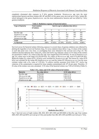
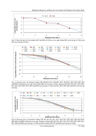
All the forty six bacterial isolates, comprising 4 genera, were screened for radiation resistance with 60
Co gamma
rays at ambient temperature (25 ± 20
C) in normal saline and percentage of bacteria survived at different doses of
gamma irradiation were calculated (Table-2). Among the 46 bacterial isolates, 45 (97.82%) isolates were
survived up to 5 kGy, 31 (67.39%) isolates were survived up to 6 kGy, 21 (45.65%) isolates were survived up to
7 kGy and 3 (6.52%) isolates were survived up to 8 kGy gamma irradiation. All the bacterial contaminants were](https://image.slidesharecdn.com/m0627984-150501063118-conversion-gate02/85/Radiation-Response-of-Bacteria-Associated-with-Human-Cancellous-Bone-2-320.jpg)
![Radiation Response of Bacteria Associated with Human Cancellous Bone
www.iosrjournals.org 83 | Page
IV. Discussion
Transmission of diseases or infections from the donor to the recipient is always a risk in allograft
transplantation. Many authors reported about infection following allograft implantation [7, 12, 16, and 17].
Wang et al. [18] reported that approximately one-half of the infections associated with human tissue transplants
were due to bacterial agents and of that, 90% were aerobic organisms. The best way to avoid infection or
diminish its incidence is careful donor selection and the application of sterile techniques in tissue procurement,
processing and storage. Varettas and Taylor [19] considered bioburden assessment as a small but integral part of
the allograft bone processing, which combine with other good tissue banking practices would provide safe
allograft bone for transplantation. Therefore, our focus was on the assessment of bacterial load associated with
unprocessed human bone and determination of radiation response of the isolates.
All the bone samples used in this experiment were collected from seronegative (HIV, HBV, and
VDRL) donor and were procured under aseptic condition. In spite of following good tissue banking practice,
donor tissues were not sterile. Average bacterial count was 9.0×10²
cfu/g (ranged from 4.1×101
to 3.1×10³
cfu/g). Many authors [19-21] had reported different rate of contamination in bone tissues. In our study, most of
bone samples were contaminated with Staphylococcus spp. Streptococcus spp. was the second highest
contaminant. Ibrahim et al. [24] also reported coagulase negative staphylococci as major contaminant in bone
allografts and twelve of their bone allografts were contaminated with streptococci. In many studies, bacterial
contaminant in bone tissues was predominantly skin flora [19-23]. Though the isolates found in this study are
comparable with the previous reported studies, it was difficult to determine the source of contamination in these
tissue samples. Microbial contamination may arise from the donor at the time of retrieval, or from the
environment and the personnel during processing [25]. Deijkers et al. [26] divided contaminating organisms into
low and high pathogenicity. They considered organisms of low pathogenicity to be skin commensals and
organisms of high pathogenicity were thought to be originated from endogenous sources in the donor, which
more likely to cause infection in the recipient.
Generally, 25 kGy gamma irradiation is used for terminal sterilization of tissue allografts. As gamma
radiation adversely affects the mechanical and biological properties of bone allografts, some tissue bank prefer
lower dose without compromising a SAL of 10-6
. Baker et al. [27] has found that sterility (10-6
SAL) of tissue
allograft could be achieved using at least 9.2 kGy gamma radiation, and in another study 15 kGy gamma
radiation was substantiated as radiation sterilization dose (RSD) for frozen bone allografts [28]. Radiation dose
necessary to achieve SAL of 10-6
depends on the initial microbial contamination level and the radiation
resistance of the contaminant. To observe the radiation effects, bacterial isolates were exposed to different doses
of gamma radiation and D10 values was calculated in this study. We found that, D10 values of bacterial isolates
ranged from 0.57 to 1.2 kGy. Streptococcus spp. was the most radioresistant isolates (D10 value 0.93-1.2 kGy)
and three of the Streptococcus spp. survived up to 8 kGy. All the bacterial isolates were killed at 9 kGy.
In a sterilization process, the nature of microbiological death or reduction is described by an
exponential function. Therefore, the number of microorganisms which survive in a sterilization process can be
expressed in terms of probability. While the probability may be reduced to a very low number, it can never be
reduced to zero. For the sterilization of medical and pharmaceutical product, maximum probability of a single
nonsterile unit in 106
units is widely accepted. Therefore, if the bioburden on an article were one million, 12 log
reduction of the initial bioburden is required to achieve a 10–6
probability of a nonsterile unit. Though the initial
bioburden of the studied bone samples ranged from 4.1×101
to 3.1×10³
cfu/g, to ensure patients safety, 12 log
reduction was calculated. In this study, twelve log reduction value (12D value) of the most resistant isolate was
14.4 kGy. From these findings, it is clear that the standard sterilization dose of 25 kGy gamma radiation was
adequate for the sterilization of these tissue allografts and a lower dose might also be acceptable. To determine
lower radiation sterilization dose (RSD), further study may therefore be suggested using radioresistant reference
strain.
V. Conclusion
Microbial contamination in musculoskeletal tissue graft is a serious concern in rehabilitative surgery.
To minimize the chance of contamination, in addition to select seronegative donor, good tissue banking practice
should be implemented in tissue procurement, processing and storage. However, aseptic processing practices
can reduce but not eliminate all microbial contaminants of tissue. To prevent the risk of diseases transmission
from donor origin, terminal sterilization is an integral part of allograft preparation. The choice of RSD depends
on the microbial contamination level and the radiation response of the contaminant. In our study, average
bioburden was 9.0×10²
cfu/g and maximum radiation resistance (D10 value) was 1.20 kGy and 12 D value of the
most resistant isolate was 14.4 kGy. Based on the average bioburden and radiation response of the contaminants,
25 kGy gamma radiation was found to be ample to ensure sterility of cancellous bone allografts.](https://image.slidesharecdn.com/m0627984-150501063118-conversion-gate02/85/Radiation-Response-of-Bacteria-Associated-with-Human-Cancellous-Bone-5-320.jpg)
![Radiation Response of Bacteria Associated with Human Cancellous Bone
www.iosrjournals.org 84 | Page
References
[1] W.W. Tomford, Transmission of disease through transplantation of musculoskeletal allografts, J Bone Joint Surg Am, 77, 1995,
1742–1754.
[2] D.P. Grogan, V. Kalen, K.J. Ross-Guidera, and L.I. Pugh, Use of allograft bone for posterior spinal fusion in idiopathic scoliosis,
Clin Orthop, 369, 1999, 273–278.
[3] D.M. Ehrler, and A.R. Vaccaro, The use of allograft bone in lumbar spine surgery, Clin Orthop, 371, 2000, 38–45.
[4] G.O. Phillips, Radiation technology in surgery and the pharmaceutical industry: an overview of applications, IAEA bulletin, 36,
1994, 19-23.
[5] N. Yusof, The use of gamma irradiation for sterilization of bone and amnion, Malaysian Journal of Nuclear Science, 12, 1994,
243-251.
[6] M.A. Kainer, J.V. Linden, D.N. Whaley, H.T. Holmes, W.R. Jarvis, D.B. Jernigan, and L.K.. Archibald, Clostridium infections
associated with musculoskeletal-tissue allografts, New Engl J Med, 350, 2004, 2564–2571.
[7] T. Eastlund, Bacterial infection transmitted by human tissue allograft transplantation, Cell Tissue Banking, 7(3), 2006, 147–166.
[8] H. Nguyen, and D.A.F. Morgan, Sterilization of allograft bone: is 25 kGy the gold standard for gamma irradiation?, Cell Tissue
Banking, 8, 2007, 81–91.
[9] C.R. Balsly, A.T. Cotter, L.A. Williams, B.D. Gaskins, M.A. Moore, and L. Wolfinbarger, Effect of low dose and moderate dose
gamma irradiation on the mechanical properties of bone and soft tissue allografts, Cell Tissue Banking, 9, 2008, 289–298.
[10] IAEA, Radiation sterilization of medical products, Proc. of an IAEA symposium, Bombay, 1974, 139-187.
[11] R.N. Mukherjee, Recommendation for the sterilization of medical products: Radiosterilization of Medical Products, Proc. of the
symposium of IAEA on ionizing radiation for sterilization of medical products and biological tissues, Bombay, India, 1974, 513-
523.
[12] M.R. Veen, R.M. Bloem, and P.L. Petit, Sensitivity and negative predictive value of swab cultures in musculoskeletal allograft
procurement, Clin Orthop Relat Res, 300, 1994, 259–263.
[13] S.B. Vehmeyer, R.M. Bloem, R.L. Deijkers, M.R. Veen, and P.L. Petit, A comparative study of blood and bone marrow cultures in
cadaveric bone donation, J Hosp Infect 43(4), 1999, 305–308.
[14] S.B. Vehmeyer, R.M. Bloem, and P.L. Petit, Microbiological screening of post-mortem bone donors—two case reports, J Hosp
Infect 47(3), 2001, 193–197.
[15] J.G. Holt, N.R. Krieg, H.A. Sneath, J.T. Staley, and S.T. William, Bergey’s Manual of Determinative Bacteriology 9th
edn.
(Baltimore: Williams and Wilkins, 1994)
[16] H.J. Mankin, F.J. Hornicek, and K.A. Raskin, Infection in massive bone allografts, Clin Ortho Relat Res, 432, 2005, 210–216.
[17] W. Tomford, J. Thongphasuk, H.J. Mankin, and M.J. Ferraro, Frozen musculoskeletal allografts: A study of the clinical incidence
and causes of infection associated with their use, J Bone Joint Surg (American Volume) 72(8), 1990, 1137–1143.
[18] S. Wang, C. Zinderman, R. Wise, and M. Braun, Infections and human tissue transplants: review of FDA MedWatch reports 2001–
2004, Cell Tissue Banking 8(3), 2007, 211–219.
[19] K. Varettas, and P. Taylor, Bioburden assessment of banked bone used for allografts, Cell Tissue Bank 12 (1), 2011, 37–43.
[20] S.F. Journeaux, N. Johnson, S.L. Bryce, S.J. Friedman, S.M.M. Sommerville, and D.A.F. Morgan, Bacterial contamination rates
during bone allograft retrieval, J Arthroplast, 14(6), 1999, 677–681.
[21] S.M.M. Sommerville, N. Johnson, S.L. Bryce, S.F. Journeaux, and D.A.F. Morgan, Contamination of banked femoral head
allograft: incidence, bacteriology and donor follow up, Aust NZJ Surg, 70, 2000, 480–484.
[22] S.B.W. Vehmeyer, R.M.S. Arnoud, R.M. Bloem, and P.L.C. Petit, Bacterial contamination of femoral head allografts from living
donors, Acta Orthop Scand, 73(2), 2002, 165–170.
[23] F. Judas, L. Teixeira, and A. Proenca, Coimbra university hospitals’ bone and tissue bank: twenty-two years of experience, Transpl
Proc, 37(6), 2005, 2799–2801.
[24] T. Ibrahim, H. Stafford, C.A.N. Esler, and R.A. Power, Cadaveric allograft microbiology. International Orthopaedics (SICOT), 28,
2004, 315–318.
[25] N. Yusof, Quality system for the radiation sterilization of tissue allografts, in G.O. Phillips, D.M. Strong, R. Von-Versen, and A.
Nather (Eds), Advances in Tissue banking, 3 (Singapore: World Scientific, 1999) 257-281.
[26] R.L.M. Deijkers, R.M. Bloem, P.L.C. Petit, R. Brand, S.B.W. Vehmeyer, and M.R. Veen, Contamination of bone allografts:
analysis of incidence and predisposing factors, J Bone Joint Surg Br, 79, 1997, 161–166.
[27] T.F. Baker, C.J. Ronholdt, and S. Bogdansky, Validating a low dose gamma irradiation process for sterilizing allografts using ISO
11137 method 2B, Cell Tissue Bank 6(4), 2005, 271-275.
[28] H. Nguyen, D.A. Morgan, L.I. Sly, M, Benkovich, S. Cull, and M.R. Forwood , Validation of 15 kGy as a radiation sterilization
dose for bone allografts manufactured at the Queensland Bone Bank: application of the VDmax 15 method, Cell Tissue Bank 9(2),
2008, 139-147.](https://image.slidesharecdn.com/m0627984-150501063118-conversion-gate02/85/Radiation-Response-of-Bacteria-Associated-with-Human-Cancellous-Bone-6-320.jpg)

The study assessed bacterial contamination in cancellous bones from live tissue donors in Bangladesh, revealing an average initial bioburden of 9.0×10² cfu/g, predominantly from Staphylococcus spp. and Streptococcus spp. Radiation resistance tests showed that all 46 bacterial isolates were eliminated at 9 kGy, with Streptococcus spp. demonstrating the highest resistance. The findings support the use of a standard radiation sterilization dose of 25 kGy for ensuring the microbiological safety of allografts.


![Polymer [ बहुलक ] Chemistry Notes PDF - Irfanullah Mehar - JJ Sir Chemistry.pdf](https://cdn.slidesharecdn.com/ss_thumbnails/polymerchemistrynotespdf-irfanullahmehar-jjsirchemistry-260210172118-3f9b37f7-thumbnail.jpg?width=640&height=640&fit=bounds)


