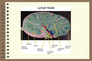
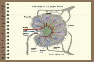
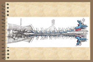
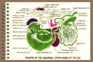
1
LYMPHATIC SYSTEM
Lymph: is the medium by which oxygen and
nutritive matter are transferred from the blood to
the body tissues and waste products are removed
Lymph fluid is similar to the plasma of the blood
but it is thinner, more watery and poorer in
protein which cannot pass readily through the
capillary wall
2
 The lymphatic begin as a network of very fine lymph
capillaries with blind extremities.
 The lymphatic capillaries are similar to veins but are
finer and have more valves. Even the largest
lymphatic vessels are very small and are difficult to
see with the naked eye. When filled with fluid (the
lymph) they have a characteristic beaded appearance
3
 all lymph vessels discharge their contents into lymph
nodes, with rear exceptions, all lymph throughout the
system passes through at least one lymph node before
it returns into the blood circulatory system.
 In every case the direction of flow of lymph in an
organ is from the center of the organ towards its
surface. The lymph nodes play an important part in
filtering out bacteria, foreign substances etc.
 Lymphatic vessels that convey lymph to a lymph node
are known as afferent lymphatics and the area
drained by the particular lymph node is known as its
drainage area
4
5
6
7
 After passing through one or more lymph nodes,
where some impurities are removed, lymph is carried
by efferent lymphatics to charge into larger lymph-
collecting vessels, which all flow towards the heart.
The largest of those is the thoracic duct, which
commences as a thin-walled dilatation about 19 mm
in width and known as receptaculum chyli (cisterna-chyli).
 This dilatation is situated in the abdomen, lying above
the aorta, and receives lymph from the lumbar and
intestinal trunks; it is the main receptacle for lymph
from the posterior part of the body, which opens into
the anterior vena cava
8
9
10
11
 Lymph from the anterior part of the body is carried
towards the heart by two tracheal lymph ducts, which
commence at the lateral retropharyneal lymph nodes
and pass down the neck on each side of the trachea
and esophagus; each duct discharges into the jugular
vein of its own side
The cavities of the serous membranes i.e. the pleura and
peritoneum are part of the lymphatic system. They
are moistened with lymph and are in communication
with lymphatic vessels.
The spleen is also part of the lymphatic system
12
13
The response of a lymph node to an irritant is
normally rapid, with enlargement, and
congestion and possibly tissue breakdown, thus
the size, colour and consistency of lymph nodes
form a valuable guide in the estimation of disease
processes in the animal body.
14
2) Lymph nodes
a- Size and shape: The size of lymph nodes varies from
that of a pinhead to that of a walnut, though the
posterior mediastinal lymph node of the ox may reach
a length of 20 cm.
 In the ruminant they are large and few in number.
The size is relatively greater in the young growing
animal than in the adult.
 In pigs: lymph nodes are lobulated.
 In camel: the superficial lymph nodes (e.g.
prepectoral, superficial inguinal) are superficially
lobated.
15
16
 b- Colour:
 The colour of lymph nodes shows considerable
variation, and may be white, grayish-blue or black.
 The mesenteric lymph nodes of cattle are always
black.
 In pig, the lymph nodes are lobulated and almost
white, except those of the head and neck, which are
reddish.
 The green colouration of lymph nodes may be due to
eosinophilia, which caused by parasitic invasion of the
drainage areas.
 The yellow colouration may be due to fatty
infiltration.
 The black or grey colouration may be due to
anthracosis in the bronchial lymph nodes.
17
18
 c- Consistency:
The consistency of lymph nodes varies in
different parts of the body. The nodes of the
abdomen being generally softer than those of
the thorax.
A physiological edema of the supramammary
and iliac lymph nodes usually encountered in
the lactating animals.
3) Functions of lymph nodes:
The lymph nodes have two functions:
1- The supply of fresh leucocytes and
2- The filtration of the lymph.
19
20
4) Haemo-lymph nodes (Haemal lymph nodes):
 These are deep red or almost black in colour, oval in
shape and up to the size of a pea, but differ from
lymph nodes in
- “accessory spleens”.
 Haemo-lymph nodes are numerous in cattle and
sheep but not found in the horse and pig.
21
In cattle, they occur especially along the course of the
aorta and in the subcutaneous fat.
In sheep and lambs, they are commonest beneath the
peritoneum sublumbar region.
they are being larger and more numerous in animals
suffering from anaemia.
5) Carcass lymph nodes:
Are those deeply seated and left attached to the carcass
after dressing. Therefore they comprise all lymph nodes
of the body with the exception of these of the head and
those removed with the thoracic and abdominal organs
during evisceration.
22
23
Carcass lymph nodes may drain:-
1-) Muscle exclusively (meat lymph nodes) as popliteal, axillary
and prescapular.
2-)Organ exclusively as the renal lymph nodes.
3-)Both muscle and organs as iliac, ischiatic and lumbar.
4-)skin exclusively: as in case of precrural (prefemoral).
6) Meat lymph nodes:
Receive lymph from muscle, tendons, skeleton and
joints.They are the only lymph nodes used in
bacteriological examination of carcasses in suspicious
cases.
In cattle: they are the prescapular, axillary precrural
(prefemoral, popliteal and ischiatic.
In pig: they are only popliteal and precrural nodes.
24
25
* Lymph nodes of camel
Generally, lymph nodes of camel are the same as
those of cattle, with some variations, that lymph
nodes which are usually present in cattle are
absent in camel such as percrural (prefemoral),
deep inguinal, anterior sternal, ventral mediastinal,
medial sacral and apical lymph node.
In camel, the superficial nodes are lobulated
surperficially.
26

Lympatic system (2).ppt

  • 1.
    1 LYMPHATIC SYSTEM Lymph: isthe medium by which oxygen and nutritive matter are transferred from the blood to the body tissues and waste products are removed Lymph fluid is similar to the plasma of the blood but it is thinner, more watery and poorer in protein which cannot pass readily through the capillary wall
  • 2.
    2  The lymphaticbegin as a network of very fine lymph capillaries with blind extremities.  The lymphatic capillaries are similar to veins but are finer and have more valves. Even the largest lymphatic vessels are very small and are difficult to see with the naked eye. When filled with fluid (the lymph) they have a characteristic beaded appearance
  • 3.
    3  all lymphvessels discharge their contents into lymph nodes, with rear exceptions, all lymph throughout the system passes through at least one lymph node before it returns into the blood circulatory system.  In every case the direction of flow of lymph in an organ is from the center of the organ towards its surface. The lymph nodes play an important part in filtering out bacteria, foreign substances etc.
  • 4.
     Lymphatic vesselsthat convey lymph to a lymph node are known as afferent lymphatics and the area drained by the particular lymph node is known as its drainage area 4
  • 5.
  • 6.
  • 7.
    7  After passingthrough one or more lymph nodes, where some impurities are removed, lymph is carried by efferent lymphatics to charge into larger lymph- collecting vessels, which all flow towards the heart. The largest of those is the thoracic duct, which commences as a thin-walled dilatation about 19 mm in width and known as receptaculum chyli (cisterna-chyli).
  • 8.
     This dilatationis situated in the abdomen, lying above the aorta, and receives lymph from the lumbar and intestinal trunks; it is the main receptacle for lymph from the posterior part of the body, which opens into the anterior vena cava 8
  • 9.
  • 10.
  • 11.
    11  Lymph fromthe anterior part of the body is carried towards the heart by two tracheal lymph ducts, which commence at the lateral retropharyneal lymph nodes and pass down the neck on each side of the trachea and esophagus; each duct discharges into the jugular vein of its own side
  • 12.
    The cavities ofthe serous membranes i.e. the pleura and peritoneum are part of the lymphatic system. They are moistened with lymph and are in communication with lymphatic vessels. The spleen is also part of the lymphatic system 12
  • 13.
    13 The response ofa lymph node to an irritant is normally rapid, with enlargement, and congestion and possibly tissue breakdown, thus the size, colour and consistency of lymph nodes form a valuable guide in the estimation of disease processes in the animal body.
  • 14.
    14 2) Lymph nodes a-Size and shape: The size of lymph nodes varies from that of a pinhead to that of a walnut, though the posterior mediastinal lymph node of the ox may reach a length of 20 cm.  In the ruminant they are large and few in number. The size is relatively greater in the young growing animal than in the adult.
  • 15.
     In pigs:lymph nodes are lobulated.  In camel: the superficial lymph nodes (e.g. prepectoral, superficial inguinal) are superficially lobated. 15
  • 16.
    16  b- Colour: The colour of lymph nodes shows considerable variation, and may be white, grayish-blue or black.  The mesenteric lymph nodes of cattle are always black.  In pig, the lymph nodes are lobulated and almost white, except those of the head and neck, which are reddish.
  • 17.
     The greencolouration of lymph nodes may be due to eosinophilia, which caused by parasitic invasion of the drainage areas.  The yellow colouration may be due to fatty infiltration.  The black or grey colouration may be due to anthracosis in the bronchial lymph nodes. 17
  • 18.
    18  c- Consistency: Theconsistency of lymph nodes varies in different parts of the body. The nodes of the abdomen being generally softer than those of the thorax. A physiological edema of the supramammary and iliac lymph nodes usually encountered in the lactating animals.
  • 19.
    3) Functions oflymph nodes: The lymph nodes have two functions: 1- The supply of fresh leucocytes and 2- The filtration of the lymph. 19
  • 20.
    20 4) Haemo-lymph nodes(Haemal lymph nodes):  These are deep red or almost black in colour, oval in shape and up to the size of a pea, but differ from lymph nodes in - “accessory spleens”.  Haemo-lymph nodes are numerous in cattle and sheep but not found in the horse and pig.
  • 21.
    21 In cattle, theyoccur especially along the course of the aorta and in the subcutaneous fat. In sheep and lambs, they are commonest beneath the peritoneum sublumbar region. they are being larger and more numerous in animals suffering from anaemia.
  • 22.
    5) Carcass lymphnodes: Are those deeply seated and left attached to the carcass after dressing. Therefore they comprise all lymph nodes of the body with the exception of these of the head and those removed with the thoracic and abdominal organs during evisceration. 22
  • 23.
    23 Carcass lymph nodesmay drain:- 1-) Muscle exclusively (meat lymph nodes) as popliteal, axillary and prescapular. 2-)Organ exclusively as the renal lymph nodes. 3-)Both muscle and organs as iliac, ischiatic and lumbar. 4-)skin exclusively: as in case of precrural (prefemoral).
  • 24.
    6) Meat lymphnodes: Receive lymph from muscle, tendons, skeleton and joints.They are the only lymph nodes used in bacteriological examination of carcasses in suspicious cases. In cattle: they are the prescapular, axillary precrural (prefemoral, popliteal and ischiatic. In pig: they are only popliteal and precrural nodes. 24
  • 25.
    25 * Lymph nodesof camel Generally, lymph nodes of camel are the same as those of cattle, with some variations, that lymph nodes which are usually present in cattle are absent in camel such as percrural (prefemoral), deep inguinal, anterior sternal, ventral mediastinal, medial sacral and apical lymph node. In camel, the superficial nodes are lobulated surperficially.
  • 26.

Editor's Notes