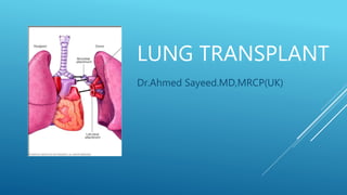
Lun g transplant indications and complications
•Download as PPTX, PDF•
4 likes•920 views

This document discusses lung transplantation, including indications, contraindications, post-transplant survival factors, and risks. It outlines criteria for different lung diseases warranting transplantation. Post-transplant risks include rejection, infection, including bacterial, viral and fungal pneumonia which are common due to immunosuppression. Factors like HLA mismatch, immunsuppression regimen, and vitamin D deficiency impact acute rejection risk. Close monitoring is required after transplantation to promptly treat potential complications.

Recommended
More Related Content
What's hot
What's hot (20)
Similar to Lun g transplant indications and complications
Similar to Lun g transplant indications and complications (20)
More from Dr Ahmed Sayeed
More from Dr Ahmed Sayeed (16)
Recently uploaded
Recently uploaded (20)
Lun g transplant indications and complications
- 2. ABSOLUTE CONTRAINDICATIONS Severe extra pulmonary organ dysfunctions Active cancer Severe psychiatric illness Infection like HIV Active or recent substance abuse
- 3. INDICATIONS FOR ADULT LUNG TRANSPLANTS BY YEAR
- 4. COPD BODE index >5 Hypertension or cor pulmonale FEv1 <20%
- 5. CYSTIC FIBROSIS Fev1 < 30% H/O ITU admission Respiratory failure/PAH Severe recurrent hemoptysis Refractory or recurrent pneumothorax
- 6. IPF 1.If Possible all patients 2.TLCO < 40% 3.Desaturation <88% on 6MWT 4.Honey combing on HRCT
- 7. IPAH NYHA class III Rapidly progressive disease 6MWT < 350m Mean Right atrial pressure >15 mmHg Cardiac index <2 L/M2
- 8. PART 1 POST TRANSPLANT SURVIVAL MEASURE: 1. FVC (Group B, D) 2. PCW pressure ≥20 (Group D) 3. Continuous mechanical ventilation 4. Age 5. Serum creatinine 6. Functional Status (NYHA class) 7. Diagnosis
- 9. PART 2 WAITING LIST URGENCY MEASURE: 1. Forced vital capacity (FVC) 2. Pulmonary artery systolic pressure (Group A, C, D)* 3. Pulmonary artery mean pressure 4. Pulmonary capillary wedge pressure 5. Supplemental O2 required at rest (Group A, C, D)* 6. Age 7. Body mass index (BMI) 8. Diabetes 9. Functional status 10. Six-minute walk distance 11. Continuous mechanical ventilation 12. Diagnosis 13. pCO2
- 10. CLASSIFICATION OF TRANSPLANT CANDIDATES Group A: COPD, alpha-1-antitrypsin, emphysema, lymphangioleiomyomatosis, bronchiectasis, sarcoidosis with a mean PA pressure ≤30 mmHg. Group B: Pulmonary hypertension (IPAH, Eisenmenger's syndrome) Group C: Cystic fibrosis, immunodeficiency disorders . Group D: IPF, other causes of pulmonary fibrosis, sarcoidosis with PA >30 mmHg, obliterative bronchiolitis (non-retransplant).
- 11. EVALUATION AND TREATMENT OF ACUTE LUNG TRANSPLANT REJECTION ●Acute cellular rejection ●Humoral rejection ●Hyperacute rejection ●Chronic lung transplant rejection
- 12. RISK FACTORS FOR ACUTE REJECTION ●Human leukocyte antigens (HLA) mismatching ●Genetic factors ●Immunosuppression regimen ●Age ●Vitamin D deficiency
- 14. BACTERIAL INFECTIONS FOLLOWING LUNG TRANSPLANTATION
- 15. BACTERIAL INFECTIONS FOLLOWING LUNG TRANSPLANTATION The high level of immunosuppression required to prevent rejection ●Adverse effects of transplantation on local pulmonary host defenses (loss of lymphatics, reduced mucociliary clearance, decreased cough) ●Constant environmental contact allowing pathogens direct access into the allograft RISK OF INFECTION
- 16. BACTERIAL INFECTIONS FOLLOWING LUNG TRANSPLANTATION Pneumonia, particularly bacterial pneumonia, is the most common type of infection in lung transplant recipients, Bloodstream, Pleural space, and Wound infections are also common
- 17. VIRAL INFECTIONS CMV pneumonitis Increases risk of bacterial and fungal superinfection Ganciclovir prohylaxis is protective
- 18. FUNGAL INFECTION Aspergillus colonization Peak after 2 month
- 19. REFERENCES Uptodate Oxford text book of respiratory medicine
- 20. THANK YOU
Editor's Notes
- ILD: interstitial lung disease; IIP: idiopathic interstitial pneumonia; CF: cystic fibrosis; A1ATD: alpha-1 antitrypsin deficiency emphysema; COPD: chronic obstructive pulmonary disease. Reproduced from: The Registry of the International Society for Heart and Lung Transplantation: Thirty-Second Annual Report. J Heart Lung Transplant 2017; 36:1037. Illustration used with the permission of Elsevier, Inc. All rights reserved. For additional and updated information, please see the International Society for Heart and Lung Transplantation slide set "Adult Lung Transplantation Statistics," at: https://www.ishlt.org/registries/slides.asp?slides=heartLungRegistry. Graphic 61485 Version 3.0
- INTRODUCTION — Acute allograft rejection is a significant problem in lung transplantation. Despite advances in induction immunosuppression and use of aggressive maintenance immunosuppression, more than a third of lung transplant recipients are treated for acute rejection in the first year after transplant [1-3]. Acute rejection is responsible for approximately 4 percent of deaths in the first 30 days following transplantation [2,3]. The clinical manifestations, evaluation, and treatment of acute cellular lung transplant rejection and the role of routine monitoring for rejection will be reviewed here. The immunobiology of transplantation, induction and maintenance immunosuppression after lung transplantation, humoral rejection, and chronic lung transplant rejection are discussed separately. (See "Transplantation immunobiology" and "Induction immunosuppression following lung transplantation" and "Maintenance immunosuppression following lung transplantation" and "Evaluation and treatment of antibody-mediated lung transplant rejection" and "Chronic lung transplant rejection: Bronchiolitis obliterans".) DEFINITIONS ●Acute cellular rejection – Acute cellular rejection is the predominant type of acute lung transplant rejection and is mediated by T lymphocyte recognition of foreign major histocompatibility complexes (MHC), also known as human leukocyte antigens (HLA) in humans, or other antigens [1,4,5]. ●Humoral rejection – Humoral rejection, which is less common than acute cellular rejection, is mediated by antibodies directed against donor HLA or other epitopes. These antibodies may have been present in the recipient at a low level prior to transplant or may develop afterwards. Generally, if HLA antibodies are identified in the potential recipient, the corresponding HLA antigens are avoided in a donor (so-called virtual cross-match). ●Hyperacute rejection is a form of humoral rejection that occurs in the first 24 hours following lung transplantation in recipients who have pre-formed anti-HLA antibodies. With improved sensitivity of HLA antibody testing, hyperacute rejection now rarely occurs. ●Chronic lung transplant rejection is manifest pathologically as dense fibrous scar tissue affecting the small airways. Clinically, chronic lung transplant rejection is known as bronchiolitis obliterans syndrome (BOS), which manifests as progressive airway obstruction that is defined as a progressive decline in forced expiratory volume in one second (FEV1). In addition, a form of restrictive Chronic Lung Allograft Dysfunction (rCLAD) has been described and appears to have significant prognostic implications [6]. Less commonly, chronic vascular rejection with atherosclerosis in the pulmonary vasculature is also present.
- RISK FACTORS FOR ACUTE REJECTION — The risk of acute lung transplant rejection is greatest in the first few months after transplant and decreases with time. Several factors have been implicated as contributing to the development of acute cellular rejection. The potential factors include: ●Human leukocyte antigens (HLA) mismatching – An increasing degree of HLA mismatch between the donor and recipient increases the risk of acute cellular rejection [2]. However, mismatch at certain HLA loci may be more important than at others. ●Genetic factors – Genetic variants in interleukin (IL)-10, multidrug resistance genotype, CCL4L chemokine, and toll-like receptor 4(TLR4) may influence the risk of acute rejection [16-19]. ●Immunosuppression regimen – In the International Society for Heart and Lung Transplantation (ISHLT) registry, the rate of acute rejection in the first year was highest among recipients on cyclosporine based regimens and lowest among those on tacrolimus based regimens [20]. In addition, induction therapy with an interleukin-2R antagonist was associated with a lower rate of acute rejection than other induction regimens. (See "Maintenance immunosuppression following lung transplantation", section on 'Calcineurin inhibitors' and "Induction immunosuppression following lung transplantation", section on 'Induction agents'.) ●Age – More rejection episodes were reported among recipients in the lowest age category (18 to 34 years) than in older age categories, although this data from the ISHLT registry was not adjusted for underlying disease or other potentially confounding factors [21]. ●Vitamin D deficiency − In a cohort of 102 lung transplant recipients, episodes of acute cellular rejection were more frequent among those with deficiency (<30 ng/mL) in 25-hydroxyvitamin D (25OHD) near the time of transplantation, than among those with normal 25OHD levels (incidence rate ratio 2.43, 95% CI 1.30 to 4.52) [22].
- CLINICAL MANIFESTATIONS — Acute cellular rejection is most likely to occur in the first six months following lung transplantation [23,24]. Often, patients are asymptomatic, and the diagnosis is made from surveillance transbronchial biopsies. When present, symptoms are nonspecific and are shared by other common complications that occur during this period. They include low-grade fever, shortness of breath, and cough with or without sputum production (table 2) [1,25]. During long-term follow-up of 120 lung transplant recipients, shortness of breath and cough were more common in those with clinically significant acute rejection (grade ≥A2) than those without (grade A0 or A1) (table 1), but comparable in frequency to those with pulmonary infection. For predicting grade >A2 rejection, the sensitivity and specificity were 68 and 50 percent for cough and 63 and 68 percent for shortness of breath, respectively. Lung examination may reveal clear lung fields, crackles, or decreased breath sounds when a pleural effusion is present as part of the acute rejection. High grade rejection may be associated with respiratory distress [25].
- Bacterial infections following lung transplantation Authors:Omar Mohamedaly, MDAimee Zaas, MD, MHSCameron Wolfe, MBBS (Hons)Scott M Palmer, MD, MHSSection Editors:Elbert P Trulock, MDKieren A Marr, MDDeputy Editor:Sheila Bond, MD Contributor Disclosures All topics are updated as new evidence becomes available and our peer review process is complete. Literature review current through: Feb 2018. | This topic last updated: Jan 11, 2017. INTRODUCTION — Lung transplantation is an effective treatment for a wide range of advanced lung diseases. While the survival of lung transplant recipients continues to improve, outcomes after lung transplantation remain inferior to other types of solid organ transplantation [1,2]. Infectious complications contribute substantially to morbidity and mortality following lung transplantation and account for over 25 percent of all posttransplant deaths [2]. This topic reviews the most common bacterial infections in lung transplant recipients. Infections caused by fungi, mycobacteria, and viruses in lung transplant recipients and general issues regarding infections in solid organ transplant recipients are discussed separately. (See "Fungal infections following lung transplantation" and "Tuberculosis in solid organ transplant candidates and recipients" and "Nontuberculous mycobacterial infections in solid organ transplant candidates and recipients" and "Prevention of cytomegalovirus infection in lung transplant recipients" and "Clinical manifestations, diagnosis, and treatment of cytomegalovirus infection in lung transplant recipients" and "Infection in the solid organ transplant recipient" and "Evaluation for infection before solid organ transplantation" and "Prophylaxis of infections in solid organ transplantation" and "Immunizations in solid organ transplant candidates and recipients".) RISK OF INFECTION — Lung transplant recipients are at increased risk for infectious complications due to the following factors: ●The high level of immunosuppression required to prevent rejection ●Adverse effects of transplantation on local pulmonary host defenses (loss of lymphatics, reduced mucociliary clearance, decreased cough) ●Constant environmental contact allowing pathogens direct access into the allograft The likelihood and type of infection varies with the degree of host immunosuppression, timing since transplantation, nature and duration of antimicrobial prophylaxis, and local hospital and regional microbiology. Pneumonia, particularly bacterial pneumonia, is the most common type of infection in lung transplant recipients, although bloodstream, pleural space, and wound infections are also common [3,4]. Bacterial, viral, fungal, and mycobacterial infections all occur at an increased frequency after lung transplantation. (See "Fungal infections following lung transplantation" and "Prevention of cytomegalovirus infection in lung transplant recipients" and "Tuberculosis in solid organ transplant candidates and recipients" and "Nontuberculous mycobacterial infections in solid organ transplant candidates and recipients".) In addition to the direct morbidity and mortality caused by infectious complications, they may also lead to loss of allograft function and contribute to the development of bronchiolitis obliterans syndrome [5,6]. (See "Infection in the solid organ transplant recipient" and "Chronic lung transplant rejection: Bronchiolitis obliterans".) TIMELINE OF INFECTION — Infections generally occur in a predictable pattern following solid organ transplantation. The posttransplant period has been divided into three stages: the first month post-transplant, one to six months post-transplant, and more than six months post-transplant. ●During the early posttransplant period, bacterial infections predominate. During the first month, there are two major causes of infection: •Infection derived from either the donor or recipient •Infectious complications of the transplant surgery and hospitalization ●From one to six months post-transplant, patients are most at risk for opportunistic infections, although residual problems from the perioperative period can persist. ●More than six months post-transplant, most patients are receiving stable and reduced levels of immunosuppression. These patients are subject to pneumonia caused by an extended range of bacteria, including pneumococcus, gram-negative bacilli, Legionella, and other common pathogens rather than opportunistic infections. The pattern of infection seen during these periods is discussed in greater detail separately. (See "Infection in the solid organ transplant recipient", section on 'Timing of infection posttransplantation'.)
- Bacterial infections following lung transplantation Authors:Omar Mohamedaly, MDAimee Zaas, MD, MHSCameron Wolfe, MBBS (Hons)Scott M Palmer, MD, MHSSection Editors:Elbert P Trulock, MDKieren A Marr, MDDeputy Editor:Sheila Bond, MD Contributor Disclosures All topics are updated as new evidence becomes available and our peer review process is complete. Literature review current through: Feb 2018. | This topic last updated: Jan 11, 2017. INTRODUCTION — Lung transplantation is an effective treatment for a wide range of advanced lung diseases. While the survival of lung transplant recipients continues to improve, outcomes after lung transplantation remain inferior to other types of solid organ transplantation [1,2]. Infectious complications contribute substantially to morbidity and mortality following lung transplantation and account for over 25 percent of all posttransplant deaths [2]. This topic reviews the most common bacterial infections in lung transplant recipients. Infections caused by fungi, mycobacteria, and viruses in lung transplant recipients and general issues regarding infections in solid organ transplant recipients are discussed separately. (See "Fungal infections following lung transplantation" and "Tuberculosis in solid organ transplant candidates and recipients" and "Nontuberculous mycobacterial infections in solid organ transplant candidates and recipients" and "Prevention of cytomegalovirus infection in lung transplant recipients" and "Clinical manifestations, diagnosis, and treatment of cytomegalovirus infection in lung transplant recipients" and "Infection in the solid organ transplant recipient" and "Evaluation for infection before solid organ transplantation" and "Prophylaxis of infections in solid organ transplantation" and "Immunizations in solid organ transplant candidates and recipients".) RISK OF INFECTION — Lung transplant recipients are at increased risk for infectious complications due to the following factors: ●The high level of immunosuppression required to prevent rejection ●Adverse effects of transplantation on local pulmonary host defenses (loss of lymphatics, reduced mucociliary clearance, decreased cough) ●Constant environmental contact allowing pathogens direct access into the allograft The likelihood and type of infection varies with the degree of host immunosuppression, timing since transplantation, nature and duration of antimicrobial prophylaxis, and local hospital and regional microbiology. Pneumonia, particularly bacterial pneumonia, is the most common type of infection in lung transplant recipients, although bloodstream, pleural space, and wound infections are also common [3,4]. Bacterial, viral, fungal, and mycobacterial infections all occur at an increased frequency after lung transplantation. (See "Fungal infections following lung transplantation" and "Prevention of cytomegalovirus infection in lung transplant recipients" and "Tuberculosis in solid organ transplant candidates and recipients" and "Nontuberculous mycobacterial infections in solid organ transplant candidates and recipients".) In addition to the direct morbidity and mortality caused by infectious complications, they may also lead to loss of allograft function and contribute to the development of bronchiolitis obliterans syndrome [5,6]. (See "Infection in the solid organ transplant recipient" and "Chronic lung transplant rejection: Bronchiolitis obliterans".) TIMELINE OF INFECTION — Infections generally occur in a predictable pattern following solid organ transplantation. The posttransplant period has been divided into three stages: the first month post-transplant, one to six months post-transplant, and more than six months post-transplant. ●During the early posttransplant period, bacterial infections predominate. During the first month, there are two major causes of infection: •Infection derived from either the donor or recipient •Infectious complications of the transplant surgery and hospitalization ●From one to six months post-transplant, patients are most at risk for opportunistic infections, although residual problems from the perioperative period can persist. ●More than six months post-transplant, most patients are receiving stable and reduced levels of immunosuppression. These patients are subject to pneumonia caused by an extended range of bacteria, including pneumococcus, gram-negative bacilli, Legionella, and other common pathogens rather than opportunistic infections. The pattern of infection seen during these periods is discussed in greater detail separately. (See "Infection in the solid organ transplant recipient", section on 'Timing of infection posttransplantation'.)