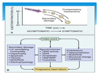
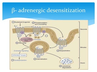
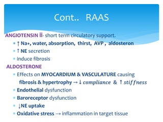
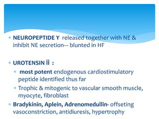
PATHOPHYSIOLOGICAL BASIS OF
HEMODYNAMICS OF LOW OUTPUT HEART
FAILURE
Aniruddha Mandal
Chair person
Dr. Dipankar Ghosh
Dastidar
 Heart failure (HF) is a clinical syndrome that
occurs in patients who, because of an
inherited or acquired abnormality of cardiac
structure and/or function, develop a
constellation of clinical symptoms (dyspnea
and fatigue) and signs (edema and rales) that
lead to frequent hospitalizations, a poor
quality of life, and a shortened life expectancy
DEFINITION
 CARDIAC OUTPUT ( C O.): Quantity of blood
pumped into the aorta each minute by the
heart
STROKE VOLUME × HEART RATE
 DEPENDS DIRECTLY ON :
(1) body metabolism
(2) exercise
(3) age
(4) Body surface area.
 CARDIAC INDEX : C O. / BODY SURFACE AREA
 Cardiac Output =
Arterial Pressure / Total
Peripheral Resistance
 Normally resistence is sum of
Blood Flow Regulation in All the
Local Tissues
 Two primary factors in cardiac
output regulation:
(1)cardiac pumping capacity
(2)venous return
CARDIAC FACTOR CONTROL OF
CARDIAC OUTPUT
HYPOEFFECTIVE HEART
Myocardial ischemia & infarction
 Myocarditis
 Cardiac metabolic derangement
 Cardiac tamponade
Arrythmia
Severe valvular heart disease
Congenital heart disease
 DETERMINANT OF VENOUS
RETURN
 Mean systemic filling
pressure (psf) --Degree of
filling of the systemic
circulation
 Right atrial pressure----
backward force
 Resistance to blood flow
VENOUS RETURN – DETERMINATION
OF CO.
INCREASED IN
 ↑ BLOOD VOLUME
 ↑ SYMPATHETIC
ACTIVITY DUE TO
CONSTRICTION OF
 Capacitance vessels
 Pulomonary vessels
 Heart chambers
 arteriole
MEAN SYSTEMIC FILLING
PRESSSURE
VENOUS RETURN = CARDIAC OUTPUT
NERVOUS & REFLEX CONTROL OF
OUTPUT
Sympathetic stimulation→effect 0n
heart
↑Na+ ,H2O
absorption-
kidney
↓ renal
blood
flow
↑AVP,RAS
↑ 𝑝𝑠𝑓.
Immediate activation of REFLEXEES
Baro Chemo
Brain
ischemia
Heart
damage
Heart suddenly severely damaged
↓ CO.
Damming of blood in
vein
Spectrum of acute heart failure
Reflex get blunted in 2-3 days.
SEMICHRONIC STAGE ensues→
Kidney
fluid
retention
↑ psf
Distended
vein→
↓ venous
resistence
Progressive recovery of
heart
Reperfusin, compensatory
hypertrophy,collateral,
Acute heart
failure
compensated
With
Sympathetic
support
After someday
symp. Support
not needed
Decompensated
Massive myocyte loss,
Overstretching,
Heart muscle edema,
PROGRESSION
 CARDIORENAL MODEL- excessive salt and water
retention caused by abnormalities of renal blood flow
 CARDIOCIRCULATORY OR HEMODYNAMIC MODEL-
abnormal pumping capacity of the heart
- not adequately explain relentleess progress
 PROGRESSIVE MODEL - primary determinant
 neurohumoral activation
 left ventricular remodeling
CHRONIC HEART FAILURE AS A
PROGRESSIVEMODEL
Activation of the Sympathetic (Adrenergic)
Nervous System
 Increased circulating Norepinephrine (NE)-2-3 times
 Heart extracts NE from the arterial blood & also
synthesized in myocardium.
 With progression cardiac depletion of NE-“exhaustion”
phenomenon
↓ myocardial tyrosine hydrxylase
 ↓ NE uptake
NEUROHUMORAL MECHANISM
β- adrenergic desensitization
 Activated comparatively later by
1. Renal hypoperfusion
2. ↓ Na delivery to macula densa
3. Sympathetic stimulation
 Angiotensin receptor- G protein coupled -2 types
 AT1 –vasoconstriction, cell growth, aldosterone and
catecholamine release-
 AT2 –vasodilation, inhibition of cell growth, natriuresis,
and bradykinin release-
Activation of the Renin-Angiotensin System
s
ANGIOTENSIN ĪĪ- short term circulatory support.
 ↑ Na+, water, absorption, thirst, AVP , aldosteron
 ↑ NE secretion
 induce fibrosis
ALDOSTERONE
 Effects on MYOCARDIUM & VASCULATURE causing
fibrosis & hypertrophy → ↓ 𝑐𝑜𝑚𝑝𝑙𝑖𝑎𝑛𝑐𝑒 & ↑ 𝑠𝑡𝑖𝑓𝑓𝑛𝑒𝑠𝑠
 Endothelial dysfunction
 Baroreceptor dysfunction
 ↓NE uptake
 Oxidative stress → inflammation in target tissue
Cont.. RAAS
 REACTIVE O2 SPECIES (ROS) ACTIVITY ↑ due to :
 Mechanical strain
 Neurohormone
 Inflammatory cytokine
 ↓ NOS activity
 EFFECT :
 Hypertrophy
 Reexpression of fetal gene programme
 Fibroblast proliferration→↑ collagen, MMP
 ↓ bioavailability of NO in peripheral vasculature
OXIDATIVE STRESS(ROS)
Neurohormonal Alterations of Renal
Function
 VASODILATTORY PROSTAGLANDIN: PGE2, PGI2
 NO, bradykinin, adrenomedullin, apelin
 NATRIURETIC PEPTIDES : ANP, BNP, CNP, DNP,
urodilantin
 Renal effects become blunted with advancing HF
COUNTER REGULATORY NEUROHORMONE
 ANP secreted in short burst in ACUTE changes
 BNP regulated transcriptionally as CHRONIC response
 PROHORMON cleaved to
 large biologically inactive N-terminal fragments (NT-ANP or NT-
BNP)
 smaller biologically active peptides (ANP or BNP)
 degraded by neutral endopeptidase
 Degraded by NEUTRAL ENDOPEPTIDASE & VASOPEPTIDASE
 Candoxatrilat endopeptidase inhibitor
 Omapatrilat inhibits both neutral endopeptidase and ACE
NATRIURETIC PEPTIDES
Neurohormonal Alterations in the
PERIPHERAL VASCULATURE
Secretion enhanced by
 Vasoactive agent (NE, angiotensin, thrombin)
 Cytokines
EFFECT
 Vasoconstriction
 cell proliferation
 pathologic hypertrophy
 Fibrosis
 Increased contractility
 ↑ Pulmonary artery pressure, resistence
ENDOTHELIN
 NEUROPEPTIDE Y released together with NE &
inhibit NE secretion--- blunted in HF
 UROTENSIN İİ :
 most potent endogenous cardiostimulatory
peptide identified thus far
 Trophic & mitogenic to vascular smooth muscle,
myocyte, fibroblast
 Bradykinin, Aplein, Adrenomedullin- offseting
vasoconstriction, antidiuresis, hypertrophy
Disrupted subcellular location
of NOS
 ↓ NOS3 in HF
 Nitroso redox imbalance–
unopposed activity of
xanthin oxidase (↓NOS1)
 Remodeling ↓ in NOS2
deficiency
NITRIC OXIDE
 TNF, PAI-1, TGF-β, resistin
and-
 Leptin : hypertension,
hypertrophy, ↑ in HF of
obese patient
 ADIPONECTIN ↓ infarct
size, apoptosis
it ↓ in hear failure
ADIPOKINES
INFLAMMATORY( IL-6, TNF) &
ANTIINFLAMMATORY(IL-10) IMBALANCE
 Traditionally described in
anatomical term
 BUT there is also
alteration in
A. Biology of cardiac
myocyte
B. Volume of myocyte &
nonmyocyte
component
C. Geometry &
architecture of
ventricular chamber
LEFT VENTRICULAR REMODELING
Alterations in the BIOLOGY
CARDIAC MYOCYTE HYPERTROPHY
Alterations in Excitation-Contraction
Coupling
↓ SERCA2
↓phospholamban
phosphorylation;
Leakage of
Ryanodine
receptor(hyperphs
phorylation)
phosphorylation&
dysfunction of L –
type Ca++ cnl.
Ca++ entry in
reverse mode by
Na+/Ca++
exchanger.
Slower delivery
of ca++ to
contractile
apparatus &
slow fall in
diastole;
change in
abundance/
phosphorylation
in regulatory
protein
Abnormal
prolongation
of A.P.
↓ force of
CONTRACTION
&
RELAXATION
Cont..
 Shift to fetal gene program – fetal isoform of myosin
heavy chain(MHC; α →β)
 ↓ Myofibrilar ATPase & Myosin ATPase
 Myocytolysis – proteolysis of myofilament
 Alteration in myofilament regulatory protein
 Altered activity of Myosin light chain ; troponin
tropomyosin complex
 CYTOSKELETAL PROTEIN (actin, desmin, dystrophin,
vinculin) altered expression
Abnormalities in Contractile and
Regulatory Proteins
 NECROSIS :
 Directly from ischemia, myocardial injury, toxin, infection
 From Neuroheumoral activation
 APOPTOSIS : induced by
 catecholamines acting through beta1-adrenergic receptor
 angiotensin II
 ROS, NO, inflammatory cytokines
 mechanical strain
 AUTOPHAGY:sequestering organelles and proteins in
a double-membrane vesicle inside the cell
(autophagosome) → subsequently delivered to the
lysosome for degradation
Alterations in the Myocardium in
Heart Failure
 Type I and type III collagen ensures
 Structural integrity of adjoining myocytes
 Interaction of collagen and integrins with the
cytoskeletal proteins --maintainin alignment of
myofibrils
 Phenotypic conversion to myofibroblast
 ↑ collagen synthesis & ↑ MMP → ↑ Turnover
 Replacement fibrosis
FIBROBLAST
Alterations in the Left Ventricular
Structure & Geometry in Heart Failure
EXTERNAL PRESSURE & CO.
HEART
FAILURE
WITH
PRESERVED
EJECTION
FRACTION
Thank you

Lowoutput cardiac failure

  • 1.
    PATHOPHYSIOLOGICAL BASIS OF HEMODYNAMICSOF LOW OUTPUT HEART FAILURE Aniruddha Mandal Chair person Dr. Dipankar Ghosh Dastidar
  • 2.
     Heart failure(HF) is a clinical syndrome that occurs in patients who, because of an inherited or acquired abnormality of cardiac structure and/or function, develop a constellation of clinical symptoms (dyspnea and fatigue) and signs (edema and rales) that lead to frequent hospitalizations, a poor quality of life, and a shortened life expectancy DEFINITION
  • 3.
     CARDIAC OUTPUT( C O.): Quantity of blood pumped into the aorta each minute by the heart STROKE VOLUME × HEART RATE  DEPENDS DIRECTLY ON : (1) body metabolism (2) exercise (3) age (4) Body surface area.  CARDIAC INDEX : C O. / BODY SURFACE AREA
  • 4.
     Cardiac Output= Arterial Pressure / Total Peripheral Resistance  Normally resistence is sum of Blood Flow Regulation in All the Local Tissues  Two primary factors in cardiac output regulation: (1)cardiac pumping capacity (2)venous return
  • 5.
    CARDIAC FACTOR CONTROLOF CARDIAC OUTPUT HYPOEFFECTIVE HEART Myocardial ischemia & infarction  Myocarditis  Cardiac metabolic derangement  Cardiac tamponade Arrythmia Severe valvular heart disease Congenital heart disease
  • 6.
     DETERMINANT OFVENOUS RETURN  Mean systemic filling pressure (psf) --Degree of filling of the systemic circulation  Right atrial pressure---- backward force  Resistance to blood flow VENOUS RETURN – DETERMINATION OF CO.
  • 7.
    INCREASED IN  ↑BLOOD VOLUME  ↑ SYMPATHETIC ACTIVITY DUE TO CONSTRICTION OF  Capacitance vessels  Pulomonary vessels  Heart chambers  arteriole MEAN SYSTEMIC FILLING PRESSSURE
  • 8.
    VENOUS RETURN =CARDIAC OUTPUT
  • 9.
    NERVOUS & REFLEXCONTROL OF OUTPUT
  • 10.
    Sympathetic stimulation→effect 0n heart ↑Na+,H2O absorption- kidney ↓ renal blood flow ↑AVP,RAS ↑ 𝑝𝑠𝑓. Immediate activation of REFLEXEES Baro Chemo Brain ischemia Heart damage Heart suddenly severely damaged ↓ CO. Damming of blood in vein Spectrum of acute heart failure
  • 11.
    Reflex get bluntedin 2-3 days. SEMICHRONIC STAGE ensues→ Kidney fluid retention ↑ psf Distended vein→ ↓ venous resistence Progressive recovery of heart Reperfusin, compensatory hypertrophy,collateral,
  • 12.
    Acute heart failure compensated With Sympathetic support After someday symp.Support not needed Decompensated Massive myocyte loss, Overstretching, Heart muscle edema, PROGRESSION
  • 13.
     CARDIORENAL MODEL-excessive salt and water retention caused by abnormalities of renal blood flow  CARDIOCIRCULATORY OR HEMODYNAMIC MODEL- abnormal pumping capacity of the heart - not adequately explain relentleess progress  PROGRESSIVE MODEL - primary determinant  neurohumoral activation  left ventricular remodeling CHRONIC HEART FAILURE AS A PROGRESSIVEMODEL
  • 15.
    Activation of theSympathetic (Adrenergic) Nervous System  Increased circulating Norepinephrine (NE)-2-3 times  Heart extracts NE from the arterial blood & also synthesized in myocardium.  With progression cardiac depletion of NE-“exhaustion” phenomenon ↓ myocardial tyrosine hydrxylase  ↓ NE uptake NEUROHUMORAL MECHANISM
  • 17.
  • 18.
     Activated comparativelylater by 1. Renal hypoperfusion 2. ↓ Na delivery to macula densa 3. Sympathetic stimulation  Angiotensin receptor- G protein coupled -2 types  AT1 –vasoconstriction, cell growth, aldosterone and catecholamine release-  AT2 –vasodilation, inhibition of cell growth, natriuresis, and bradykinin release- Activation of the Renin-Angiotensin System
  • 19.
  • 20.
    ANGIOTENSIN ĪĪ- shortterm circulatory support.  ↑ Na+, water, absorption, thirst, AVP , aldosteron  ↑ NE secretion  induce fibrosis ALDOSTERONE  Effects on MYOCARDIUM & VASCULATURE causing fibrosis & hypertrophy → ↓ 𝑐𝑜𝑚𝑝𝑙𝑖𝑎𝑛𝑐𝑒 & ↑ 𝑠𝑡𝑖𝑓𝑓𝑛𝑒𝑠𝑠  Endothelial dysfunction  Baroreceptor dysfunction  ↓NE uptake  Oxidative stress → inflammation in target tissue Cont.. RAAS
  • 21.
     REACTIVE O2SPECIES (ROS) ACTIVITY ↑ due to :  Mechanical strain  Neurohormone  Inflammatory cytokine  ↓ NOS activity  EFFECT :  Hypertrophy  Reexpression of fetal gene programme  Fibroblast proliferration→↑ collagen, MMP  ↓ bioavailability of NO in peripheral vasculature OXIDATIVE STRESS(ROS)
  • 22.
  • 23.
     VASODILATTORY PROSTAGLANDIN:PGE2, PGI2  NO, bradykinin, adrenomedullin, apelin  NATRIURETIC PEPTIDES : ANP, BNP, CNP, DNP, urodilantin  Renal effects become blunted with advancing HF COUNTER REGULATORY NEUROHORMONE
  • 24.
     ANP secretedin short burst in ACUTE changes  BNP regulated transcriptionally as CHRONIC response  PROHORMON cleaved to  large biologically inactive N-terminal fragments (NT-ANP or NT- BNP)  smaller biologically active peptides (ANP or BNP)  degraded by neutral endopeptidase  Degraded by NEUTRAL ENDOPEPTIDASE & VASOPEPTIDASE  Candoxatrilat endopeptidase inhibitor  Omapatrilat inhibits both neutral endopeptidase and ACE NATRIURETIC PEPTIDES
  • 25.
    Neurohormonal Alterations inthe PERIPHERAL VASCULATURE
  • 26.
    Secretion enhanced by Vasoactive agent (NE, angiotensin, thrombin)  Cytokines EFFECT  Vasoconstriction  cell proliferation  pathologic hypertrophy  Fibrosis  Increased contractility  ↑ Pulmonary artery pressure, resistence ENDOTHELIN
  • 27.
     NEUROPEPTIDE Yreleased together with NE & inhibit NE secretion--- blunted in HF  UROTENSIN İİ :  most potent endogenous cardiostimulatory peptide identified thus far  Trophic & mitogenic to vascular smooth muscle, myocyte, fibroblast  Bradykinin, Aplein, Adrenomedullin- offseting vasoconstriction, antidiuresis, hypertrophy
  • 28.
    Disrupted subcellular location ofNOS  ↓ NOS3 in HF  Nitroso redox imbalance– unopposed activity of xanthin oxidase (↓NOS1)  Remodeling ↓ in NOS2 deficiency NITRIC OXIDE
  • 29.
     TNF, PAI-1,TGF-β, resistin and-  Leptin : hypertension, hypertrophy, ↑ in HF of obese patient  ADIPONECTIN ↓ infarct size, apoptosis it ↓ in hear failure ADIPOKINES
  • 30.
    INFLAMMATORY( IL-6, TNF)& ANTIINFLAMMATORY(IL-10) IMBALANCE
  • 31.
     Traditionally describedin anatomical term  BUT there is also alteration in A. Biology of cardiac myocyte B. Volume of myocyte & nonmyocyte component C. Geometry & architecture of ventricular chamber LEFT VENTRICULAR REMODELING
  • 33.
    Alterations in theBIOLOGY CARDIAC MYOCYTE HYPERTROPHY
  • 34.
  • 35.
    ↓ SERCA2 ↓phospholamban phosphorylation; Leakage of Ryanodine receptor(hyperphs phorylation) phosphorylation& dysfunctionof L – type Ca++ cnl. Ca++ entry in reverse mode by Na+/Ca++ exchanger. Slower delivery of ca++ to contractile apparatus & slow fall in diastole; change in abundance/ phosphorylation in regulatory protein Abnormal prolongation of A.P. ↓ force of CONTRACTION & RELAXATION Cont..
  • 36.
     Shift tofetal gene program – fetal isoform of myosin heavy chain(MHC; α →β)  ↓ Myofibrilar ATPase & Myosin ATPase  Myocytolysis – proteolysis of myofilament  Alteration in myofilament regulatory protein  Altered activity of Myosin light chain ; troponin tropomyosin complex  CYTOSKELETAL PROTEIN (actin, desmin, dystrophin, vinculin) altered expression Abnormalities in Contractile and Regulatory Proteins
  • 37.
     NECROSIS : Directly from ischemia, myocardial injury, toxin, infection  From Neuroheumoral activation  APOPTOSIS : induced by  catecholamines acting through beta1-adrenergic receptor  angiotensin II  ROS, NO, inflammatory cytokines  mechanical strain  AUTOPHAGY:sequestering organelles and proteins in a double-membrane vesicle inside the cell (autophagosome) → subsequently delivered to the lysosome for degradation Alterations in the Myocardium in Heart Failure
  • 38.
     Type Iand type III collagen ensures  Structural integrity of adjoining myocytes  Interaction of collagen and integrins with the cytoskeletal proteins --maintainin alignment of myofibrils  Phenotypic conversion to myofibroblast  ↑ collagen synthesis & ↑ MMP → ↑ Turnover  Replacement fibrosis FIBROBLAST
  • 39.
    Alterations in theLeft Ventricular Structure & Geometry in Heart Failure
  • 42.
  • 43.
  • 45.

Editor's Notes

  • #6 Frank starling law
  • #7  normal unstressful conditions, the cardiac output is controlled almost entirely by peripheral factors that determine venous return
  • #9 Rotate the curve downward :::::simultaneous CO & venous return curve – equlibrium point
  • #10 HIGH PRESSURE RECEPTOR --BARORECEPTOR : short term control--CHEMORECEPTOR : not a powerful arterial pressure controller LOW PRESSURE RECEPTOR: present in ATRIA & PULMONARY ARTERY minimizing arterial pressure changes in response to changes in blood volume Reflex dilatation afferent arteriol ↓ ADH secretion ↑ ANP secretion Bainbridge reflex ↑ SA node firing
  • #16 term neurohormone is largely a historical term, - biologically active molecule ::::: inhibitory inputs from the high-pressure carotid sinus and aortic arch baroreceptors and the low-pressure cardiopulmonary mechanoreceptors.::nonbaroreflex peripheral chemoreceptors and muscle metaboreceptors are the major excitatory inputs
  • #18 β1 - receptor density Adenylyl cyclase activity Contractile response
  • #19 Added later on in kidney phylogenetically in higher primate::::abundant in nerve distribution-AT1:::in fibroblast & interstitium- AT2:::There is RENIN dependent & independent pathway; and also ACE dependent & independeent pathway
  • #23 inhibition of V1 receptors increased cardiac output without affecting electrolytes or hormone levels inhibition of V2 receptors increased serum sodium concentration, plasma renin activity, and plasma AVP levels but did not affect hemodynamics
  • #24 Secreted in response to CARDIAC WALL TENSION modulated by neurohormone, age, sex reasons for this blunting include low renal perfusion pressure, relative deficiency or altered molecular forms of the natriuretic peptides, and decreased levels of natriuretic peptide receptors.
  • #28 urotensin II mediated a dose-dependent vasodilator response in normal subjects, whereas urotensin II mediated a dose-dependent vasoconstrictor response in patients with HF
  • #29  Adipokines include adiponectin, tumor necrosis factor, plasminogen activator inhibitor type 1, transforming growth factor-β, and resistin decreased adiponectin levels correlates with the development of obesity-linked HF
  • #34 preserved cellular organization:::addition of new contractile elements in localized areas of the :::nuclei with highly lobulated membranes, accompanied by the displacement of adjacent myofibrils with loss of the normal registration of the Z-bands::::loss of contractile elements (myocytolysis) with marked disruption of Z-bands and severe disruption of the normal parallel arrangement of the sarcomeres::HDAC –histone deacetylase complex
  • #35 ryanodine receptor extends from the membrane of the SR toward the T tubule to constitute the junctional calcium release complex that bridges the gap between the SR and the T tubule
  • #38 cell fate in the failing heart is the intensity or rapidity of the injury, the expression levels of the downstream proapoptotic and antiapoptotic proteins, and the extent of the calcium overload and the intracellular ATP levels
  • #42 CSD = cardiac support device