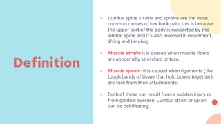
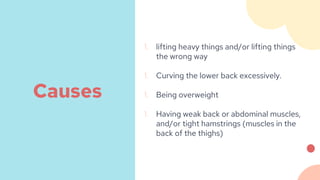
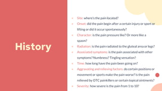
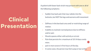
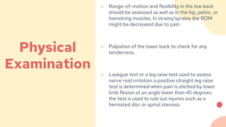
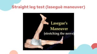
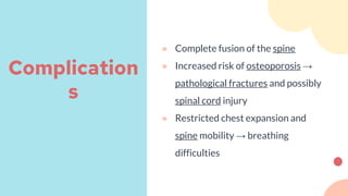
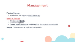
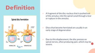
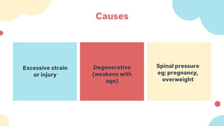
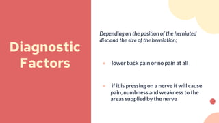
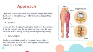
2. ● Illustrate a focused history, physical exam, diagnostic testing, management plan, complications/red
flags and indications for referral for the following:
1. Lower back strain/sprain
2. Spondylosis
3. Spondylolysis
4. Spondylolisthesis
5. Ankylosing Spondylitis
6. Spinal Stenosis
7. Compression Fracture
8. Herniated Disc
● Recognize the systemic causes of back pain.
Objectives
3. Abdulmajeed is a 27 year old male who presented with localized lower back
pain not radiating anywhere else the patient states he “was playing a football
game and accidentally tripped over” on physical exam there was no restriction
of motion and a negative straight leg raise test however there was tenderness
in the lower right side of his back. What is the most likely diagnosis?
6. ● Lumbar spine strains and sprains are the most
common causes of low back pain, this is because
the upper part of the body is supported by the
lumbar spine and it’s also involved in movement,
lifting and bending.
● Muscle strain: it is caused when muscle fibers
are abnormally stretched or torn.
● Muscle sprain: it is caused when ligaments (the
tough bands of tissue that hold bones together)
are torn from their attachments.
● Both of these can result from a sudden injury or
from gradual overuse. Lumbar strain or sprain
can be debilitating..
Definition
7. 1. lifting heavy things and/or lifting things
the wrong way
1. Curving the lower back excessively.
1. Being overweight
1. Having weak back or abdominal muscles,
and/or tight hamstrings (muscles in the
back of the thighs)
Causes
8. ● Site: where’s the pain located?
● Onset: did the pain begin after a certain injury or sport or
lifting or did it occur spontaneously?
● Character: is the pain pressure like? Or more like a
spasm?
● Radiation: is the pain radiated to the gluteal area or legs?
● Associated symptoms: is the pain associated with other
symptoms? Numbness? Tingling sensation?
● Time: how long have the pain been going on?
● Aggravating and relieving factors: do certain positions or
movement or sports make the pain worse? is the pain
relieved by OTC painkillers or certain topical ointments?
● Severity: how severe is the pain from 1 to 10?
History
9. A patient with lower back strain may present with one or all of
the following symptoms :
● Sudden low back pain that may radiate into the
buttocks, but NOT the legs and worsens with movement
.
● Stiffness in the low back area and/ or restricting range of
motion.
● Inability to maintain normal posture due to stiffness
and/or pain
● Muscle spasms either with activity or at rest
● Pain that persists for a maximum of 10-14 days rarely
more.
● pain is more severe in first hours of the day.
● In some cases, the person may feel a pop or tear at the
Clinical
Presentation
10. ● Range-of-motion and flexibility in the low back
should be assessed as well as in the hip, pelvic, or
hamstring muscles. In strains/sprains the ROM
might be decreased due to pain.
● Palpation of the lower back to check for any
tenderness.
● Lasègue test or a leg raise test used to assess
nerve root irritation a positive straight leg raise
test is determined when pain is elicited by lower
limb flexion at an angle lower than 45 degrees.
the test is used to rule out injuries such as a
herniated disc or spinal stenosis
Physical
Examination
12. ● Diagnostic testing is usually not necessary, unless pain has lasted for more than six
weeks and has not improved as expected following physical therapy or If symptoms are
persistent for longer than six weeks and physical therapy has not improved the
condition, or a disc injury is suspected then the following tests may be ordered by a
doctor.
● X-ray: an x-ray should be the initial modality.
● Magnetic resonance imaging (MRI): should be saved for conditions of which there is
suspicion on x-ray or soft tissue injury.
Investigation
13. Non-pharmacological:
● Staying active and exercise and avoiding bed rest
● Warm/cold packs
● Physical therapy
● Maintaining a healthy weight.
Pharmacological:
● Non-steroidal anti-inflammatory medications such as aspirin
● OTC painkillers such as acetaminophen
● Muscle relaxant: short-term basis to reduce spasms
Management
14. If untreated or recurring, a strain/sprain may
lead to:
● Decreased physical activity and therefore
weight gain
● Decrease of muscular strength in adjacent
muscles
● Decrease in bone density
Complication
s
15. ● Pain is persisting for more than 6 weeks
● Pain radiating beyond gluteal area (one or both lower limbs)
● Unexplainable fever (=/>38)
● More frequent muscle spasms
● Severe abdominal pain
● Loss of bladder and/or bowel control (immediately refer to ER)
● Inability to maintain normal posture due to stiffness
Referral
16. Marwa is 70 year old female complaining of mild lower back pain that is
intermittent and associated with certain movements she also states that she
feels that her spine is “ stiff and rigid” . Physical exam is unremarkable. Lateral
spine x-ray shows disc degeneration and osteophyte formation. What is the
most likely diagnosis?
19. Risk Factors
● Advanced age
● Smoking
● Atherosclerosis
● DM
● Obesity
● Occupation
● Heavy objects lifting
20. What are the current symptoms of this patient? If
there’s pain use the SOCRATES mnemonic to help you
assess the pain that the patient is experiencing.
● Site: where’s the pain located?
● Onset: did the pain begin after a certain injury or
sport or lifting or did it occur spontaneously?
● Character: is the pain pressure like? Or more like
a spasm?
● Radiation: is the pain radiated to the gluteal area
or legs?
History
21. ● Associated symptoms: is the pain associated
with other symptoms? Numbness? Tingling
sensation?
● Time: how long have the pain been going on?
● Aggravating and relieving factors: do certain
positions or movement or sports make the
pain worse? is the pain relieved by OTC pain
killers or certain topical ointments?
● Severity: how severe is the pain from 1 to 10?
History
Cont.
22. ● People who are of older age usually
present asymptotically.
● However, if pain is experienced then the
pain is usually of mild nature and is
associated with specific movements or
long term inactivity.
● Pain is worse with activity, although it
may be increased with certain positions
e.g sitting .
● Another symptom that might be present
is stiffness.
Clinical
Presentation
23. ● The physician should look for any abnormalities
during inspection this includes and a normal
curvature or shape.
● Palpation then follows to detect any spinal
tenderness ,bumps ,inflammatory regions and
spasms
● The range of motion (ROM) should be
assessed in different position when bending
forward and backward and side to side.
Findings in spondylosis:
● Mild tenderness on palpations might be
present.
● Decreases range of motion
● Pain on flexion suggest disc degeneration
while pain on extension suggests
osteophyte formation
Physical
Examination
24. ● X-Ray: is initially used in case there are other symptoms such as anesthesia to detect
bone spurs and loss of disc height
● CT/MRI: to observe any degeneration in the soft tissues of the spine. Finding might
include hypertrophy of facets and T2 signal decrease in joint which might suggest
annular tear and end plate collapse.
Investigation
26. Non-pharmacological:
● Physical therapy include ice, heat, massage, and ultrasound.
● Strengthening exercises
● Weight loss
● Maintain correct posture when sitting and standing.
● Quit smoking. Smoking is a risk factor for atherosclerosis (hardening of the arteries),
which can cause lower back pain
Pharmacological:
● Analgesics
● Anti-inflammatory
● Muscle relaxants
● Corticosteroids injected in epidural space if the pain is acute and radicular (radiates to a
limb)
Management
27. If untreated, spondylosis may lead to:
● Radiculopathy
● Neurological dysfunction due to nerve
compression
● Spinal stenosis
● Cervical spondylosis
Complication
s
28. ● Pain is unmanageable with analgesics
● Loss of balance and difficulty walking
● Loss of bladder or bowel control (immediately refer to ER)
● Development of acute nerve dysfunction such as sudden weakness in a
limb or more
● If the pain is associated with loss of weight or fever (>/=38.0 C)
Referral
29. A 23-year-old man presents to his primary health care physician with low back
pain and stiffness that has persisted for more than 3 months. There is no history of
obvious injury. His back symptoms are worse when he awakes in the morning, and
the stiffness lasts more than 1 hour. His back symptoms improve with exercise. He
normally takes an anti-inflammatory drug during the day, and finds his stiffness is
worse when he misses a dose. He has had 2 bouts of uveitis in the past.
31. ● Ankylosing spondylitis
(spondyloarthritis):a type of
seronegative spondyloarthropathy, is a
chronic inflammatory disease of the
axial skeleton that leads to partial or
even complete fusion and rigidity of the
spine.
Definition
32. Epidemiology:
•Sex: ♂ > ♀ (3:1)
•Age: 15–40 years
Etiology:
•Genetic predisposition: 90–95% of
patients are HLA-B27 positive
EPIDEMIOLOGY
AND ETIOLOGY:
33. 1. Articular symptoms
Most common presenting symptoms:
● back and neck pain
● Gradual onset of dull pain that progresses
slowly
● Morning stiffness that improves with
activity
● Pain is independent of positioning; at night
● Tenderness over the sacroiliac joints
● Limited mobility of the spine (especially
reduced forward lumbar flexion)
● Inflammatory enthesitis
● Dactylitis
● Arthritis outside the spine (hip, shoulder,
knee joint)
Clinical
Presentation
34. 2. Extra-articular manifestations
● Most common: acute, unilateral anterior
uveitis (∼ 25% of cases)
● Fatigue
● Restrictive pulmonary disease due to
decreased mobility of the spine and thorax
● Gastrointestinal symptoms
● Rare
● Cardiac: aortic root inflammation and
subsequent aortic valve insufficiency,
atrioventricular blocks
Clinical
presentation
Cont.
35.
36. Physical
Examination
Chest expansion measurement: to monitor
disease severity
•Method: measure chest circumference in full expiration
and inspiration
Examination of the hip
•Mennell sign: tenderness to percussion and pain on
displacement of the sacroiliac joints
•FABER test: FABER (Flexion, ABduction, and External
Rotation) provokes pain in the ipsilateral hip
37. Schober test
•The Schober test is used to measure spine mobility.
The first sacral vertebra (S1) and another point 10 cm
above are marked (using a pen or sticker) while the
patient is standing. The patient bends forward with
straight knees as far as possible. Normally, the
distance between these two points increases by ≥ 4 cm
(green). A smaller increase in distance between these
two points (red) is pathological.
38. Laboratory findings
•↑ CRP and ESR
•Auto-antibodies (e.g., rheumatoid factor, antinuclear antibodies) are negative
•HLA-B27 positive in 90–95% of cases
Investigation
39. Imaging
1. X-Ray
● Helps confirm a diagnosis and evaluate the severity of disease
● The changes usually occur symmetrically.
Pelvis (best initial test): to examine the sacroiliac joints
● Signs of sacroiliitis, including ankylosis (fusion of the articular surfaces)
Spine
● Loss of lordosis
● Syndesmophytes resulting in a so-called 'bamboo spine' in anteroposterior radiograph in the later
stages
Thorax: ankylosis of costosternal and costovertebral joints
1. MRI: Best method for early detection
Investigation
40. •CT scan of the thoracic spine (sagittal
plane)
•Characteristic changes in ankylosing
spondylitis with syndesmophytes (1),
subchondral sclerosis (red overlay), disc
space narrowing (2), and ossification of
the supraspinous ligament (3).
CT scan
41. ● Bilateral sacroiliitis in ankylosing
spondylitis
● Native axial CT scan of the pelvis,
bone window, at the level of the
sacroiliac joint: The image shows
typical changes in ankylosing
spondylitis with erosions (green
areas) and sclerotization (red
areas). The joint cavity can best
be guessed and is essentially
completely destroyed (green
broken line). (1 = sacral foramina,
2 = sacral canal
CT scan
45. Physical therapy
● Consistent and rigorous physical therapy
Medical therapy
● First choice: NSAIDs
● Additional options
● Tumor necrosis factor-α inhibitors (e.g., etanercept, adalimumab)
Surgery: in severe cases to improve quality of life
Management
47. ● A fragment of the disc nucleus that is pushed out
of the annulus, into the spinal canal through a tear
or rupture in the annulus.
● Discs that become herniated are usually in an
early stage of degeneration
● Due to this displacement, the disc presses on
spinal nerves, often producing pain, which may be
severe.
Definition
49. Depending on the position of the herniated
disc and the size of the herniation;
● lower back pain or no pain at all
● if it is pressing on a nerve it will cause
pain, numbness and weakness to the
areas supplied by the nerve
Diagnostic
Factors
50. Typically, a herniated disc is preceded by an episode of low
back pain or a long history of intermittent episodes of low
back pain.
● Sciatica:
Sharp electric like pain, tingling and numbness that radiates
from the buttock into the leg and sometimes into the foot,
increase with standing, walking and straightening the leg.
● Cervical spine:
Dull sharp pain in the neck or between the shoulders,
radiates down the arm, hand and fingers, increase with
movement of the neck.
Approach
51. Neurological examination:
• Reflexes
• Muscle strength
• Walking ability
• Ability to feel light touches,
pinpricks or vibration
• Range of motion
• +ve leg raise
*In most cases of herniated disk, a
physical exam and a medical history are
all that's needed for a diagnosis.
Physical
Examination
53. ● The initial treatment for a herniated disc is usually conservative and nonsurgical.
● Low, painless activity helps the spinal nerve inflammation to decrease.
● Bedrest is not recommended.
● Mild to moderate pain : NSAIDs or epidural steroids injection.
● Physiotherapy
Management
56. ● Neurological deficit
● Bone cement emboli following vertebroplasty
kyphoplasty
● Heat or pressure damage to spinal cord or nerve
roots
● Continued morbidity from osteoporosis
● Progressive kyphosis
● Decreased mobility
Complication
s
57. Referral to a spine specialist, such as a neurosurgeon, is also
recommended if symptoms persist for greater than four weeks.
A specialist will often want advanced imaging, such as the MRI,
completed prior to the appointment.
Referral
58. Systemic disorders:
● Primary or metastatic neoplasms
● Osseous, diskal, or epidural infections
● Inflammatory spondyloarthropathy
● Metabolic bone diseases, including osteoporosis
● Vascular disorders (eg, atherosclerosis, vasculitis)
Referred pain:
● Gastrointestinal disorders (eg, pancreatitis, pancreatic cancer, cholecystitis)
● Cardiorespiratory disorders (eg, pericarditis, pleuritis, pneumonia)
● Disorders of the ribs or sternum
● Genitourinary disorders (eg, nephrolithiasis, prostatitis, pyelonephritis)
● Thoracic or abdominal aortic aneurysms
● Hip disorders (eg, injury, inflammation, or end-stage degeneration of the joint and associated
soft tissues [tendons, bursae, ligaments])
Systemic causes of back pain:
59. 1. Age <18 or >50
● Younger patients with acute back pain should be evaluated for congenital
defects, spondylolysis, or vertebral fracture
● In adolescents,night pain and weight loss necessitate an evaluation for
malignancy
● New-onset back pain in patients over age 50 years is concerning for tumor and
infection, as well as intra-abdominal processes such as abdominal aortic
aneurysm, pancreatitis, or nephrolithiasis
● Maintain a high clinical suspicion of vertebral fracture in older adults who
present with acute onset of low back pain.
Red Flags and when to refer in back pain:
60. 2. Anticoagulation or antiplatelet use
● The incidence of spontaneous epidural hematoma is low; however, patients on
anticoagulants or antiplatelets and those with thrombophilia are at a greater
risk, hence it can result in spinal and nerve root compression
3. Fever
● Can be an indicator of infection and has a reported incidence of 66% to 83% in
patients with spinal epidural abscesses.
Red Flags and when to refer in back pain:
61. 4. Genitourinary symptoms
● Urinary retention and bowel incontinence frequently are associated with cauda
equina syndrome or nerve root impingement.
5. Immunocompromised
● Other conditions such as alcohol abuse, malnutrition, chronic corticosteroid use,
and diabetes can increase patient susceptibility to spinal infection and vertebral
compression fractures.
Red Flags and when to refer in back pain:
62. 6. IV drug abuse
● Recurrent bacteremia may cause hematogenous seeding of the spine,
resulting in spondylodiscitis and epidural abscess.
7. Recent surgery or spinal injection
● A history of lumbar puncture may be significant and increase patient risk for
epidural hematoma or infection.
8. Trauma
● Major trauma (or minor trauma in older adults after a fall) can result in Acute
spinal cord compression or hematomas
Red Flags and when to refer in back pain:
63. 9. History of malignancy and unexpected weight loss:
● metastatic epidural spinal cord compression correlated with prostate, breast,
and lung cancers.
10. Neurological symptoms
Red Flags and when to refer in back pain:
64. References
● BMJ best practice
● American academy of orthopaedic surgeons
● UpToDate
● https://www.nhs.uk/conditions/spondylolisthesis/
● https://journals.lww.com/jaapa/fulltext/2020/08000/red_flags_of_low_back_pain.1.aspx
● •1.Hsiang JK. Spinal Stenosis. In: Spinal Stenosis. New York, NY:
WebMD.http://emedicine.medscape.com/article/1913265-overview#a4. February 13, 2017. Accessed
February 16, 2017.