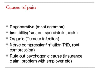
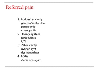
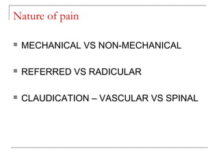
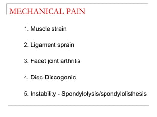
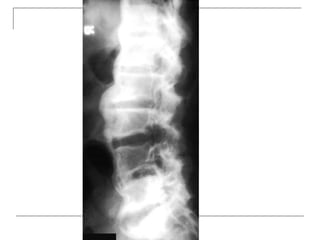
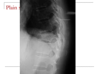
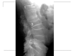
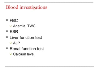
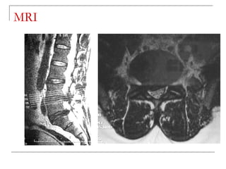
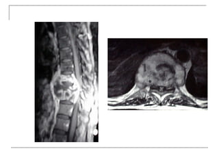
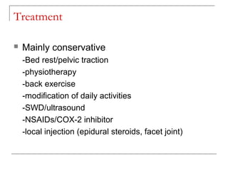
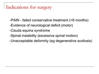
Low Back Pain
Dr Herman Gofara, SpOT (K) Spine
Incidence
 Very common among working group
 90% in pt >45years old
 80% resolves with conservative treatment (in
<3 months)
 Only 5-10% may require operation
Implication
 Work & productivity loss
Anatomical consideration
 Commonly at lumbosacral junction (L4/L5,
L5/S1)
 Why?
 Most mobile region of the spine
 Therefore prone to degeneration (wear &
tear)
Causes of pain
 Degenerative (most common)
 Instability(fracture, spondylolisthesis)
 Organic (Tumour,infection)
 Nerve compression/irritation(PID, root
compression)
 Rule out psychogenic cause (insurance
claim, problem with employer etc)
Referred pain
1. Abdominal cavity
gastritis/peptic ulcer
pancreatitis
cholecystitis
2. Urinary system
renal calculi
UTI
3. Pelvic cavity
ovarian cyst
dysmenorrhea
4. Aorta
Aortic aneurysm
Nature of pain
 MECHANICAL VS NON-MECHANICAL
 REFERRED VS RADICULAR
 CLAUDICATION – VASCULAR VS SPINAL
MECHANICAL PAIN
1. Muscle strain
2. Ligament sprain
3. Facet joint arthritis
4. Disc-Discogenic
5. Instability - Spondylolysis/spondylolisthesis
NON-MECHANICAL PAIN
 Infection – PYOGENIC VS TB
 Tumour – PRIMARY VS SECONDARY
 Primary - BENIGN VS MALIGNANT
Common causes of low back pain
Pathology Age Pain nature Assoc pain Assoc sx
DEGENERA
TIVE
Spondylosis
>40y mechanical Distance
claudication
Active pt
Spondylolisth
esis
<20y
>40y
mechanical extension Hyperextensi
on activity
Trauma Any age mechanical - Trauma
Infection Any age non-
mechanical
Rest pain Fever
Mets >50y Non-
mechanical
Rest pain Primary +
LOW
LOA
Osteoporosis >60y mechanical - Trivial trauma
RED FLAGS
 Constitutional symptoms
 LOW, LOA, fever
 AGE(>50)
 IMMUNOCOMPROMISED,
 TB CONTACT
 KNOWN CANCER
 NEUROLOGICAL DEFICIT (CAUDA EQUINA SYN)
Physical findings
 General examination
 Age
 Ill looking
 Local examination – DO NOT MISS A
GIBBUS
 Deformity
 Scoliosis/kyphosis
 Step deformity
 Local tenderness/paraspinal spasm
 Limited ROM
 Full neurological examination
 ANAL TONE / PERIANAL SENSATION
 DERMATOME & MYOTOME
Investigations
Plain radiograph
 AP
-loss of lumbar lordosis
-reduced disc space
-osteophytes
-deformity
-fracture (increase interpedicular distance)
-osteoporosis
-pedicle disruption
 Lateral
-fracture/wedging
-kyphosis
-spondylolisthesis
 Oblique
-spondylolysis (SCOTTIE DOG)
Plain x-rays
Blood investigations
 FBC
 Anemia, TWC
 ESR
 Liver function test
 ALP
 Renal function test
 Calcium level
CT Scan
 better visualization of bone pathology (eg.
cortical destruction)
 fracture
 tumor
MRI
 -better soft tissue visualization
 -disc
 -ligaments (ALL,PLL)
 -nerves (spinal cord, roots)
 -bone marrow
 -pus collection
MRI
CT myelogram
 role replaced by MRI
 for delineation of neural structures where MRI
is not available/contraindicated
CT Myelogram
Bone scan
 Suspicious of multiple bone mets
 Eg. with history of untreated/treated CA
 Negative in Multiple myeloma
Treatment
 Mainly conservative
-Bed rest/pelvic traction
-physiotherapy
-back exercise
-modification of daily activities
-SWD/ultrasound
-NSAIDs/COX-2 inhibitor
-local injection (epidural steroids, facet joint)
Pelvic traction
Surgery
Indications for surgery
-PAIN - failed conservative treatment (>6 months)
-Evidence of neurological deficit (motor)
-Cauda equina syndrome
-Spinal instability (excessive spinal motion)
-Unacceptable deformity (eg degenerative scoliosis)
Surgery
1. DECOMPRESSION of spinal nerves (BURST
FRACTURE, Spinal stenosis, PID)
2. Fusion & Stabilization (Instrumentation)
3. Correction of deformity
Non Surgery Treatment
THANK YOU

Low back pain