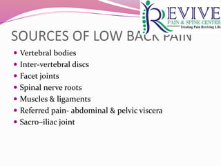
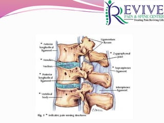
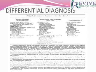
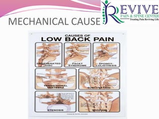
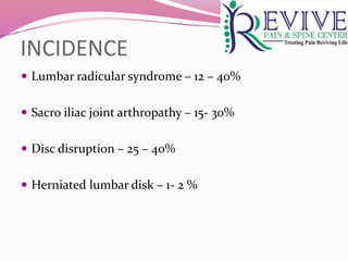
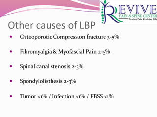
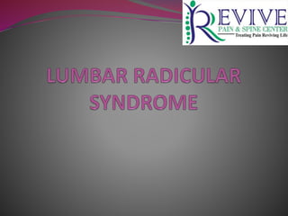
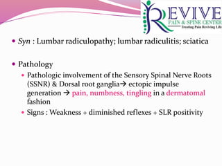
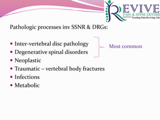
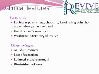
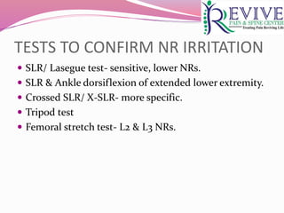
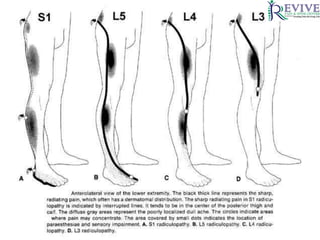
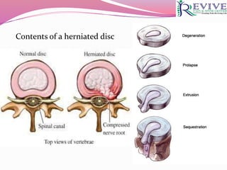
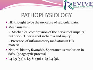
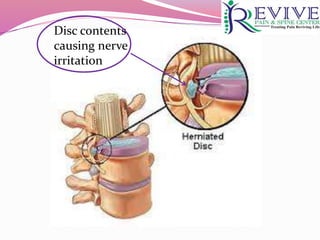
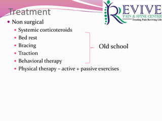
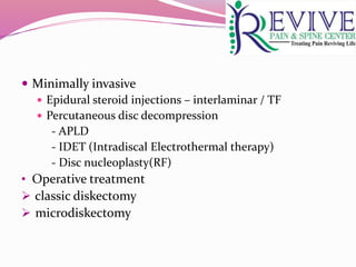
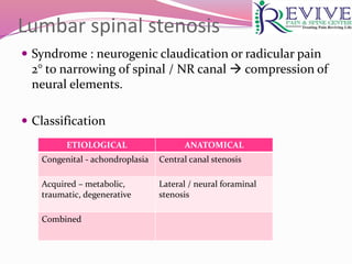
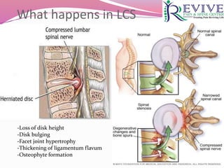
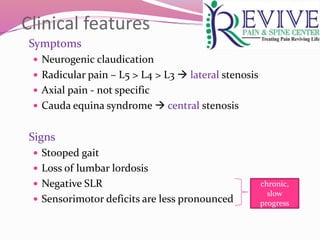
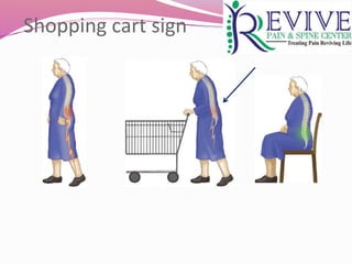
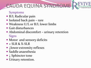
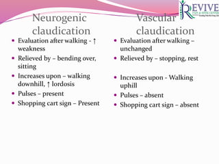
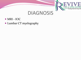
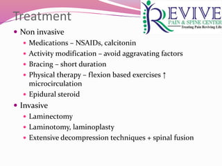
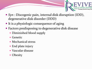
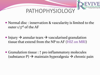
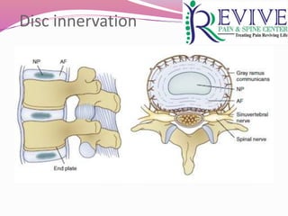
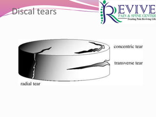
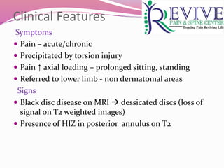
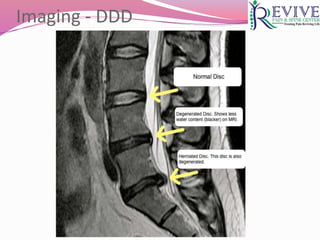
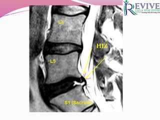
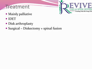
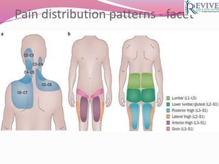
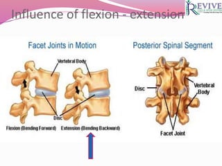
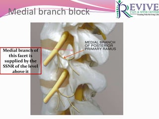
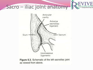
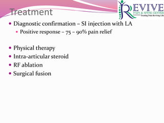
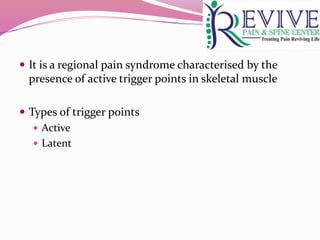
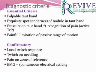
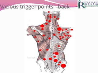
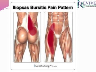
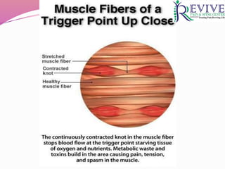
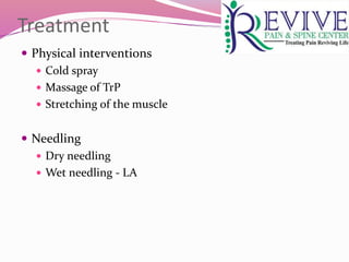
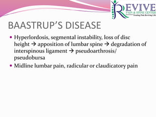
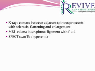
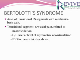
DR. VARUN SINGLA
MD (PGI Chandigarh)
PDCC-Pain Management (SGPGI Lucknow)
Fellow FPCI (Mumbai)
Member, Indian Society for Study of Pain
Peer Reviewer, Cochrane PaPaS, UK
INTRODUCTION
 Low back pain is the most common source of pain in
modern society
 Incidence of 1st episode – 6.3 – 15.4%
 Incidence of recurrent LBP – 1.5 – 36%*
 Identifying the source of the low back pain is of
paramount importance to a pain physician.
* Best Pract Res Clin Rheumatol, 2010 Dec;24 (6):769 -81
 One of the most common reasons for seeking medical
attention, second only to respiratory issues.
 84% of adults will have low back pain at some point
 Wide variety of approaches for treatment
 Suggests that optimal approach is unsure
 Most episodes are self-limited
 Some suffer from chronic or recurrent courses, with
substantial impact on quality of life
SOURCES OF LOW BACK PAIN
 Vertebral bodies
 Inter-vertebral discs
 Facet joints
 Spinal nerve roots
 Muscles & ligaments
 Referred pain- abdominal & pelvic viscera
 Sacro–iliac joint
DIFFERENTIAL DIAGNOSIS
MECHANICAL CAUSES
INCIDENCE
 Lumbar radicular syndrome – 12 – 40%
 Sacro iliac joint arthropathy – 15- 30%
 Disc disruption – 25 – 40%
 Herniated lumbar disk – 1- 2 %
Other causes of LBP
 Osteoporotic Compression fracture 3-5%
 Fibromyalgia & Myofascial Pain 2-5%
 Spinal canal stenosis 2-3%
 Spondylolisthesis 2-3%
 Tumor <1% / Infection <1% / FBSS <1%
 Syn : Lumbar radiculopathy; lumbar radiculitis; sciatica
 Pathology
 Pathologic involvement of the Sensory Spinal Nerve Roots
(SSNR) & Dorsal root ganglia ectopic impulse
generation  pain, numbness, tingling in a dermatomal
fashion
 Signs : Weakness + diminished reflexes + SLR positivity
Pathologic processes inv SSNR & DRGs:
 Inter-vertebral disc pathology
 Degenerative spinal disorders
 Neoplastic
 Traumatic – vertebral body fractures
 Infections
 Metabolic
Most common
Clinical features
Symptoms
 Radicular pain- sharp, shooting, lancinating pain that
travels along a narrow band.
 Paresthesias & numbness
 Weakness in territory of inv NR
Objective Signs
 Gait disturbances
 Loss of sensation
 Reduced muscle strength
 Diminished reflexes
TESTS TO CONFIRM NR IRRITATION
 SLR/ Lasegue test- sensitive, lower NRs.
 SLR & Ankle dorsiflexion of extended lower extremity.
 Crossed SLR/ X-SLR- more specific.
 Tripod test
 Femoral stretch test- L2 & L3 NRs.
Differential diagnoses
 Entrapment neuropathy of sciatic nerve
 Piriformis muscle
 Ischial tunnel syndrome
Pain & paresthesia along its distribution, often involving
multiple dermatomes
 Pain from IVD, SI joint and myofascial pain can also be
referred to LL- d/t Interneuronal Convergence within
spinal cord.
 Non dermatomal
 Deep aching
 Lacks objective signs of nerve root irritation
DIAGNOSTIC TESTS
 Not recommended for LBP & radicular pain of < 4-6
wks.  because of favorable natural history & often
spontaneous resolution of symptoms.
 Also, common +nce of abnormal diagnostic findings in
asymptomatic individuals.
IMAGING STUDIES
 MRI
 CT
 CT-Myelography
 Plain radiography
 Electro-diagnostic studies –
EMG, NCV, SSEP- routine use not recommended
Contents of a herniated disc
PATHOPHYSIOLOGY
 HD thought to be the mc cause of radicular pain.
 Mechanisms :
- Mechanical compression of the nerve root impairs
nutrition  nerve root ischemia and injury.
- Presence of inflammatory mediators in HD
material.
 Natural history favorable. Spontaneous resolution in
60%. (phagocytic process)
 L4-L5 (59) > L5-S1 (30) > L3-L4 (9).
Disc contents
causing nerve
irritation
Treatment
 Non surgical
 Systemic corticoteroids
 Bed rest
 Bracing
 Traction
 Behavioral therapy
 Physical therapy – active + passive exercises
Old school
 Minimally invasive
 Epidural steroid injections – interlaminar / TF
 Percutaneous disc decompression
- APLD
- IDET (Intradiscal Electrothermal therapy)
- Disc nucleoplasty(RF)
• Operative treatment
 classic diskectomy
 microdiskectomy
Lumbar spinal stenosis
 Syndrome : neurogenic claudication or radicular pain
2° to narrowing of spinal / NR canal  compression of
neural elements.
 Classification
ETIOLOGICAL ANATOMICAL
Congenital - achondroplasia Central canal stenosis
Acquired – metabolic,
traumatic, degenerative
Lateral / neural foraminal
stenosis
Combined
What happens in LCS
-Loss of disk height
-Disk bulging
-Facet joint hypertrophy
-Thickening of ligamentum flavum
-Osteophyte formation
Clinical features
Symptoms
 Neurogenic claudication
 Radicular pain – L5 > L4 > L3  lateral stenosis
 Axial pain - not specific
 Cauda equina syndrome  central stenosis
Signs
 Stooped gait
 Loss of lumbar lordosis
 Negative SLR
 Sensorimotor deficits are less pronounced
chronic,
slow
progress
Shopping cart sign
CAUDA EQUINA SYNDROME
Symptoms
 B/L Radicular pain
 Isolated back pain – rare
 Weakness U/L or B/L lower limbs
 Gait disturbances
 Abdominal discomfort – urinary retention
Signs
 Motor and sensory deficits
 + SLR & X-SLR
 ↓lower extremity reflexes
 Saddle anaesthesia
 ↓ Sphincter tone
 Urinary retention.
Neurogenic
claudication
 Evaluation after walking - ↑
weakness
 Relieved by – bending over,
sitting
 Increases upon – walking
downhill, ↑ lordosis
 Pulses – present
 Shopping cart sign – Present
Vascular
claudication
 Evaluation after walking –
unchanged
 Relieved by – stopping, rest
 Increases upon - Walking
uphill
 Pulses – absent
 Shopping cart sign – absent
DIAGNOSIS
 MRI - IOC
 Lumbar CT myelography
Treatment
 Non invasive
 Medications – NSAIDs, calcitonin
 Activity modification – avoid aggravating factors
 Bracing – short duration
 Physical therapy – flexion based exercises ↑
microcirculation
 Epidural steroid
 Invasive
 Laminectomy
 Laminotomy, laminoplasty
 Extensive decompression techniques + spinal fusion
 Syn : Discogenic pain, internal disk disruption (IDD),
degenerative disk disorder (DDD)
 It is a physiologic consequence of aging
 Factors predisposing to degenerative disk disease
 Diminished blood supply
 Genetic
 Mechanical stress
 End plate injury
 Vascular disease
 Obesity
PATHOPHYSIOLOGY
 Normal disc : innervation & vascularity is limited to the
outer 1/3rd of the AF
 Injury  annular tears  vascularised granulation
tissue that extend from the NP to AF (HIZ on MRI)
 Granulation tissue : ↑ pro inflammatory molecules
(substance P)  maintain hyperalgesia  chronic pain
Disc innervation
Discal tears
Clinical Features
Symptoms
 Pain – acute/chronic
 Precipitated by torsion injury
 Pain ↑ axial loading – prolonged sitting, standing
 Referred to lower limb - non dermatomal areas
Signs
 Black disc disease on MRI  dessicated discs (loss of
signal on T2 weighted images)
 Presence of HIZ in posterior annulus on T2
Imaging - DDD
HIZ
Treatment
 Mainly palliative
 IDET
 Disk arthroplasty
 Surgical – Diskectomy + spinal fusion
Lumbar facet – anatomy
Causes – Facet syndrome
 Inflammatory arthritides – RA, AS
 Degenerative
 Synovial cysts
 Infections
 Microtrauma
 Meniscoid & synovial entrapments
 Joint subluxation
Clinical features
Symptoms
 Low back pain – U/L or B/L
 Referral to groin, hip, posterior thigh
 Pain ↑ on twisting, extension, sitting, standing
 Pain ↓ on forward flexion, rest, walking
Signs
 Localized tenderness over facets
 No neurological deficits
 Normal SLR
Pain distribution patterns - facet
Influence of flexion - extension
What clinches diagnosis
Analgesic response to targeted, low volume LA
injections – only accepted standard for diagnosis !!
Treatment
Diagnostic facet injections – followed by
 Intra-articular LA + steroid
 Facet denervation – lesioning of the MB at the same
vertebral level & 1 level above
Medial branch block
Medial branch of
this facet is
supplied by the
SSNR of the level
above it
Sacro – iliac joint anatomy
Factors predisposing to SIJD
 Trauma
 Leg length discrepancy
 Spinal deformity
 Surgery
 Lumbar facet syndrome
 Pregnancy
 Inflammatory arthritides – AS
 Infections
Clinical features
Symptoms
 Pain – low back – max at the affected site
 Radiation – buttock, groin & posterior thigh
 Hypomobility
Signs
 Tender SI sulcus
 Positive clinical tests – FABER, Yeoman, Gaenslen’s
 Normal lab findings & imaging
Treatment
 Diagnostic confirmation – SI injection with LA
 Positive response – 75 – 90% pain relief
 Physical therapy
 Intra-articular steroid
 RF ablation
 Surgical fusion
 It is a regional pain syndrome characterised by the
presence of active trigger points in skeletal muscle
 Types of trigger points
 Active
 Latent
Diagnostic criteria
Essential Criteria
 Palpable taut band
 Exquisite spot tenderness of nodule in taut band
 Pressure on taut band  recognition of pain (active
TrP)
 Painful limitation of passive range of motion
Confirmatory
 Local twitch response
 Twitch on needling
 Pain on zone of reference
 EMG – spontaneous electrical activity
Various trigger points - back
Treatment
 Physical interventions
 Cold spray
 Massage of TrP
 Stretching of the muscle
 Needling
 Dry needling
 Wet needling - LA
BAASTRUP’S DISEASE
 Hyperlordosis, segmental instability, loss of disc
height  apposition of lumbar spine  degradation of
interspinous ligament  pseudoarthrosis/
pseudobursa
 Midline lumbar pain, radicular or claudicatory pain
 X-ray : contact between adjacent spinous processes
with sclerosis, flattening and enlargement
 MRI- edema interspinous ligament with fluid
 SPECT scan Tc : hyperemia
BERTOLOTTI’S SYNDROME
 Asso. of transitional LS segments with mechanical
back pain.
 Transitional segment a/w axial pain, related to
- neoarticulation
- C/L facet at level of asymmetric neoarticulation
- IDD in the at-risk disk above.
Lbp differentials

Lbp differentials

  • 1.
    DR. VARUN SINGLA MD(PGI Chandigarh) PDCC-Pain Management (SGPGI Lucknow) Fellow FPCI (Mumbai) Member, Indian Society for Study of Pain Peer Reviewer, Cochrane PaPaS, UK
  • 2.
    INTRODUCTION  Low backpain is the most common source of pain in modern society  Incidence of 1st episode – 6.3 – 15.4%  Incidence of recurrent LBP – 1.5 – 36%*  Identifying the source of the low back pain is of paramount importance to a pain physician. * Best Pract Res Clin Rheumatol, 2010 Dec;24 (6):769 -81
  • 3.
     One ofthe most common reasons for seeking medical attention, second only to respiratory issues.  84% of adults will have low back pain at some point  Wide variety of approaches for treatment  Suggests that optimal approach is unsure  Most episodes are self-limited  Some suffer from chronic or recurrent courses, with substantial impact on quality of life
  • 4.
    SOURCES OF LOWBACK PAIN  Vertebral bodies  Inter-vertebral discs  Facet joints  Spinal nerve roots  Muscles & ligaments  Referred pain- abdominal & pelvic viscera  Sacro–iliac joint
  • 6.
  • 7.
  • 8.
    INCIDENCE  Lumbar radicularsyndrome – 12 – 40%  Sacro iliac joint arthropathy – 15- 30%  Disc disruption – 25 – 40%  Herniated lumbar disk – 1- 2 %
  • 9.
    Other causes ofLBP  Osteoporotic Compression fracture 3-5%  Fibromyalgia & Myofascial Pain 2-5%  Spinal canal stenosis 2-3%  Spondylolisthesis 2-3%  Tumor <1% / Infection <1% / FBSS <1%
  • 11.
     Syn :Lumbar radiculopathy; lumbar radiculitis; sciatica  Pathology  Pathologic involvement of the Sensory Spinal Nerve Roots (SSNR) & Dorsal root ganglia ectopic impulse generation  pain, numbness, tingling in a dermatomal fashion  Signs : Weakness + diminished reflexes + SLR positivity
  • 12.
    Pathologic processes invSSNR & DRGs:  Inter-vertebral disc pathology  Degenerative spinal disorders  Neoplastic  Traumatic – vertebral body fractures  Infections  Metabolic Most common
  • 13.
    Clinical features Symptoms  Radicularpain- sharp, shooting, lancinating pain that travels along a narrow band.  Paresthesias & numbness  Weakness in territory of inv NR Objective Signs  Gait disturbances  Loss of sensation  Reduced muscle strength  Diminished reflexes
  • 14.
    TESTS TO CONFIRMNR IRRITATION  SLR/ Lasegue test- sensitive, lower NRs.  SLR & Ankle dorsiflexion of extended lower extremity.  Crossed SLR/ X-SLR- more specific.  Tripod test  Femoral stretch test- L2 & L3 NRs.
  • 17.
    Differential diagnoses  Entrapmentneuropathy of sciatic nerve  Piriformis muscle  Ischial tunnel syndrome Pain & paresthesia along its distribution, often involving multiple dermatomes  Pain from IVD, SI joint and myofascial pain can also be referred to LL- d/t Interneuronal Convergence within spinal cord.  Non dermatomal  Deep aching  Lacks objective signs of nerve root irritation
  • 18.
    DIAGNOSTIC TESTS  Notrecommended for LBP & radicular pain of < 4-6 wks.  because of favorable natural history & often spontaneous resolution of symptoms.  Also, common +nce of abnormal diagnostic findings in asymptomatic individuals.
  • 19.
    IMAGING STUDIES  MRI CT  CT-Myelography  Plain radiography  Electro-diagnostic studies – EMG, NCV, SSEP- routine use not recommended
  • 21.
    Contents of aherniated disc
  • 22.
    PATHOPHYSIOLOGY  HD thoughtto be the mc cause of radicular pain.  Mechanisms : - Mechanical compression of the nerve root impairs nutrition  nerve root ischemia and injury. - Presence of inflammatory mediators in HD material.  Natural history favorable. Spontaneous resolution in 60%. (phagocytic process)  L4-L5 (59) > L5-S1 (30) > L3-L4 (9).
  • 23.
  • 24.
    Treatment  Non surgical Systemic corticoteroids  Bed rest  Bracing  Traction  Behavioral therapy  Physical therapy – active + passive exercises Old school
  • 25.
     Minimally invasive Epidural steroid injections – interlaminar / TF  Percutaneous disc decompression - APLD - IDET (Intradiscal Electrothermal therapy) - Disc nucleoplasty(RF) • Operative treatment  classic diskectomy  microdiskectomy
  • 27.
    Lumbar spinal stenosis Syndrome : neurogenic claudication or radicular pain 2° to narrowing of spinal / NR canal  compression of neural elements.  Classification ETIOLOGICAL ANATOMICAL Congenital - achondroplasia Central canal stenosis Acquired – metabolic, traumatic, degenerative Lateral / neural foraminal stenosis Combined
  • 28.
    What happens inLCS -Loss of disk height -Disk bulging -Facet joint hypertrophy -Thickening of ligamentum flavum -Osteophyte formation
  • 29.
    Clinical features Symptoms  Neurogenicclaudication  Radicular pain – L5 > L4 > L3  lateral stenosis  Axial pain - not specific  Cauda equina syndrome  central stenosis Signs  Stooped gait  Loss of lumbar lordosis  Negative SLR  Sensorimotor deficits are less pronounced chronic, slow progress
  • 30.
  • 31.
    CAUDA EQUINA SYNDROME Symptoms B/L Radicular pain  Isolated back pain – rare  Weakness U/L or B/L lower limbs  Gait disturbances  Abdominal discomfort – urinary retention Signs  Motor and sensory deficits  + SLR & X-SLR  ↓lower extremity reflexes  Saddle anaesthesia  ↓ Sphincter tone  Urinary retention.
  • 32.
    Neurogenic claudication  Evaluation afterwalking - ↑ weakness  Relieved by – bending over, sitting  Increases upon – walking downhill, ↑ lordosis  Pulses – present  Shopping cart sign – Present Vascular claudication  Evaluation after walking – unchanged  Relieved by – stopping, rest  Increases upon - Walking uphill  Pulses – absent  Shopping cart sign – absent
  • 33.
    DIAGNOSIS  MRI -IOC  Lumbar CT myelography
  • 34.
    Treatment  Non invasive Medications – NSAIDs, calcitonin  Activity modification – avoid aggravating factors  Bracing – short duration  Physical therapy – flexion based exercises ↑ microcirculation  Epidural steroid  Invasive  Laminectomy  Laminotomy, laminoplasty  Extensive decompression techniques + spinal fusion
  • 36.
     Syn :Discogenic pain, internal disk disruption (IDD), degenerative disk disorder (DDD)  It is a physiologic consequence of aging  Factors predisposing to degenerative disk disease  Diminished blood supply  Genetic  Mechanical stress  End plate injury  Vascular disease  Obesity
  • 37.
    PATHOPHYSIOLOGY  Normal disc: innervation & vascularity is limited to the outer 1/3rd of the AF  Injury  annular tears  vascularised granulation tissue that extend from the NP to AF (HIZ on MRI)  Granulation tissue : ↑ pro inflammatory molecules (substance P)  maintain hyperalgesia  chronic pain
  • 38.
  • 39.
  • 41.
    Clinical Features Symptoms  Pain– acute/chronic  Precipitated by torsion injury  Pain ↑ axial loading – prolonged sitting, standing  Referred to lower limb - non dermatomal areas Signs  Black disc disease on MRI  dessicated discs (loss of signal on T2 weighted images)  Presence of HIZ in posterior annulus on T2
  • 42.
  • 43.
  • 44.
    Treatment  Mainly palliative IDET  Disk arthroplasty  Surgical – Diskectomy + spinal fusion
  • 46.
  • 47.
    Causes – Facetsyndrome  Inflammatory arthritides – RA, AS  Degenerative  Synovial cysts  Infections  Microtrauma  Meniscoid & synovial entrapments  Joint subluxation
  • 48.
    Clinical features Symptoms  Lowback pain – U/L or B/L  Referral to groin, hip, posterior thigh  Pain ↑ on twisting, extension, sitting, standing  Pain ↓ on forward flexion, rest, walking Signs  Localized tenderness over facets  No neurological deficits  Normal SLR
  • 49.
  • 50.
  • 51.
    What clinches diagnosis Analgesicresponse to targeted, low volume LA injections – only accepted standard for diagnosis !!
  • 52.
    Treatment Diagnostic facet injections– followed by  Intra-articular LA + steroid  Facet denervation – lesioning of the MB at the same vertebral level & 1 level above
  • 53.
    Medial branch block Medialbranch of this facet is supplied by the SSNR of the level above it
  • 55.
    Sacro – iliacjoint anatomy
  • 56.
    Factors predisposing toSIJD  Trauma  Leg length discrepancy  Spinal deformity  Surgery  Lumbar facet syndrome  Pregnancy  Inflammatory arthritides – AS  Infections
  • 57.
    Clinical features Symptoms  Pain– low back – max at the affected site  Radiation – buttock, groin & posterior thigh  Hypomobility Signs  Tender SI sulcus  Positive clinical tests – FABER, Yeoman, Gaenslen’s  Normal lab findings & imaging
  • 58.
    Treatment  Diagnostic confirmation– SI injection with LA  Positive response – 75 – 90% pain relief  Physical therapy  Intra-articular steroid  RF ablation  Surgical fusion
  • 60.
     It isa regional pain syndrome characterised by the presence of active trigger points in skeletal muscle  Types of trigger points  Active  Latent
  • 61.
    Diagnostic criteria Essential Criteria Palpable taut band  Exquisite spot tenderness of nodule in taut band  Pressure on taut band  recognition of pain (active TrP)  Painful limitation of passive range of motion Confirmatory  Local twitch response  Twitch on needling  Pain on zone of reference  EMG – spontaneous electrical activity
  • 62.
  • 65.
    Treatment  Physical interventions Cold spray  Massage of TrP  Stretching of the muscle  Needling  Dry needling  Wet needling - LA
  • 66.
    BAASTRUP’S DISEASE  Hyperlordosis,segmental instability, loss of disc height  apposition of lumbar spine  degradation of interspinous ligament  pseudoarthrosis/ pseudobursa  Midline lumbar pain, radicular or claudicatory pain
  • 67.
     X-ray :contact between adjacent spinous processes with sclerosis, flattening and enlargement  MRI- edema interspinous ligament with fluid  SPECT scan Tc : hyperemia
  • 68.
    BERTOLOTTI’S SYNDROME  Asso.of transitional LS segments with mechanical back pain.  Transitional segment a/w axial pain, related to - neoarticulation - C/L facet at level of asymmetric neoarticulation - IDD in the at-risk disk above.