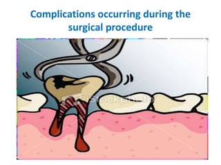
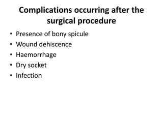
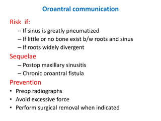
2. Soft tissue injuries
a. Tear of a flap
b. Puncture wound
c. Stretch or abrasion
Problems with tooth being extracted
a. Root fracture
b. Root displacement
c. Tooth lost into pharynx
Injuries to adjacent teeth
a. Fracture or dislodgment of restoration
b. Luxation of adjacent tooth
c. Extraction of wrong tooth
3. • Injuries to osseous structures
a. Fracture of alveolar process
b. Fracture of tuberosity
c. Fracture of mandible
Injuries to adjacent structure
a. Injury to nerves
b. Injury to TMJ
Oroantral communications
4. Complications occurring after the
surgical procedure
• Presence of bony spicule
• Wound dehiscence
• Haemorrhage
• Dry socket
• Infection
5. Injuries to adjacent nerves
• Branches of 5th cranial nerve
– Mental nerve
– Nasoplatine
– Buccal
– Lingual
– Inferior alveolar
Mental nerve
Injured during:
• surgical removal of premolar roots
• impacted mand premolar
• periapical surgery in the area
Cause anesthesia or paresthesia of the lip and chin
6. Prevention
– Be aware of nerve anatomy
– Avoid releasing incision b/w canine and 2nd
premolar
– Refer to oral and maxillofacial surgeon if needed
Buccal and Nasopalatine nereves:
– Frequently sectioned
– Supply relatively small area
– Reinnervation usu occurs rapidly
Lingual nerve
Located directly against the lingual aspect of mand in
retromolar pad area. Injured during impacted mand
3rd molar surgery
Prevention
– Incision for impacted 3rd molar made buccally
– Avoid stretching or excessive dissection lingually
– Rarely regenerates if excessively traumatized
7. Inferior alveolar nerve
– Injured during extraction of impacted mand 3rd
molar
– Inform patient preoperatively about the possibility
– Take precaution to avoid injuring the nerve
8. Injury to TMJ
Injured during extraction of mand molars due to
excessive force
Prevention
– Avoid excessive force
– Support the jaw during extraction
– Use of bite block
Treatment
– Rest for jaw, apply moist heat, soft diet
– 600-800 ibuprofen every 4 hours
– 500-1000 acetaminophen if pt cannot tolerate
NSAIDs
9. Oroantral communication
Risk if:
– If sinus is greatly pneumatized
– If little or no bone exist b/w roots and sinus
– If roots widely divergent
Sequelae
– Postop maxillary sinusitis
– Chronic oroantral fistula
Prevention
• Preop radiographs
• Avoid excessive force
• Perform surgical removal when indicated
10. Diagnosis
– Examine the tooth if bone is adhered to root
– Nose blowing test
– Radiographs
– Do not probe
Treatment
Depends on the size of communication
Size is estimated from the bone piece attached
to the root. If no bone piece less than 2mm
If less than 2mm:
• Ensure quality clot formation
• Sinus precautions
11. If 2 to 6mm
Ensure clot maintenance
– Figure of eight suture
– Clot promoting substances (gelfoams)
– Sinus precautions
– Antibiotics ( amoxicillin, cephalexin, clindamycin)
– Nasal decongestant spray
If 7 or more
If no pre-existing sinusitis
– Sinus closure with a flap
– Same medication and sinus precaution
12. If pre-existing sinusitis
– Even a small communication can progress to
chronic fistula
– Refer to oral and maxillofacial surgeon
Follow up
– If communication exist for more than 2 wks refer
13. Postoperative bleeding
Reasons
– Highly vascular tissue
– Open wound
– Impossible to apply pressure dressings
– Patient may dislodge blood clot
– Salivary enzymes may lyse clot
Prevention
Detailed history
– Previous extraction or surgery
– Family history
– Any medication or liver disease
– Lab investigations and hematologist consultation
wherever necessary
14. • Atraumatic surgery
• Good hemostasis
• Patients instructions
Primary control of bleeding
• Atraumatic surgery
• Smooth sharp bony spicules
• Remove all granulation tissue
• inspect for bleeders if present then pressure pack or
ligation.
• Check for bleeding from bone if present crush foramen if
bleeding from foramen.
• Bite on damp gauze sponge for 30mins.
• Remove gauze inspect for bleeding /persistent ooze
– if controlled pack again with damp gauze for 30mins.
– If not then additional measures to achieve hemostasis
15. – Absorbable gelatin sponge (e.g
gelfoam)provides scaffold for formation of clot.
– Oxidized regenerated cellulose (e.g surgicel)
promotes coagulaion, can be packed under
pressure, reserved for more persistent bleeding as
it cause delayed healing.
– Topical thrombin saturated onto a gelatin sponge
bypasses steps in coagulation cascade
covert fibrinogen to fibrin enzymatically which
forms clot.
– Collagen promotes platelet aggregation and
accelerate blood coagulation.
• Microfibular collagen (avitene)
• Collaplug
• Collatape ( highly crosslinked but more expensive)
16. Secondary bleeding
In case of secondary bleeding ask pt to:
– Rinse gently with chilled water and bite on a damp
gauze for 30mins
– If persist rinse again and bit on a teabag. Tannin
help stop bleeding.
– If still not stopped then:
– Blood saliva and fluid suctioned and liver clots
removed and bleeding site examined carefully
– If generalized ooze then pack and apply pressure
with finger for 5mins.
– If not controlled, give block, remove old clot and
identify source of bleeding
17. – Same measures applied as for primary bleeding
– Monitor for 30mins before discharging the patient
– If local measures fail then consult hematologist for
assessment of bleeding profile
19. Wound dehiscence
Causes
• Inadequate bony support
• Suturing under tension
• Internal oblique ridge area
– Exposed bone
• Leave as such
• Smooth it with bone file under L.A.
20. Dry socket
• One of the most common post operative
complications following the extraction of
permanent teeth is a condition known as “DRY
SOCKET”.
• Dry socket :
• post operative pain in and around the extraction
site , which increases in Severity at any time
between 1 and 3 days after the extraction
• Accompanied by a partially or totally
disintegrated blood clot within
• the alveolar socket with or without halitosis.
21. Sign and symptoms
• The denuded alveolar bare bone may be
painful and tender.
• Some patients may also complain of ‘intense
continuous pain’ irradiating to the ipsilateral
ear, temporal region or the eye.
• Regional lymphadenopathy (occasionally)
• Unpleasant taste (occasionally)
• Trismus is a rare occurrence in mandibular
third molar extractions probably due to
lengthy and traumatic surgery.
22. Incidence
• 3-4% following routine dental extractions
• 1% to 45% after the removal of mandibular
third molars.
• 25-30% after the removal of impacted
mandibular third molars.
• Occurs 10 times more frequently following the
removal of 3rd molars than from all other
locations.
23. Onset
• Mostly 1-3 days after tooth extraction.
• Within a week in 95 %and 100% of all cases of
dry socket.
• The duration of dry socket varies to some
degree, depending on the severity of the
disease, but it usually ranges from 5-10 days.
24.
25. Etiology
Multifactorial origin
• 1. Oral micro-organisms
• 2. Difficulty & trauma during surgery
• 3. Roots or bone fragments remaining in the wound
• 4. Excessive irrigation or curettage of the alveolus after
extraction.
• 5. Physical dislodgement of the clot
• 6. Local blood perfusion & anesthesia
• 7. Oral contraceptive
• 8. Smoking
26. Microorganism
• Increased frequency of dry socket in patients
with
• 1, Poor oral hygiene
• 2, Pre-existing local infection such as
pericoronitis and advanced periodontal
disease
• Reduced incidence of dry socket in
conjunction with anti bacterial measures.
27. Pathogenesis
• Partial or complete lysis and destruction of the
blood clot was caused by tissue kinases
liberated during inflammation by a direct or
indirect activation of plasminogen in the
blood.
29. Treatment
• With references in the literature correlating to
the prevention of dry socket can be divided
into
• 1. Non- pharmacological and
• 2. Pharmacological preventive measures.
30. Non pharmacological
• 1. Use of good quality current preoperative
radiographs.
• 2. Careful planning of surgery.
• 3. Use of good surgical principles.
• 4. Extractions should be performed with
minimum amount of trauma and maximum
amount of care.
• 5. Confirm presence of blood clot subsequent to
extraction.
31. • 6. Preoperative oral hygiene measures.
• 7. Encourage the patient to stop/limit smoking
in immediate postoperative period.
• 8. Avoid vigorous mouth rinsing for the first 24
hours of post extraction
• 9.Comprehensive pre and postoperative
verbal instructions should be given.
33. Antibacterial
• Prophylactic antibacterials, either given systemically or
used locally. Systemic anti bacterials (controversial)
• penicillin
• clindamycin
• erythromycin
• metronidazole
• Preoperative administration of antibacterial agents is
more effective.
• A significantly reduced incidence of dry socket
following light socket irrigation with Betadine & topical
application of Clindamycin in Gelfoam.
34. Antiseptic
• Chlorhexidine (CHX) is antiseptic with anti
microbial properties.
• USE OF WHITEHEAD’S VARNISH:
• Whitehead’s varnish is a combination of
‘iodoform, balsam tolutan, styrax liquid I a
base liquid.
• RESULT: Significant decrease in incidence of
postoperative pain.Haemorrhage and
swelling.
35. Alveogyl
• Has been widely used in the management of
dry socket and is frequently mentioned in the
literature.
• It contains: butamben(anesthetic)
• eugenol(analgesic)
• iodoform(antimicrobial)
• Topical use of ‘para-hydroxybenzoic
acid(PHBA) in extraction wounds as Anti-
fibrinolytic agents.
36. • Steroidal anti-inflammatory agents
• Topical use of corticosteroids in the prevention of
dry socket –
• decreases immediate post – operative
complications failed to reduce the occurrence of
dry socket
• Immediate placement of eugenol containing
dressing into the extraction socket is beneficial in
the prevention of postextraction complication.
• Use of clot supporting agents such as ‘polylactic
acid(PLA)’ was widely promoted as ultimate
solution for preventing dry socket.
37. • 1. Remove any suture to allow adequate
exposure of extraction site.
• 2. Irrigate the socket with isotonic saline
gently,careful suctioning of all excess irrigation.
• 3. Do not attempt to curette the socket.
• 4. Prescription of potent oral analgesics.
• 5. Patient is given with a ‘plastic syringe with
curved tip for home irrigation’ with chlorhexidine
solution.
38. surgical management
• • Under block anesthesia
• • Sharp margins were trimmed, rounded
• • Any foreign bodies present were thoroughly
removed
• • Detached gingival margins are also scraped.
• • Desired medications as well as precautions
• • Patients was not only without pain but was also
comfortable both physically as well as
psychologically from the very next day.