The document provides guidelines for the diagnosis and management of systolic heart failure in low- and middle-income countries. It discusses the clinical assessment of patients with heart failure, including taking a thorough history focusing on symptoms, etiology, functional status, comorbidities, and medications. Physical examination and various diagnostic tests are also outlined, along with goals of therapy and pharmacological and surgical management approaches. Comorbid conditions that often accompany heart failure like kidney disease, angina, and sleep disorders are also addressed.
![Practice Guidelines for the Diagnosis and Management of
Systolic Heart Failure in Low- and Middle-Income
Countries
Ragavendra R. Baliga*, G. William Decy
, Jagat Narulaz
Columbus, OH, USA; Boston, MA, USA; and New York, NY, USA
TABLE OF CONTENTS
Clinical assessment..........................................141
Symptoms of HF and relevant facets in history ...141
Physical examination ..................................144
Diagnostic tests ..............................................150
Laboratory...............................................150
BNP and amino-terminal proBNP ...................150
ECG ......................................................150
Chest x-ray ..............................................152
Echocardiography and Doppler ......................152
Stress testing ............................................153
Six-minute walk test, cardiopulmonary stress test/
regular stress test .....................................154
Right heart catheterization ............................155
Left heart catheterization and coronary
angiography ...........................................156
Endomyocardial biopsy ...............................156
Cardiac magnetic resonance imaging ...............156
Management of HF .........................................157
Goals of therapy........................................157
Lifestyle changes .......................................157
Pharmacological therapy of HF ......................157
ACE inhibitors......................................157
ARB ..................................................158
Beta-blockers........................................159
ACE inhibitors or beta-blockers first ............159
Aldosterone antagonists ...........................159
Diuretic therapy ....................................161
Digoxin ..............................................164
Hydralazine and isosorbide dinitrate
combination.......................................164
Anticoagulant/antiplatelet therapy ...............164
Prophylactic implantable cardioverter-defibrillator
placement .............................................164
Cardiac resynchronization ............................165
Surgical approaches....................................165
Coronary artery bypass graft .....................165
LV remodeling surgery or mitral valve repair..165
LVAD ................................................165
Cardiac transplantation............................165
Comorbidities................................................165
HF and kidney disease ................................165
HF and angina..........................................166
Sleep disordered breathing ...........................166
HF in the elderly .......................................166
Conclusions ..................................................166
References ....................................................166
Heart failure (HF) occurs when the cardiac output is
not adequate to meet the metabolic demands of the tissues
or is able to do so only at an elevated ventricular filling
pressure.
CLINICAL ASSESSMENT
Symptoms of HF and relevant facets in history
Compiling the history of a patient with heart failure should
focus on establishing the diagnosis; determining the
etiology; evaluating functional status including shortness of
breath, dizziness, history of hospitalizations, and fluid
status; determining precipitating factors; and assessing
comorbidities including thyroid function, sleep apnea,
arthritis, and reviewing all medications.
History taking can often separate heart failure into
ischemic cardiomyopathy and nonischemic cardiomyop-
athy, and the latter includes that due to hypertension,
rheumatic heart disease, peripartum cardiomyopathy,
human immunodeficiency virus (HIV) cardiomyopathy,
alcoholic cardiomyopathy, and rarely chemotherapy-
induced cardiomyopathy. The natural history of cardio-
myopathy depends on the etiology (Figure 1) [1], with
peripartum cardiomyopathy having the best prognosis and
HIV cardiomyopathy having the worst prognosis.
Symptoms of heart failure such as edema, weight gain,
and shortness of breath generally precede heart failure
hospitalizations (Figure 2) [2]. Shortness of breath and
orthopnea suggest left-sided heart failure. The presence of
paroxysmal nocturnal dyspnea is due to alveolar edema and
typically occurs 1 to 3 h after the patient retires to bed and
resolves 10 to 30 min after the patient arises. In the EPICA
(Epidemiologia da Insuficiência Cardiaca e Aprendizagem
[Epidemiology of Heart Failure and Learning]) registry, the
presence of paroxysmal nocturnal dyspnea, orthopnea, and
shortness of breath suggested a high specificity (w99%) for
heart failure [3]. Orthopnea has a sensitivity of 90% and
specificity of 95% for elevated left ventricular (LV) filling
pressure. In the ADHERE (Acute Decompensated Heart
Failure National Registry) and OPTIMIZE-HF (Organized
Program to Initiate Lifesaving Treatment in Hospitalized
Patients with Heart Failure) registries, approximately 90% of
patients reported shortness of breath and about one-third of
the patients had shortness of breath [4,5]. The severity of
From the *Division of
Cardiovascular Medicine,
Wexner Medical Center,
The Ohio State University,
Columbus, OH, USA;
yCardiology Division,
Massachusetts General
Hospital Heart Center, Har-
vard Medical School,
Boston, MA, USA; and the
zCardiovascular Imaging
Program, Zena and Michael
A. Wiener Cardiovascular
Institute, Icahn School of
Medicine at Mount Sinai,
New York, NY, USA. Corre-
spondence: R. Baliga
(baliga.3@osu.edu).
GLOBAL HEART
© 2013 Published by
Elsevier Ltd. on behalf of
World Heart Federation
(Geneva).
All rights reserved.
VOL. 8, NO. 2, 2013
ISSN 2211-8160/$36.00.
http://dx.doi.org/10.1016/
j.gheart.2013.05.002
GLOBAL HEART, VOL. 8, NO. 2, 2013 141
June 2013: 141-170
WHITE PAPER gRECS j](https://image.slidesharecdn.com/lmicwhitepaperforsystolichf-160210084559/85/Lmic-white-paperforsystolichf-1-320.jpg)
![shortness of breath is used to determine the functional class.
The New York Heart Association (NYHA) functional class
sheds light not only on the prognosis, but it also determines
therapy for systolic heart failure [6]. In the SOLVD (Studies of
Left Ventricular Dysfunction) database, 2-year mortality on
optimal angiotensin-converting enzyme (ACE) inhibitor therapy
in systolic heart failure patients with NYHA functional class IV
was 40% to 50%, class III heart failure 30% to 40%, class II
heart failure 20%, and NYHA class I was 10% (Table 1).
Cheyne-Stokes respiration, or periodic breathing, is common
in advanced HF, is usually associated with low-output states,
and may be perceived by the patient (and the patient’s family)
as either severe shortness of breath or transient cessation of
breathing (often mistaken for sleep apnea). Shortness of
breath can also be used to monitor response to therapy using
the self-reported 7-point Likert dyspnea scale [7] (Table 2). A
recent study using invasive hemodynamic measurements
found that the short-term improvement of shortness of
breath during therapy with vasodilators and diuretics
depends on 2 hemodynamic variables: pulmonary capillary
wedge pressure and mean pulmonary artery pressure
(PAP). The improvement in shortness of breath correlated
with both the absolute level and the magnitude of reduction
of these 2 variables [8]. And the likelihood of achieving
improvement in shortness of breath is particularly high
when both pulmonary capillary wedge pressure and mean
PAP were effectively reduced. However, there is no corre-
lation between shortness of breath and improvements in
other hemodynamic variables such as cardiac index and
systemic vascular resistance. When patients are able to
perform moderate levels of activity, shortness of breath is
a relatively sensitive symptom of HF. However, it may not
be prominent in patients who are inactive, and the diag-
nosis of HF is often delayed or overlooked. Also, shortness
of breath may become less prominent with the onset of
right ventricular (RV) failure and tricuspid regurgitation,
which may lead to lower pulmonary venous pressures. It
must be remembered that shortness of breath is also
a common symptom of patients with pulmonary disease,
obesity, or anemia and of sedentary individuals.
Chest pain is an important symptom that needs to be
evaluated in all HF patients. The RESOLVD (Randomized
Evaluation of Strategies for Left Ventricular Dysfunction)
investigators [9] found that myocardial ischemia was the
cause for hospitalization in 12% of the patients with
systolic dysfunction, and in another study, the chest pain
in HF patients was the presenting symptom for acute
coronary syndrome in nearly one-third of the patients [10].
The presence of chest pain suggests demand ischemia, or
in those without coronary artery disease, it suggests
myocarditis, pulmonary embolism, pulmonary hyperten-
sion, or significant limitations in pericardial blood flow.
Fatigue in HF suggests low-flow state, but it may also
be due to the presence of associated sleep apnea or
depression. Patients with HF can be screened for depres-
sion with 2 questions: 1) Over the past 2 weeks, have you
felt down, depressed, or hopeless? and 2) Over the past 2
weeks, have you felt little interest or pleasure in doing
things [11,12]? According to the U.S. Preventive Services
Task Force Recommendation Statement, these 2 simple
questions are more sensitive and specific than lengthy
statements or questionnaires [12]. The presence of
depression is associated with increased likelihood of
recurrent hospitalizations or mortality due to HF [13]. The
presence of fatigue should also prompt evaluation for sleep
apnea. Patients with HF may manifest either obstructive
sleep apnea or central sleep apnea. The former is a
comorbidity usually seen in overweight or obese patients,
whereas central sleep apnea is a clinical manifestation of
HF. Identification of central sleep apnea is important
because treatment may benefit patients [14,15].
Lightheadedness or dizziness suggests that pulse
pressure and/or systolic blood pressure (BP) is low or that
Proportion of Patients Surviving
Time
0
5
10
15
Cardiomyopathy due to HIV infection
Cardiomyopathy due to infiltrative myocardial disease
Cardiomyopathy due to doxorubicin therapy
Cardiomyopathy due to ischemic heart disease
Idiopathic cardiomyopathy
Peripartum cardiomyopathy
0.00 0.25 0.50 0.75 1.00
FIGURE 1. Adjusted Kaplan-Meier estimates of survival
according to the underlying cause of cardiomyopathy.
HIV, human immunodeficiency virus. Adapted, with
permission, from Felker GM et al. [1]. Redrawn figure by
Anthony Baker.
jgRECS
142 GLOBAL HEART, VOL. 8, NO. 2, 2013
June 2013: 141-170](https://image.slidesharecdn.com/lmicwhitepaperforsystolichf-160210084559/85/Lmic-white-paperforsystolichf-2-320.jpg)
![the patient has orthostatic dizziness or severe valvular
aortic stenosis. Abdominal distension and fluid retention
suggests associated right-sided heart failure.
Dietary indiscretion including fluid intake and sodium
intake needs to be assessed. Fluid intake exceeding 3 to 4 l
a day can contribute significantly to fluid retention. Three
simple questions [16] regarding sodium intake include the
following: 1) Is salt added at the table after the food is
served? 2) Does the patient eat processed foods particularly
pickles, canned foods, potato chips, TV dinners? 3) How
often does the patient eat out for meals including cafeterias
at work, or restaurants [16]?
History taking should include assessment of frailty,
particularly physical exhaustion and weight loss [17]. One
question to determine physical exhaustion is: Over the past
2 weeks have you been bothered by feeling tired or having
little energy? Patients who answered “more than half the
days” or “nearly every day” should be considered as
experiencing physical exhaustion [17]. Unintentional
weight loss can be assessed by the following question: In
the past year, have you lost any weight unintentionally
(without trying)? A response of “10 pounds or more”
should be considered as unintentional weight loss. If this
assessment suggests that patient may be frail, then they
should be formally tested for weak grip strength, slowness,
and low physical activity. Patients should be considered as
frail if they met 3 or more of the following criteria: weak
grip strength; physical exhaustion; slowness; low physical
activity; and unintentional weight loss. Intermediate frailty
was defined as meeting 1 or 2 criteria [17].
Drugs such as nonsteroidal anti-inflammatory drugs
and rosiglitazone are associated with sodium retention and
may precipitate acute HF. Calcium channel blockers such
as diltiazem and verapamil are negative inotropes that are
best avoided in chronic HF patients.
History regarding substance abuse including tobacco
and alcohol [18] should be obtained. Observational studies
FIGURE 2. Number of days from onset of worsening of selected symptoms of heart failure to admission to hospital:
cumulative percentage of patients. Adapted, with permission, from Schiff et al. [2].
TABLE 1. NYHA class and 2-year mortality
NYHA
Class Physical Activity
2-Year
Mortality (%)
on ACE-I
I Asymptomatic (no limitation of
physical activity; there is no
shortness of breath, fatigue,
or palpitations with ordinary
physical activity)
10
II Slight limitation (shortness of
breath, fatigue, or palpitations
with ordinary physical activity)
20
III Marked limitation (shortness of
breath, fatigue, or palpitations
with activities of daily living)
30e40
IV Symptoms at rest (shortness of
breath, fatigue, or palpitations
at rest)
40e50
ACE-I, angiotensin-converting enzyme inhibitors; NYHA, New York
Heart Association.
TABLE 2. 7-Point Likert dyspnea scale
Markedly better
Moderately better
Minimally better
No change
Markedly worse
Moderately worse
Minimally worse
gRECS j
GLOBAL HEART, VOL. 8, NO. 2, 2013 143
June 2013: 141-170](https://image.slidesharecdn.com/lmicwhitepaperforsystolichf-160210084559/85/Lmic-white-paperforsystolichf-3-320.jpg)
![suggest that tobacco cessation is associated with decreased
mortality and morbidity [19,20]. Alcohol is responsible for
21% to 36% of all cases of nonischemic dilated cardio-
myopathy. Patients who consume >90 g of alcohol a day
(w7 to 8 standard drinks per day) for >5 years are at risk
for the development of asymptomatic LV dysfunction.
Those who continue to drink may become symptomatic
and develop signs and symptoms of overt HF. Without
complete abstinence, the 4-year mortality for alcoholic
cardiomyopathy approaches 50%. Therefore, accurate and
detailed assessment of alcohol use in chronic heart failure
(CHF) is essential.
Physical examination
Physical examination includes assessment of outer
appearance, height, weight, body mass index (BMI), waist
hip ratio, vital signs (including pulse, BP, respiration and
temperature), neck veins including hepatojugular venous
reflux, chest and lungs, precordial and cardiovascular
examination, abdomen, and the extremities [21] (Figure 3).
The physical exam also involves supplemental maneuvers
including Valsalva maneuver.
The outer appearance gives signs of distress, depres-
sion, hygiene, cachexia, and obesity that are important
prognostic markers and are usually determined by a quick
look at the patient. Measurements of height, weight, waist
circumference, and hip circumference are useful to deter-
mine BMI and waist hip ratio. Generally, the waist
circumference should be less than one-half the patient’s
height. Obesity is more frequently associated with diastolic
HF, diabetes mellitus, and ischemic heart disease. The
Framingham Heart Study findings indicate that obesity and
being overweight are strong predictors of subsequent
clinical HF. The study reported that for every 1 kg/m2
increase in BMI, the risk of HF during a 14-year follow-up
increased by 5% in men and 7% in women, with graded
increases in the risk of HF noted across all BMI categories
[22]. However, the relationship between HF and obesity is
complex; in several studies, obesity appears to be associ-
ated with a better overall clinical prognosis—the so-called
obesity paradox [23]. Conversely, the presence of cachexia
indicates that the HF is more advanced and is associated
with worse prognosis [24].
Examination of the pulse for rate and rhythm is
important in the evaluation of a HF patient. An elevated
heart rate of >110 beats/min for prolonged periods should
suggest a tachycardia-induced cardiomyopathy. In the
ADHERE registry [25], about 30% of the patients had atrial
fibrillation and was present on the electrocardiogram (ECG)
in 20% of the patients in the OPTIMIZE-HF trial [26]. The
presence of pulsus alternans (Figure 4) suggests that the ejection
fraction (EF) is low and the LV volumes are large [27].
The systolic blood pressure (SBP) can be helpful in
determining the cardiac reserve. For example, in 2 patients
with similar EF, if one’s SBP is 140 mm Hg and the other’s
is low, then the former has a much better cardiac reserve.
The LVEF is, therefore, best interpreted in the context of
SBP. When patients with severely depressed LV systolic
dysfunction have SBP <80 mm Hg, the prognosis is worse
even when they are asymptomatic. In the Framingham
study, all 3 BP parameters were related to the risk for HF,
but the relation was strongest for systolic and pulse pres-
sure [28]. A 1-SD (20 mm Hg) increment in systolic
pressure conferred a 56% increased risk for HF (hazard
ratio [HR]: 1.56, 95% confidence interval [CI]: 1.37 to
1.77); similarly, a 1-SD (16 mm Hg) increment in pulse
pressure conferred a 55% increased risk for CHF (HR:
1.55, 95% CI: 1.37 to 1.75). Pulse pressure is an important
predictor of risk of HF in the elderly because arterial
stiffness increases with age. For every 10 mm Hg elevation
in pulse pressure, there is a 14% increase in risk of CHF
(95% CI: 1.05 to 1.24; p ¼ 0.003) [29]. Those in the
highest tertile of pulse pressure (>67 mm Hg) have a 55%
increased risk of CHF (p ¼ 0.02) compared with those in
the lowest tertile (<54 mm Hg). Pulse pressure is more
FIGURE 3. Physical exam in heart failure. CHF, chronic
heart failure. Adapted, with permission, from Runge MS
et al. [21].
jgRECS
144 GLOBAL HEART, VOL. 8, NO. 2, 2013
June 2013: 141-170](https://image.slidesharecdn.com/lmicwhitepaperforsystolichf-160210084559/85/Lmic-white-paperforsystolichf-4-320.jpg)
![predictive than SBP alone and was independent of diastolic
blood pressure (DBP) in the elderly population. A low
pulse pressure is also a risk predictor in both ischemic and
nonischemic heart failure [30,31]. In nonischemic HF, low
pulse pressure is an independent predictor of overall
mortality (HR: 0.84 per 10 mm Hg; p ¼ 0.036), whereas
mean arterial pressure is not. In addition, higher NYHA
class and lower pulse pressure (HR: 0.87 per 10 mm Hg;
p ¼ 0.002) were the only independent predictors for first
HF hospitalization in both ischemic and nonischemic
patients [31]. Proportional pulse pressure [(SBP e DBP)/
SBP] correlates better with cardiac index (r ¼ 0.82);
a proportional pulse pressure—(SBP e DBP)/SBP of <25%—
suggests cardiac index <2.2 l/min/m2
with 91% sensitivity and
83% specificity [32]. The presence of hypertension has
a sensitivity of 60% to 81% and a specificity of 59% to 70%
for detecting diastolic dysfunction [33].
Jugular venous distension (Figures 5 and 6), reflecting
the hemodynamic changes in the right atrium, is an impor-
tant marker of both right and left HF [34e36]. Studies have
suggested that estimations of right atrial (RA) pressure by
the bedside do not necessarily correlate with invasive
A
B
C
FIGURE 6. Abnormal jugular venous waveforms [34]. (A)
Large A waves associated with reduced right ventricular
compliance or elevated right ventricular end-diastolic
pressure. The phonocardiographic tracing (bottom of
image) shows timing of the corresponding right-sided S4.
(B) Normal jugular venous waveform (bottom), mild
TR (middle), and severe TR (top), with corresponding
phonocardiogram. With severe TR, there is “ventricula-
rization” of the jugular venous waveform, with a prom-
inent V-wave and rapid Y descent. The X descent is
absent. (C) Jugular venous waveform in constrictive
pericarditis with a prominent Y descent. Note the timing
of the pericardial knock (K) relative to S2. The abrupt rise
in pressure after the nadir of the Y descent is caused by
the rapid rise in venous pressure with ventricular filling.
ECG, electrocardiograph; JVP, jugular venous pulse; TR,
tricuspid regurgitation. Adapted, with permission, from
Abrams [36].
FIGURE 4. Pulsus alternans in a patient with severe left
ventricular systolic dysfunction. The systolic pressure
varies from beat to beat independently of the respiratory
cycle. The rhythm is sinus throughout. Adapted, with
permission, from Braunwald E and Bonow RO [34].
FIGURE 5. Thenormaljugularvenous waveform recorded
at cardiac catheterization. Note the inspiratory fall in
pressure and the dominant X/X0
descent. Adapted, with
permission, from Braunwald E and Bonow RO [34].
gRECS j
GLOBAL HEART, VOL. 8, NO. 2, 2013 145
June 2013: 141-170](https://image.slidesharecdn.com/lmicwhitepaperforsystolichf-160210084559/85/Lmic-white-paperforsystolichf-5-320.jpg)
![measurements of RA pressure [37]. However, an elevated
jugular venous pulse (JVP) has been shown to be a reflection
of left-sided filling pressures [38] and prognosis [39]. The
presence of jugular venous distension, at rest or inducible, has
a sensitivity (81%), specificity (80%), and predictive accuracy
(81%) for elevation of the pulmonary capillary wedge pressure
(!18 mm Hg) [38]. In the SOLVD study, an elevated JVP was
associated with an increased risk of hospitalization for HF
(relative risk [RR]: 1.32, 95% CI: 1.08 to 1.62; p < 0.01),
death or hospitalization for HF (RR: 1.30; 95% CI: 1.11 to
1.53; p < 0.005), and death from pump failure (RR: 1.37,
95% CI: 1.07 to 1.75; p < 0.05) [39] but not from cardiac
arrhythmia. When the jugular vein is not readily visible
(Tables 3 and 4), the hepato-jugular reflex maneuver should
be performed to determine whether, indeed, it is elevated or
distended. The hepato-jugular reflux is elicited by gentle
continuous pressure on the abdomen for about 10 s, and an
increase in JVP by this maneuver reliably predicts a pulmonary
capillary wedge pressure of greater than 15 mm [40] in the
absence of isolated RV dysfunction. Examination of jugular
venous, therefore, can guide therapy of LV filling pressures
in approximately 80% of chronic HF patients without
obvious noncardiac disease and is an important prognostic
marker of mortality and rehospitalizations.
Palpation is useful to determine LV hypertrophy and RV
enlargement. A sustained apical impulse greater than 3 cm in
diameter suggests LV hypertrophy, and an impulse displaced
lateral to left mid-clavicular line suggests LV enlargement.
Experienced clinicians are able to estimate the EF by gauging the
strength of the apical impulse [41]; for example, normal or mildly
hypokinetic, moderately, or markedly hypokinetic corresponds to
EF >50%, 30% to 50%, and <30%, respectively [41,42]
(Figure 7). An abnormal apical impulse has a sensitivity of
31% to36% and a specificity of 89% to95% for detectinga LV
EF less than 40% [33]. A left-sided parasternal heave suggests
RV enlargement.
Auscultation in HF should focus on presence or
absence of third heart sound, loud second heart sound, and
for signs of valvular heart disease. The third heart sound is
a low-frequency sound that is heard in diastole about 120
to 180 ms after the second heart sound [43]. It is best
heard with the bell of the stethoscope at the point of
maximal impulse and with the patient in the lateral
decubitus position. Interobserver variability in detecting
the third heard sound is significant [44], because it can be
difficult to hear. A third heart sound in HF is due to
decreased ventricular compliance, increased filling pressures
(Figure 8), or increased early diastolic filling rates [45e49].
Very occasionally, the third heart sound may be “palpable”
(Figure 7). In the SOLVD database, the presence of a third
heart sound was associated with higher risk of all-cause
mortality (RR: 1.35, 95% CI: 1.17 to 1.55; p < 0.001),
hospitalization for HF (RR: 1.70, 95% CI: 1.46 to 1.97; p <
0.001), the composite endpoint of death or hospitalization
for HF (RR: 1.42, 95% CI: 1.26 to 1.60; p < 0.001), and
death from pump failure (RR: 1.77, 95% CI: 1.46 to 2.15;
p < 0.001) but not death from arrhythmia (RR: 1.22, 95%
CI: 0.90 to 1.65; p ¼ 0.20). The presence of third heart sound
is associated with poor outcomes including progression of HF.
Cardiac examination should, therefore, require careful auscul-
tation to determine whether third heart sound is audible [50]
(Figure 9).
TABLE 4. Distinguishing jugular venous pulse from carotid pulse
Feature Internal Jugular Vein Carotid Artery
Appearance of pulse Undulating 2 troughs and 2 peaks for every cardiac
cycle (biphasic)
Single brisk upstroke (monophasic)
Response to inspiration Height of column falls and troughs become more
prominent
No respiratory change to contour
Palpability Generally not palpable (except in severe TR) Palpable
Effect of pressure Can be obliterated with gentle pressure at base of
vein/clavicle
Cannot be obliterated
Reproduced, with permission, from Abrams J [34].
TABLE 3. Tips to assist the clinician in the assessment of the jugular venous pressure
1. Begin with the patient sitting upright at 90
. If no pulsation is visible, lower the patient gradually toward the supine position
until a pulsation is seen. If no pulsation is seen whether the patient is upright, supine, or at a reclining angle between upright
and supine, it will not be possible to estimate the jugular venous pressure.
2. Examine both sides of the neck.
3. Assess both the internal and external jugular veins. If only the external jugular vein is visible, confirm that there are
respirophasic changes in the venous pulsation before using it to estimate right atrial pressure.
4. Compress inferior to an identified pulsation to distinguish the noncompressible arterial pulsation from the compressible
venous pulsation.
Reproduced, with permission, from Vader et al. [52].
jgRECS
146 GLOBAL HEART, VOL. 8, NO. 2, 2013
June 2013: 141-170](https://image.slidesharecdn.com/lmicwhitepaperforsystolichf-160210084559/85/Lmic-white-paperforsystolichf-6-320.jpg)
![Pulmonary artery systolic (PAS) pressures tend to track
elevated left-sided filling pressures in the absence of mitral
valve disease or lung disease. In patients with suitable chest
anatomy for auscultation, pulmonary artery systolic pres-
sure can be estimated by the bedside by the distance of both
components of second heart sound from the left second
intercostal space (Figure 10). For detection of pulmonary
capillary wedge pressure (PCWP) !22 mm Hg, positive
predictive value was 95% for PAS !60 mm Hg [35].
Pulmonary artery systolic pressure correlated very closely with
PCWP (r ¼ 0.79) and could be estimated as 2 Â PCWP [35].
The presence of crackles at the lung bases is a sign of
elevated left-sided filling pressures [51] (Table 5). Chest
examination is best performed after the patient has coughed
so that crackles due to stasis are cleared. In chronic HF,
crackles are often absent even in patients known to have PCWP
20 mm Hg (normal is 12 mm Hg). In the ADHERE and
OPTIMIZE-HF registries, crackles was present in only about
two-thirds of the patients [4]. LV failure, therefore, cannot
be excluded by the absence of crackles. The sensitivity of
crackles for elevated LV filling pressures (PCWP 18 mm
Hg) is 15% to 65%, specificity about 90%, positive
predictive value of 100%, and negative predictive value of
35% [38]. Pleural effusion in HF is usually right-sided,
reflecting the greater pleural surface area of the right lung
and careful percussion of the chest and auscultation is an
important part of the clinical examination.
Examination of the abdomen and extremities for hepa-
tomegaly, ascites, and bilateral pitting leg edema should be
done to determine the presence of right-sided HF. The
presence of ascites is uncommon in HF unless there is asso-
ciated tricuspid regurgitation. An acute congested liver may
present with right upper quadrant pain. The presence of
a pulsatile liver indicates the presence of significant tricuspid
regurgitation. Peripheral edema is a sign that tends to alert the
physician of the presence of HF and was present in about two-
thirdsofthe patients with decompensated HFinthe ADHERE
and OPTIMIZE-HF registries [4,52]. However, bilateral leg
edema is seen in a variety of other conditions including
cirrhosis of the liver and nephrotic syndrome.The accuracyof
FIGURE 7. Apical impulse and palpable third heart
sound. RFW, rapid filling wave. Adapted, with permission,
from Leier CV and Chatterjee K [41] and Leier CV and
Chatterjee K [42].
mm Hg
Left Ventricular End-Diastolic Pressure
S3
and/or S4
S3
and S
4
S3
alone
S4
alone
No
S3
or S4
5 010 15 20 25 30 35
Percentage
Left Ventricular Ejection Fraction
S3
and/or S4
S3
and S
4
S3
alone
S4
alone
No
S3
or S4
0706050403020 01 80 90
A B
P = 0.003
P = 0.04
P = 0.002
P 0.001
P = 0.003
P = 0.04
P = 0.002
P 0.001
FIGURE 8. Median LVEDP (A) and LVEF (B) for patients based on phonocardiographic presence of a third and/or
fourth heart sound [34]. Median, interquartile ranges, error bars, and outlier values (circles) are shown. The p values
are compared with first column. LVEDP, left ventricular end-diastolic pressure; LVEF, left ventricular ejection fraction.
Adapted, with permission, from Marcus GM et al. [45]. Redrawn figure by Anthony Baker.
gRECS j
GLOBAL HEART, VOL. 8, NO. 2, 2013 147
June 2013: 141-170](https://image.slidesharecdn.com/lmicwhitepaperforsystolichf-160210084559/85/Lmic-white-paperforsystolichf-7-320.jpg)
![edema for elevated LV filling pressure has a sensitivity of 25%
to 67% and specificity of 95%. The presence of an elevated
jugular venous pressure increases the specificity [32] of peripheral
edema as a sign in decompensated HF. Examination of the
peripheries should also focus on signs of poor perfusion
including cool lower extremities (Figures 11and 12), which is
seen in low outpatient failures such as cardiogenic shock,
although the latter is uncommon [4].
The Valsalva maneuver is sometimes done to differ-
entiate whether the shortness of breath is due to lung
disease or due to CHF [53,54]. Patients are instructed to
perform a Valsalva breath at the end of normal respiratory
cycle and then hold it for 10 s followed by normal
breathing. In normal individuals or patients with mildly
abnormal LV function, the Valsalva maneuver results in
a brief rise in BP with the onset of strain and then falls
below baseline until the strain is released. On releasing the
strain, there is an overshoot response in BP, which then
returns to baseline. Therefore, in such individuals, the first
Korotkoff sound is heard for 1 to 3 s in early Valsalva
(phase 1), completely fades away during the last 6 to 7 s of
Valsalva breath (phase 2), and then returns much louder
(phase 4) just after the release (phase 3) of the held breath
and then followed by normal breathing [41,55]
(Figure 13). In patients with mild HF, the overshoot is
absent and, therefore, the Korotkoff sound is softer or may
not be heard in the initial 1 to 3 s of Valsalva, then it also
FIGURE 9. Kaplan-Meier plots demonstrating the prog-
nostic value of an elevated jugular venous pressure [34]
and S3 in asymptomatic (A, B) heart failure patients with
systolic dysfunction. Adapted, with permission, from
Drazner et al. [50]. Redrawn figure by Anthony Baker.
FIGURE 10. Estimating the pulmonary artery systolic
pressure by the location of the pulmonic component of
the second heart sound [41,42]. Adapted, with permis-
sion, from Leier CV and Chatterjee K [42].
TABLE 5. Accuracy of clinical examination for elevated LV filling
pressure [51]
Finding Sensitivity, % Specificity, %
Rales 15e65 90
Edema 25e67 95
Orthopnea 90 95
Elevated JVP 80 90
LV, left ventricular; JVP, jugular venous pulse.
Reproduced, with permission, from King M et al. [51].
jgRECS
148 GLOBAL HEART, VOL. 8, NO. 2, 2013
June 2013: 141-170](https://image.slidesharecdn.com/lmicwhitepaperforsystolichf-160210084559/85/Lmic-white-paperforsystolichf-8-320.jpg)
![fades away during the last 6 to 7 s of Valsalva and does not
return or does so weakly on release of the held breath. In
patients with severe HF, on release of strain, it returns to
baseline, resulting in the “square-wave” response; the
Korotkoff sound is soft and does not fade away during
Valsalva, but will do so during the post-Valsalva period.
Apart from difficulty in eliciting this sign, it may not be
useful in patients on beta-blocker therapy.
The accuracy of clinical findings in the diagnosis of HF
is shown in Table 6.
FIGURE 11. Clinical classification of the mode of heart failure (Forrester classification). H-I to H-IV refer to hemo-
dynamic severity, with reference figures for CI and pulmonary capillary pressures shown on the vertical and horizontal
axes, respectively. C-I to C-IV refer to clinical severity. CI, cardiac index. Adapted, with permission, from Forrester JS,
Diamond GA, Swan HJ. Correlative classification of clinical and hemodynamic function after acute myocardial infarction.
Am J Cardiol 1977;39:137-45.
FIGURE 12. Two minute hemodynamic assessment. Adapted, with permission, from Braunwald E and Bonow RO [34].
gRECS j
GLOBAL HEART, VOL. 8, NO. 2, 2013 149
June 2013: 141-170](https://image.slidesharecdn.com/lmicwhitepaperforsystolichf-160210084559/85/Lmic-white-paperforsystolichf-9-320.jpg)
![DIAGNOSTIC TESTS
Laboratory
The initial workup includes hematological studies (complete
blood count, hemoglobin, hematocrit, white blood cell
count, platelets, and serum ferritin), chemistries (blood urea
nitrogen, serum creatinine and estimated glomerular filtra-
tion rate [GFR], serum sodium, and potassium), thyroid-
stimulating hormone, B-type natriuretic peptide (BNP),
HIV, and factors for connective tissue disorders. A very low
hematocrit may be seen with hyperdynamic HF. Anemia is
more common in the presence of comorbid conditions such
as diabetes mellitus, advanced age, and renal disease. The
relative risk of death increases by a factor of 1.6 in anemic
patients with HF who also have chronic kidney disease.
Fasting glucose is worth measuring not only because HF is
associated with diabetes, but also because it is a prognostic
marker of 30-day mortality in acute HF independent of
diabetes [56]. The presence of hyponatremia portends a
poor prognosis. The presence of hypokalemia may suggest
that secondary hyperaldosteronism and these patients are
most likely to benefit from therapy with mineralocorticoid
receptor antagonists. Elevated serum ferritin suggests iron
overload, and in such instances, hemochromatosis must be
suspected. Serum liver function tests at baseline are useful if
amiodarone or warfarin therapy is contemplated.
BNP and amino-terminal proBNP
BNP and amino-terminal (NT) proBNP are relatively
sensitive and specific markers for clinically confirmed heart
failure [57]. However, levels of both BNP and NT-pro-BNP
increase with age in the absence of clinical HF, particularly
in women, probably reflecting increased ventricular stiffness
associated with aging and hypertension. Elevated natriuretic
peptide levels portend a worse prognosis. Serial measure-
ments may be of value in guiding treatment. BNP levels may
also be increased in a variety of conditions including chronic
obstructive pulmonary disease (COPD) in patients in whom
elevations may reflect diastolic dysfunction or RV dysfunc-
tion, but that nevertheless may lead to a false-positive clinical
diagnosis of HF. The half-life of BNP is 22 min and
NT-proBNP is 60 to 120 min. The cut point for diagnosing
HF in acute dyspnea is 100 pg/ml (clinical range 0 to 5,000
pg/ml), whereas for NT-proBNP the cutoff to rule out HF is
300 pg/ml (clinical range 0 to 35,000), whereas to rule in
HF, it is 900 pg/ml (Table 7). NT-proBNP is cleared by the
kidney, and, therefore, the levels strongly correlate with
GFR, whereas BNP is cleaved by neutral endopeptidases
and, therefore, has only a moderate correlation with GFR
(Table 7). Because there is significant variation in BNP levels
between individuals, studies have suggested that a 50% to
70% increase in levels from “dry” BNP (i.e., BNP levels when
the patient is euvolemic) is suggestive of acute exacerbation
of HF in chronic HF.
ECG
ECG is useful to determine rate (to confirm the severity of
tachycardia) and rhythm (atrial fibrillation). LV hypertrophy
on ECG suggests that HF is associated with diastolic
dysfunction. The presence of Q waves indicates loss of viable
myocardium. The presence of left bundle branch block
(LBBB) with QRS duration 140 ms suggests mechanical
dyssynchrony and identifies a patient who may be a candi-
date for cardiac resynchronization therapy (CRT) if optimal
medical management fails to improve symptoms. The
presence of low-voltage complexes should prompt investi-
gation for infiltrative disease, particularly cardiac amyloid.
For the diagnosis of HF, an abnormal electrocardiogram has
a sensitivity of 81% and negative predictive value of 75%
[58]. The presence of anterior Q waves has a sensitivity of
32% to 44% and specificity of 89%, whereas LBBB has
a sensitivity of 18% and specificity of 95% for detecting an EF
40% [33]. When the ECG is completely normal in a patient
presenting acutely with shortness of breath, HF is unlikely
(likelihood 2%), whereas in patients presenting nonacutely,
ArterialBP(mmHg)
Phase 1
Phase 2
Phase 3
Phase 4
Korotkoff sounds heard
StopStart Valsalva
Sinusoidal
response
Square-
wave
response
Absent
overshoot
FIGURE 13. BP tracing with Valsalva maneuver [34] in
a normal, healthy person (top line). Briefly audible
sounds during initial strain phase in a patient who has
severe heart failure with a “square-wave” response (middle
line). A square-wave response is indicative of pulmonary
capillary wedge pressure 25 mm Hg. Normal sinusoidal
responsewithKorotkoffsoundsintermittentduringstrainand
release in (top line) a patient who has mild heart failure with
“absent overshoot” (bottom line). That is, it is intermediate
between normal and the square wave and is reflective of
modestly elevated filling pressures. Persistence of Korotkoff
soundsthroughoutstrainphase.BP,bloodpressure.Adapted,
with permission, from Shamsham F and Mitchell J [55].
Redrawn figure by Anthony Baker.
jgRECS
150 GLOBAL HEART, VOL. 8, NO. 2, 2013
June 2013: 141-170](https://image.slidesharecdn.com/lmicwhitepaperforsystolichf-160210084559/85/Lmic-white-paperforsystolichf-10-320.jpg)
![TABLE 6. Accuracy of initial findings in diagnosis of HF
Ruling in HF Ruling out HF
Finding Has Conclusive
Effect
Positive
Likelihood
Ratio 10 Specificity
Finding Has Conclusive
Effect
Negative
Likelihood
Ratio 0.1 Sensitivity
Displaced cardiac apex* 16 0.95 Framingham criteria for
systolic HF
0.04 0.97
Third heart sound 11 0.99
Chest radiography
Interstitial edema 12 0.97
Venous congestion 12 0.96
Finding Has Moderate
Effect
Positive
Likelihood
Ratio of 5 to 10 Specificity
Finding Has Moderate
Effect
Negative
Likelihood
Ratio of 0.1 to 0.2 Sensitivity
History of HF 5.8 0.90 Framingham criteria
Hepatojugular reflex 6.4 0.96 For HF 0.1 0.92
Jugular venous distention 5.1 0.92 For diastolic HF 0.13 0.89
Reduced BNP level 0.1 0.94
Reduced NT-proBNP 0.14 0.92
Finding Has Small Effect
Positive
Likelihood
Ratio of 2 to 5 Specificity
Finding Has Small
Effect
Negative
Likelihood
Ratio of 0.2 to 0.5 Sensitivity
Framingham criteria Dyspnea on exertion 0.48 0.84
For systolic HF 4.57 0.79 Chest radiography
For HF 4.35 0.79 Cardiomegaly 0.33 0.97
For diastolic HF 4.21 0.79 Venous congestion 0.48 0.96
Initial clinical judgment 4.4 0.86 ECG: normal* 0.27 0.84
History of myocardial
infarction
3.1 0.87
RALES (crackles) 2.8 0.78
Murmur 2.6 0.90
Paroxysmal nocturnal
dyspnea
2.6 0.84
Peripheral edema 2.3 0.78
Orthopnea 2.2 0.77
Elevated BNP level 2.92 0.66
Elevated NT-proBNP 2.67 0.65
Chest radiography
Cardiomegaly 3.3 0.78
Pleural effusion 3.2 0.92
ECG
Atrial fibrillation 3.8 0.93
New T-wave change 3.0 0.92
Any abnormality 2.2 0.78
Note: Items are listed by the following groups: clinical criteria; history; physical examination; laboratory tests; chest radiography; and ECG.
BNP, B-type natriuretic peptide; ECG, electrocardiogram; HF, heart failure; NT-proBNP, amino (N)-terminal proeB-type natriuretic peptide; RALES,
Randomized Aldactone Evaluation Study.
*Findings from research evaluating only systolic HF.
Reproduced, with permission, from King M et al. [51].
gRECS j
GLOBAL HEART, VOL. 8, NO. 2, 2013 151
June 2013: 141-170](https://image.slidesharecdn.com/lmicwhitepaperforsystolichf-160210084559/85/Lmic-white-paperforsystolichf-11-320.jpg)
![the negative predictive value is lower (likelihood 10% to
14%) [59e62]. One study suggested that changes in QRS
duration are as useful as BNP is in monitoring HF [63].
Therefore, ECG is useful in assessing heart rate, rhythm, and
conduction abnormalities; detection of LV hypertrophy, low-
voltage complexes, and chamber enlargement; detection of
myocardial ischemia or infarction; determination of electrical
dyssynchrony; and assessment of QT interval corrected for
heart rate.
Chest x-ray
Evidence of pulmonary venous hypertension (upper lobe
redistribution, enlarged pulmonary veins), interstitial edema
(haziness of the central vascular shadows or increased central
interstitial lung markings), or pulmonary edema (perihilar or
patchy peripheral infiltrates) is seen more commonly in acute
HF patients and only in a minority of chronic HF patients. The
absence of these findings in chronic HF reflects the increased
capacity of the lymphatics to remove interstitial and alveolar
fluid and possibly the subjectivity of interpretation. This
absenceoffindingsonchestx-rayisconsistentwiththeabsence
of crackles in most patients with chronic HF despite markedly
elevated pulmonary venous pressures. Pleural effusions are
more common and larger on the right side than on the left side.
Cardiomegaly as evidenced by a cardiothoracic ratio 0.60
suggests HF or valvular heart disease. However, in about one-
half of patients with HF, the cardiothoracic ratio is 0.50. For
the diagnosis of HF, an abnormal chest x-ray had an estimated
sensitivity of 57% and a negative predictive value of 83% [58].
Vascular redistribution, cardiomegaly, and interstitial edema
can be useful to detect elevated filling pressures and to detect
reduced LVEF [33,38] (Table 8). Chest x-ray, both poster-
oanterior and lateral views, is, therefore, helpful to determine
cardiac size, evidence of pulmonary edema, and determination
of location of cardiac devices such as a pacemaker. Presence of
hilar adenopathy should raise the suspicion of cardiac sarcoid.
It must be noted thatsignificant LVsystolic dysfunction maybe
present in the absence of cardiomegaly on chest x-ray. Overall,
chest x-ray is of limited utility in the diagnostic workup of
patients suspected to have HF.
Echocardiography and Doppler
Echocardiography with Doppler examination has replaced
the chest x-ray in the evaluation of HF, and it can provide
useful hemodynamic measurements (Table 9) without
cardiac catheterization [64,65]. The assessment is directed
to determine cardiac anatomy (LV size, RV size, LV wall
thickness), cardiac function (LVEF), severity of valvular
disease, presence of pulmonary hypertension, RV function,
estimation of RA pressure (Table 9) and Doppler indices of
diastolic dysfunction, and the presence of pericardial
TABLE 7. Comparison of BNP and NT-pro-BNP
BNP NT-proBNP
Half-life, min 22 60e120
Clearance
Primary mechanism Neutral endopeptidase Renal
Hemodialysis No No
Cut point for diagnosing HF in acute dyspnea 100 pg/ml 300 pg/ml to rule out, 900 pg/ml to
rule in, or age-adjusted cut points
Recommended fluctuation for diagnosing
acute or chronic HF
50% to 70% increase from
baseline
25% increase from baseline
Correlation with GFR Moderate Strong
Clinical range, pg/ml 0e5,000 0e35,000
GFR, glomerular filtration rate; BNP, B-type natriuretic peptide; HF, heart failure; NT-proBNP, amino (N)-terminal proeB-type natriuretic peptide.
Reproduced, with permission, from Daniels LB, Maisel AS. Natriuretic peptides. J Am Coll Cardiol 2007;50:2357-68.
TABLE 8. Accuracy of chest x-ray findings for detection of elevated LV filling pressures or reduced EF
Vascular Redistribution
Cardiomegaly or Vascular
Redistribution Interstitial Edema
Detection of elevated LV filling pressures
Sensitivity 10%e58% [33]; 65% [38] 27% [38]
Specificity 79%e100% [33]; 80% [38] 87% [38]
Positive predictive value 89% [38] 83% [38]
Negative predictive value 48% [38] 33% [38]
Detection of LVEF 30%
Sensitivity 4%e33% [34]
Specificity 87%e100% [34]
EF, ejection fraction; LV, left ventricular.
jgRECS
152 GLOBAL HEART, VOL. 8, NO. 2, 2013
June 2013: 141-170](https://image.slidesharecdn.com/lmicwhitepaperforsystolichf-160210084559/85/Lmic-white-paperforsystolichf-12-320.jpg)
![effusion. To detect asymptomatic LV dysfunction, echo-
cardiography should be considered to assess cardiac
structure and function in patients with coronary artery
disease, particularly after myocardial infarction or revas-
cularization, valvular heart disease, first-degree relatives of
familial cardiomyopathy, cardiomegaly, atrial fibrillation or
flutter, ECG evidence of LV hypertrophy, LBBB, pathologic
Q waves, or complex ventricular arrhythmias. Parameters
related to systolic function include LVEF (reduced when
50%), LV fractional shortening (reduced when 25%),
LV regional function (dyskinesia, hypokinesia, or akinesia),
increased LV end-diastolic size (diameter !60 mm,
32 mm/m2
, volume 97 ml/m2
), and increased LV end-
systolic size (diameter 45 mm, 25 mm/m2
, volume
43 ml/m2
), and reduced LV outflow tract velocity time
integral (15 cm). A lower LVEF is associated with
a poorer prognosis (Figure 14). Parameters related to dia-
stolic function (Table 10) include abnormalities of mitral
inflow pattern and tissue velocities or e0
(an E:e0
ratio 15
suggests increased elevated left atrial pressure and can be
helpful in determining whether the shortness of breath is
due to left-sided HF) (Table 10), increased left atrial
volume index (a volume 34 ml/m2
), and increased LV
mass index (95 g/m2
in women or 115 g/m2
in men).
Other parameters include RV function (reduced when
TAPSE [Tricuspid Annular Plane Systolic Excursion]
16 mm), tricuspid regurgitation peak velocity (increased
when 3.4 m/s), systolic PAP (increased when 50 mm Hg),
or dilated inferior vena cava (assessment of inferior vena cava
diameter not only allows estimation of RA pressure, but it is
also helpful in predicting the prognosis; in patients with
chronic HF with or without a reduced LVEF, an increase in
the inferior vena cava’s diameter identifies patients with an
adverse outcome [66]) (Table 11). Often, echocardiography
can be used in conjunction with serum BNP to make the
diagnosis in difficult cases [67,68] (Figure 15).
Although transesophageal echocardiography is not
routinely useful in assessment of HF, it may be useful when
cardiac magnetic resonance imaging (CMR) cannot be
done, as in ventilated patients, or in the absence of good
windows for transthoracic echocardiography, and when
CMR is not available, as in obese or COPD patients. It is also
useful in those with complex valvular heart disease or those
with underlying congenital heart disease.
Stress testing
For ischemia in patients with significant LV dysfunction
and large hypocontractile LV, stress testing is better done
with stress radionuclide angiography than stress echocar-
diography. As radionuclide angiography is based on count
statistics, it is a better method to assess LV function in large
hypocontractile ventricle, where both end-diastolic and
end-systolic counts tend to be large when CMR is not
available. This allows high-quality assessment of LV func-
tion in regular sinus rhythm. Whereas in patients with
borderline decreases in LV function, stress echocardiog-
raphy is a better tool because it allows excellent discrimi-
nation of excellent LV function in hyperdynamic ventricles,
with markedly exaggerated motion. The low count density
in end systole with radionuclide angiography results in
overestimation of LV function. Use of radionuclide
TABLE 9. Hemodynamic parameters and their echocardiographic correlates
Hemodynamic Parameter Echocardiographic Correlate Comment
RAP IVC diameter and respirophasic
variation
Not valid in positive pressure ventilation except
to exclude high RAP
Pulmonary artery systolic
pressure
4(VTR)2
þ RAP See Table 11 for RAP estimation
Pulmonary artery diastolic
pressure
4(VPR)2
þ RAP See Table 11 for RAP estimation
PCWP E/Em E/Em 12 (TDE of septal mitral annulus) predicts
PCWP 15 mm Hg in patients who have
reduced EF
Left atrial pressure SBP e 4(VMR)2
Exclude patients with acute MR, prosthetic mitral
valve, and LVOT obstruction or peripheral arterial
disease of the arm
Cardiac output HR ÂVTI Â area Accurate measure of LVOT diameter is critical
dP/dt 32/Dt Dt measured from continuous wave Doppler of
MR jet
Pulmonary vascular resistance 10(VTR/RVOT VTI) þ 0.16
dP/dT, first derivative of left ventricular pressure; EF, ejection fraction; HR, heart rate; IVC, inferior vena cava; LVOT, left ventricular outflow tract;
MR, mitral regurgitation; PCWP, pulmonary capillary wedge pressure; RAP, right atrial pressure; RVOT, right ventricular outflow tract; SBP, systolic
blood pressure; TDE, tissue Doppler echocardiography; VMR, peak velocity of mitral regurgitant jet; VPR, peak velocity of pulmonary regurgitant jet;
VTI, velocity time integral; VTR, peak velocity of tricuspid regurgitant jet.
Reproduced, with permission, from Abraham J and Abraham TP [64].
gRECS j
GLOBAL HEART, VOL. 8, NO. 2, 2013 153
June 2013: 141-170](https://image.slidesharecdn.com/lmicwhitepaperforsystolichf-160210084559/85/Lmic-white-paperforsystolichf-13-320.jpg)
![angiography for serial assessment requires careful consid-
eration of exposure to excessive radiation.
Six-minute walk test, cardiopulmonary stress test/
regular stress test
Recent data suggests that the 6-minute walk (6MW)
test provides prognostic information comparable to
cardiopulmonary exercise testing in ambulatory patients
with systolic HF [69]. 6MW test can be easily performed in
these patients even when they have advanced symptoms of
HF and are physically deconditioned, frail, or elderly.
6MW test can be used for serial determinations of func-
tional class and exercise capacity to determine disease
severity and to monitor response to therapy and progres-
sion of HF. The cardiopulmonary stress is useful for
Ejectionfraction(%)
Number of deaths Number in group
Adjusted hazard ratio
compared to EF ≥60%
20
20-29
30-39
40-49
50-59
60
1563
2527
2433
1131
794
1237
4035
8567
9749
5854
3868
4798
Death From Any Cause
Ejectionfraction(%)
Number of deaths Number in group
Adjusted hazard ratio
compared to EF ≥60%
20
20-29
30-39
40-49
50-59
60
175
212
366
956
1232
454
2934
3031
4825
7825
6965
8837
Cardiovascular Death
FIGURE 14. Adjusted hazard ratios (HR) comparing death from any cause and cardiovascular death by groups of left
ventricular ejection fraction (with LVEF ‡60% as the reference group). The HR for death in patients with an EF 50%-
59% and in those with an EF 40%-49% was not increased compared with patients with an EF of !60%; however, the HR
for death increased steadily with an EF of 40%. (Adapted, with permission, from Meta-analysis Global Group in Chronic
Heart Failure (MAGGIC). The survival of patients with heart failure with preserved or reduced left ventricular ejection
fraction: an individual patient data meta-analysis. Eur Heart J 2012;33:1750-7.) Redrawn figure by Anthony Baker.
jgRECS
154 GLOBAL HEART, VOL. 8, NO. 2, 2013
June 2013: 141-170](https://image.slidesharecdn.com/lmicwhitepaperforsystolichf-160210084559/85/Lmic-white-paperforsystolichf-14-320.jpg)
![identifying candidacy for heart transplantation, determining
functional status and disability, and formulating an exercise
regimen, but it is more demanding and more expensive than
6MW. When cardiopulmonary stress testing is not available,
data from regular Bruce treadmill test are the best option
to determine functional status [70] (Tables 12 and 13).
Generally, patients with NYHA class I functional status
should be able to complete stage 2 on the Bruce protocol,
which corresponds to 7.5 metabolic equivalents (METs) or
a peak oxygen consumption (VO2) of 26.3 cc/min/kg.
NYHA class II functional status patients should be able to
attain stages 1 to 2 on the Bruce protocol, which corre-
sponds to 6 METs or a peak VO2 of 21.0 cc/min/kg.
NYHA class III functional status patients should be able to
complete stage 1 on the Bruce protocol, which corre-
sponds to 4.5 METs or a peak VO2 of 15.8 cc/min/kg.
NYHA class IV functional status patients will not be able
to complete stage 1 on the Bruce protocol, which corre-
sponds to 3.0 METs or a peak VO2 of 10.5 cc/min/kg.
(1 MET ¼ 3.5 cc O2
/min/kg.) It has been argued that
neither the 6MW test nor the cardiopulmonary stress test
adds incremental prognostic value to clinical variables
such as NYHA class, the LVEF and biomarker BNP [71].
The cardiopulmonary stress test is useful in detecting candi-
dacy for heart transplantation or mechanical circulatory
support, determining prescription for cardiac rehabilitation,
or determining functional capacity for employment capabil-
ities.There ispoorcorrelation,however,betweenexercise capacity
and resting hemodynamic parameters including EF.
Right heart catheterization
Indications for right heart catheterization include patients
with uncertain hemodynamics, suspected low cardiac
output, optimization of outpatient medications, evaluation
of potential cardiac transplant candidate, presence of
associated significant renal dysfunction, or low or unstable
BP (Table 14). Right heart catheterization provides infor-
mation regarding RA pressure, PAP, PCWP, and cardiac
output (Table 13). From these parameters, cardiac index,
pulmonary vascular resistance, systemic vascular resis-
tance, and transpulmonary gradient can be calculated.
A transpulmonary gradient 15 mm Hg is associated with
poor prognosis. PCWP approximates left atrial pressure,
which in turn approximates LV end-diastolic pressure
(EDP) in the absence of significant mitral valve pathology.
An important technical consideration in the measurement
of PCWP is when is the PCWP measured in the respiratory
cycle? In the intensive care units, PCWP is measured at
end-expiration, whereas in the cardiac catheterization lab,
it is recorded throughout the respiratory cycle. It must also
be remembered that thermodilution cardiac output may
not be accurate in low cardiac output states, intracardiac
shunting, severe tricuspid regurgitation, or in the presence
of cardiac arrhythmias. The Fick method of calculating
cardiac output is the most accurate method if the heart rate
or rhythm is irregular, for example, atrial fibrillation.
Complications of right heart catheterization include tran-
sient arrhythmias, particularly when passing the catheter
through the RV outflow tract, induction of right bundle
branch block or complete heart block in those with pre-
existing LBBB, bleeding at site of insertion, arterial punc-
ture during insertion, nerve injury, pneumothorax, air
embolism, pulmonary infarction, formation of thrombi in
the veins, or intracardiac and endocarditis.
TABLE 10. Grading of diastolic dysfunction
Pathophysiology Normal
Grade I
Y Relaxation
Grade II
Y Relaxation
[ LVEDP
Grade III (Reversible)
Y Relaxation
Y Compliance
[ LVEDP
Grade III (Irreversible)
Y Relaxation
YY Compliance
[[ LVEDP
E/A 0.75e1.5 0.75 0.75e1.5 1.5 1.5
DT, ms 150e200 200 200 150e200 150
IRT, ms 50e100 100 50e100 Y Y
PVs/PVd 1 PVs PVd PVs PVd PVs PVd PVs PVd
PVa, m/s 0.35 0.35 !0.35 !0.35 !0.35
adureAdur 20 20 !20 !20 !20
E/Em 10 10 !10 !10 !10
DT, deceleration time; dur, duration; IRT, inter-response time; LVEDP, left ventricular end-diastolic pressure; PVa, pulmonary venous atrial; PVd,
pulmonary venous diastolic; PVs, pulmonary venous systolic.
Reproduced, with permission, from Abraham J and Abraham TP [64].
TABLE 11. Estimation of RAP
IVC Diameter, cm
Respiratory
Variation
Estimated RAP,
mm Hg
Small, 1.5 Collapse 0e5
Normal, 1.5e2.5 Decrease by 50% 5e10
Normal Decrease by 50% 10e15
Dilated, 2.5 Decrease by 50% 15e20
Dilated No change 20
RAP, right atrial pressure.
Reproduced, with permission, from Abraham J and Abraham TP [64].
gRECS j
GLOBAL HEART, VOL. 8, NO. 2, 2013 155
June 2013: 141-170](https://image.slidesharecdn.com/lmicwhitepaperforsystolichf-160210084559/85/Lmic-white-paperforsystolichf-15-320.jpg)
![Left heart catheterization and coronary
angiography
Coronary angiography should be considered when an
ischemic etiology is suspected, including in patients with
new onset heart failure or any unexplained decrease in LV
systolic function, when the risk factor profile suggests an
ischemic etiology or a history of cardiac arrest. It should also
be considered in acute cardiogenic shock, acute pulmonary
edema, or when surgical therapy is being considered for
valvular disease. LV hemodynamics (e.g., LVEDP) should be
obtained during left heart catheterization. Right and left
catheterization is helpful to diagnose restrictive cardiomy-
opathy. Criteria for restrictive cardiomyopathy include
greater than 5-mm difference between LVEDP and RV dia-
stolic pressure, pulmonary artery systolic pressure 55 mm
Hg, RVEDP less than one-third the RV systolic pressure, LV
rapid filling wave 7 mm Hg, and normal 3 mm Hg
respiratory variation in mean RA pressure.
Endomyocardial biopsy
Endomyocardial biopsy is occasionally useful when
restrictive or infiltrative cardiomyopathy (e.g., amyloid) or
myocarditis is suspected and may help establish the
diagnosis. The utility of this test has been described in
detail in other guidelines [72].
Cardiac magnetic resonance imaging
CMR is useful for serial assessment of ventricular
morphology and function [73,74], evaluation of ischemic
heart disease [75], determination of myocardial viability and
revascularization [76], identifying the etiology of HF [77],
evaluation of hypertrophic and infiltrative cardiomyopathies
[78], assessment of pericardial disease and intracardiac
thrombus [79], valvular and hemodynamic assessment [80],
and risk stratification [81]. CMR is the gold standard for the
assessment of cardiac morphology as CMR images can
accurately and reproducibly assess LV and RV chamber sizes,
wall thickness, mass, and wall motion [82]. CMR’s ability
to determine the composition of abnormal tissue allows
the detection of infiltrative cardiomyopathies (such as
amyloidosis) or constrictive pericarditis. It is useful in
FIGURE 15. Combining BNP and echocardiography for diagnosis [67]. Algorithm for integrated use of B-type natriuretic
peptide levels and echocardiography for diagnosis of acute heart failure. Use of age-stratified values for amino-terminal pro-B-
type natriuretic peptide (NT-proBNP) provides more accurate test performance: 50 years, use NT-proBNP 450 pg/ml; 50 to
75 years, use NT-proBNP 900 pg/ml; 75 years, use NT-proBNP 1,800 pg/ml. Adapted, with permission, from Troughton
RW and Richards AM [68].
TABLE 12. Using Bruce Treadmill Test to estimate peak VO2
NYHA Class METs VO2 Bruce
I 7.5 26.3 z2
II 6.0 21.0 1e2
III 4.5 15.8 z1
IV 3.0 10.5 1
METs, metabolic equivalents; NYHA, New York Heart Association;
VO2, oxygen consumption. Permission from James Fang, MD.
TABLE 13. Functional classification of patients with CHF
Class Severity
VO2max,
ml/kg/min
Anaerobic
Threshold,
ml/kg/min
Maximal
Cardiac
Index,
l/min/m2
A None to mild 20 14 8
B Mild to
moderate
16e20 11e14 6e8
C Moderate to
severe
10e15 8e11 4e6
D Severe 6e9 5e8 2e4
E Very severe 6 3e4 2
VO2max, maximum oxygen consumption.
Reproduced, with permission, from Weber KT and Janicki JS [70].
jgRECS
156 GLOBAL HEART, VOL. 8, NO. 2, 2013
June 2013: 141-170](https://image.slidesharecdn.com/lmicwhitepaperforsystolichf-160210084559/85/Lmic-white-paperforsystolichf-16-320.jpg)
![differentiating patients who have significant pericardial
constriction and might benefit after pericardiectomy from
those with primary restrictive physiology. CMR is the
imaging of choice in those with complex congenital heart
disease. CMR is also useful for LV and RV functional
assessment because faster image acquisition allows better
cine assessment and better spatial resolution (making
planimetry of interface between the LV cavity and the
myocardium accurate). The myocardial tagging technique is
useful in determining regional function, assessing rotation,
contraction, and strain in the subendocardial, midwall, and
subepicardial layers of the LV wall. CMR can also determine
myocardial perfusion, including contractile reserve and
perfusion reserve. Adenosine stress perfusion imaging allows
early detection of subendocardial perfusion defects that are
not detected by conventional methods, whereas dobutamine
stress cine CMR is used to detect ischemia-induced wall
motion abnormalities. The high-spatial resolution provided
by delayed-enhancement CMR, along with its ability to
detect nonviable myocardium, makes it useful for differen-
tiating ischemic from nonischemic myocardial disorders and
detecting myocardial scarring. The epicardium typically is
involved in ischemic heart disease because coronary
artery disease progresses as a wave front from the epi-
cardium to the endocardium. Isolated midwall or epicardial
hyperenhancement strongly suggests a nonischemic
etiology. The ability to detect scarring by delayed-
enhancement technique makes it useful in the early nonin-
vasive detection of sudden death in patients with dilated,
hypertrophic, and restrictive cardiomyopathies. Also, with
the development of CMR-compatible pacemakers and defi-
brillators, CMR could likely be used increasingly in HF
patients. Limitations of CMR include lack of ready avail-
ability and that they cannot be used in a variety of clinical
conditions, including patients with claustrophobia, poor
kidney function (GFR 30 ml/min/m2
) due to risk of
progressive systemic fibrosis when linear gadolinium
chelates are used, and those with metallic objects in the body
including shrapnel and pacemakers/defibrillators.
MANAGEMENT OF HF
Goals of therapy
The goals of therapy in HF are to reduce symptoms, delay
the progression of HF, and prolong survival. Achievement
of these goals depends on a combination of lifestyle
changes and medical and surgical therapy.
Lifestyle changes
The most important lifestyle changes include dietary sodium
restriction, limitation of water intake in decompensated HF,
weight loss in obese individuals, exercise, and smoking
cessation. Patients should be encouraged to restrict their
daily dietary sodium intake to less than 2,000 mg a day and
restrict daily fluid intake to 2 l (64 oz.). A daily morning
weight is recommended; any increments of weight 2 lbs.
from dry weight may require incremental diuretic therapy.
Pharmacological therapy of HF
ACE inhibitors ACE inhibitors should be considered in
NYHA class I to IV patients with LV systolic dysfunction.
In asymptomatic LV dysfunction, ACE inhibitors prevent
recurrent rehospitalizations as demonstrated by the
SOLVD prevention arm [83]. The CONSENSUS (The
Effects of Enalapril on Mortality in Severe Congestive
Heart Failure) and SOLVD trials were among the first
studies that demonstrated that ACE inhibitors improve
survival in patients with symptomatic LV systolic
dysfunction. The number needed to treat (NNT) ¼ 27
(i.e., 27 patients needed to be treated to save 1 life). ACE
inhibitors, therefore, should be considered in all NYHA
functional classes of systolic HF. Angiotensin receptor
blockers (ARB) are a reasonable alternative to suppress
the renin-angiotensin system as evidenced by the findings
of the CHARM (Candesartan in Heart Failure) trial in
those intolerant to ACE inhibitors or as first-line therapy.
TABLE 14. Normal values for heart pressures
Chamber Average
Right atrium 5 Æ 2
Right ventricle 25 Æ 5/5 Æ 2
Pulmonary artery 25 Æ 5/10 Æ 2
Left atrium 10 Æ 2
Left ventricle 120 Æ 20/15 Æ 5
A B C
FIGURE 16. Mode of death in heart failure. (A) NYHA class II; (B) NYHA class III; (C) NYHA class IV. CHF, congestive heart
failure; NYHA, New York Heart Association. Adapted, with permission, from [90].
gRECS j
GLOBAL HEART, VOL. 8, NO. 2, 2013 157
June 2013: 141-170](https://image.slidesharecdn.com/lmicwhitepaperforsystolichf-160210084559/85/Lmic-white-paperforsystolichf-17-320.jpg)
![Adverse effects with ACE inhibitors can be classified
into 2 groups: 1) those related to suppression of angio-
tensin: hypotension and worsening renal function; and 2)
those related to kinin production: cough and angioedema.
A dry, hacking cough will require discontinuation therapy
in 5% to 10% of the patients. It is important to determine
that the cough is not due to increased LVEDP due to
congestion of HF before discontinuing therapy. ARB
should be started in those who cannot tolerate ACE
inhibitors due to cough. Azotemia, when due to mild or
moderate HF, usually improves with initiation of ACE-
inhibitor therapy. Worsening kidney function, defined as
an increase in creatinine of 0.3 mg/dl within the first
2 weeks, is not uncommon (12.0%). Worsening renal
failure with initiation of ACE inhibitor therapy suggests
significant and usually bilateral renal artery stenosis and
may require discontinuation. When blood urea nitrogen
exceeds 50 mg/dl or serum creatinine is 2 mg/dl,
adjustment of ACE-inhibitor therapy is best done in
collaboration with a nephrologist and cardiologist.
The use of ACE inhibitors during the first [84], second,
and third trimesters of pregnancy is contraindicated because
of their association with an increased risk of fetal malfor-
mations. Pregnancy is an absolute contraindication to initiation
or continuation of ACE-inhibitor therapy. ACE inhibitors have
a small molecular size and therefore readily transfer into
breast milk. Captopril, enalapril, and quinapril are the only
ACE inhibitors that have been designated safe in breast-
feeding mothers. Generally, ACE inhibitors are best avoi-
ded in the first few weeks after delivery to avoid neonatal
hypotension. The other ACE inhibitors and ARB currently
remain contraindicated while breast-feeding because of lack
of data. Captopril, however, is not routinely used because it
contains a sulfhydryl group that is associated with rash,
neutropenia, and nephrotic syndrome. All these side effects
are dose-dependent and neutropenia tends to occur in those
with underlying collagen vascular disease.
Diuretic dosage may sometimes decrease with initia-
tion of ACE-inhibitor therapy. It is best to avoid increasing
doses of both diuretics and ACE inhibitors simultaneously
to avoid the risk of hypotension or pre-renal azotemia.
ACE-inhibitor doses are best increased when the patient is
“wet” (as opposed to beta-blockers where it is better to
increase the dose or initiate therapy when the patient is
relatively “dry”); increasing ACE-inhibitor dose when the
patient is dry often results in azotemia.
ARB ARB are useful in patients who cannot tolerate ACE
inhibitors due to cough because the former do not produce
dough as a side effect. The CHARM Alternative study found
that ARB therapy resulted in a RR reduction of 23% of the
primary composite outcome of cardiovascular death or
hospitalization in the HF patients on candesartan (absolute
risk reduction of 7 fewer patients experiencing this outcome
per 100 treated) [85]. In the Val-HeFT (Valsartan Heart
Failure Trial) and CHARM Added [86] trials, addition of
ARB to ACE-inhibitor therapy was found to be beneficial.
In the Val-HeFT study, when valsartan was added to
therapy (93% were already on ACE inhibitors and 35%
on beta-blockers), there was significant reduction of
combined endpoint of HF hospitalization and mortality
although it had no effect on mortality [87]. In the CHARM
Added trial, there was a 15% RR reduction with NNT ¼
27 for cardiovascular death or hospitalization for HF in
patients on candesartan in addition to ACE-inhibitor
therapy. ARB are therefore useful in patients who are
intolerant to ACE inhibitors or in patients with HF who
are still symptomatic despite treatment with optimal
40 8 12 16
Mortality, %
Placebo
6.25 mg
bid
12.5 mg
bid
25 mg
bid
Carvedilol Mortality dose response P 0.001
* P 0.05 vs placebo
† P = 0.07 vs placebo
*
*
†
FIGURE 17. Dose-dependent carvedilol effects on
mortality. bid, twice a day. Adapted, with permission,
from Colucci WS [92]. Redrawn figure by Anthony Baker.
TABLE 15. Impact of beta blockers on mortality and hospitalizations
Study Drug Heart Failure Severity Mortality, % Hospitalization, %
US Carvedilol Carvedilol Mild-moderate Y65 Y27
CIBIS-II Bisoprolol Moderate-severe Y34 Y20
MERIT-HF Metoprolol CR/XL Mild-moderate Y34 Y18
BEST Bucindolol Moderate-severe Y10 (p ¼ 0.13) Y8 (p ¼ 0.08)
COPERNICUS Carvedilol Severe Y35 Y20
SENIORS Nebivolol Mild-moderate Y12 (p ¼ 0.21) Y10
BEST, Beta-Blocker Evaluation of Survival Trial; CIBIS-II, The Cardiac Insufficiency Bisoprolol Study II; COPERNICUS, Effect of Carvedilol on Survival in
Severe Chronic Heart Failure; MERIT-HF, Metoprolol CR/XL Randomized Intervention Trial in Congestive Heart Failure; SENIORS, Study of Effects of
Nebivolol Intervention on Outcomes and Rehospitalisation in Seniors With Heart Failure.
jgRECS
158 GLOBAL HEART, VOL. 8, NO. 2, 2013
June 2013: 141-170](https://image.slidesharecdn.com/lmicwhitepaperforsystolichf-160210084559/85/Lmic-white-paperforsystolichf-18-320.jpg)
![therapy with beta-blockers and aldosterone antagonist.
Using maximum dose ACE inhibitors with ARB increases
risk of renal dysfunction including the need for renal
replacement therapy. Also, the safety of using combination
therapy of ACE inhibitors, ARB, and aldosterone
antagonists is uncertain and is best avoided.
Beta-blockers Beta-blockers [88] are useful because
increased adrenergic activity occurs even in asymptomatic
LV dysfunction and the MERIT-HF (Metoprolol CR/XL
Randomized Intervention Trial in Congestive Heart
Failure) found that sudden death due to cardiac
arrhythmias is more likely in less symptomatic patients
with systolic dysfunction (Figure 16). Although all beta-
blockers are not similar, the CIBIS-II (The Cardiac
Insufficiency Bisoprolol Study II) [89], MERIT-HF [90],
and COPERNICUS (Effect of Carvedilol on Survival in
Severe Chronic Heart Failure) [91] trials are randomized
controlled trials that have shown approximately one-third
reduction in mortality in patients with systolic HF with
bisoprolol, extended release metoprolol, and carvedilol,
respectively [92] (Figure 17, Table 15). Beta-blockers also
result in reverse remodeling and improve LV function
(Figure 18). The SENIORS (Study of Effects of Nebivolol
Intervention on Outcomes and Rehospitalisation in
Seniors With Heart Failure) trial has shown that
nebivolol therapy was associated with significant
reduction in composite outcome of mortality or
cardiovascular hospitalizations in elderly patients with
HF [93]. Beta-blockers, therefore, are recommended for
all hemodynamically stable patients with HF due to
systolic dysfunction, unless contraindicated by a history
of asthma, heart block, or symptomatic hypotension.
NNT with beta-blocker (vs. no beta-blocker) ranges from
15 to 43 when normalized for 1 year [94] (Table 16). It
is not required to be on maximum dose ACE-inhibitor
therapy to initiate beta-blockers, as most patients
enrolled in clinical trials were not on high-dose ACEI.
Dosing should “start low and go slow” with dosing
increments. It is desirable to achieve target doses if
tolerated by the patient. Side effects of beta-blockers
include bradycardia, hypotension, impotence, fluid
retention, and fatigue. Relative contraindications to beta-
blockers include decompensated HF (it is best to start
beta-blockers when the patient is dry), bronchial asthma,
bradycardia, beta-agonists therapy (such as dobutamine),
and hypotension.
ACE inhibitors or beta-blockers first It has been
argued that beta-blockers as monotherapy may be adequate
for management of systolic HF. Others have argued that
beta-blockers needed to be started first [95,96]. With the
current data it is recommended that all patients be on
ACE-inhibitor therapy and beta-blockers [97]. It is unlikely
trials will be conducted in HF without ACE-inhibitor
therapy because of ethical issues to give a final answer on
this ongoing controversy. It has been suggested that
initiation of therapy with carvedilol before an ACE-inhibitor
therapy results in higher tolerable doses of carvedilol and
better improvements in LV function [98].
Aldosterone antagonists The RALES (Randomized
Aldactone Evaluation Study) showed that the addition of
spironolactone (analdosterone receptor antagonist) to anACE
+20 +4 +6 +8
Change in LVEF, %
+10
Placebo
6.25 mg
bid
12.5 mg
bid
25 mg
bid
Carvedilol
EF dose response P 0.001
* P 0.05 vs placebo
*
*
*
FIGURE 18. Dose-dependent carvedilol effects on ejec-
tion fraction [92]. Abbreviations as in Figures 8 and 17.
Adapted, with permission, from Colucci WS [92]. Redrawn
figure by Anthony Baker.
TABLE 16. Number needed to treat for mortality
Guideline Recommended
Therapy
Relative Risk
Reduction
in Mortality
Number Needed to
Treat for Mortality
Number Needed to
Treat for Mortality
(Standardized to
36 Months)
Relative Risk
Reduction in HF
Hospitalizations
ACEI/ARB 17% 22 over 42 months 26 31%
Beta-blocker 34% 28 over 12 months 9 41%
Aldosterone antagonist 30% 9 over 24 months 6 35%
Hydralazine/nitrate 43% 25 over 10 months 7 33%
CRT 36% 12 over 24 months 8 52%
ICD 23% 14 over 60 months 23 NA
Reproduced, with permission, from Fonarow GC, et al. Am Heart J 2011;161:1024-30.
gRECS j
GLOBAL HEART, VOL. 8, NO. 2, 2013 159
June 2013: 141-170](https://image.slidesharecdn.com/lmicwhitepaperforsystolichf-160210084559/85/Lmic-white-paperforsystolichf-19-320.jpg)
![inhibitor reduced all-cause mortality by 30% (NNT ¼ 9) and
cardiac mortality by 31% in systolic HF [99]. When compared
with the placebo group, the spironolactone group was
associated with a 35% lower frequency of hospitalization for
worsening HF. Spironolactone therapy may result in
gynecomastia, hyperkalemia, and azotemia. Aldosterone
receptor blockers should be considered in all patients with NYHA
class II to IV HF due to LV systolic dysfunction and creatinine
2.5 mg/dl.
The EPHESUS (Eplerenone Post-AMI Heart Failure
Efficacy and Survival Study) showed that eplerenone
(also an aldosterone receptor antagonist) improved
survival in systolic HF following myocardial infarction
[100]. In the EPHESUS study, there was a 13% reduc-
tion in mortality from cardiovascular causes or hospi-
talization for cardiovascular events in patients on
eplerenone therapy (NNT ¼ 30). There was also a 21%
relative reduction in the rate of sudden death (NNT ¼
83). Eplerenone, therefore, is recommended as additional
therapy in LV systolic dysfunction and HF to be started 3 to
14 days following myocardial infarction unless complicated
by azotemia or hyperkalemia.
TABLE 17. Guidelines for minimizing the risk of hyperkalemia in patients treated with aldosterone antagonists
1. Impaired renal function is a risk factor for hyperkalemia during treatment with aldosterone antagonists. The risk of hyper-
kalemia increases progressively when serum creatinine exceeds 1.6 mg/dl.* In elderly patients or others with low muscle mass
in whom serum creatinine does not accurately reflect glomerular filtration rate, determination that glomerular filtration rate or
creatinine clearance exceeds 30 ml/min is recommended.
2. Aldosterone antagonists should not be administered to patients with baseline serum potassium in excess of 5.0 mEq/l.
3. An initial dose of spironolactone 12.5 mg or eplerenone 25 mg is recommended, after which the dose may be increased to
spironolactone 25 mg or eplerenone 50 mg if appropriate.
4. The risk of hyperkalemia is increased with concomitant use of higher doses of ACE inhibitors (captopril !75 mg daily; enalapril
or lisinopril !10 mg daily).
5. Nonsteriodal anti-inflammatory drugs and cyclo-osygenase-2 inhibitors should be avoided.
6. Potassium supplements should be discontinued or reduced.
7. Close monitoring of serum potassium is required; potassium levels and renal function should be checked in 3 days and at
1 week after initiation of therapy and at least monthly for the first 3 months.
8. Diarrhea or other causes of dehydration should be addressed emergently.
ACE, angiotensin-converting enzyme.
*Although the entry criteria for the trials of aldosterone antagonists included creatinine 2.5 mg/dl, the majority of patients had creatinine much
lower. In 1 trial [98], 95% of patients had creatinine 1.7 mg/dl.
Reproduced, with permission, from Pitt B et al. [100].
FIGURE 19. Cumulative benefits of medical therapy on mortality. Adapted, with permission, from Fonarow GC, et al.
Potential impact of optimal implementation of evidence-based heart failure therapies on mortality. Am Heart J
2011;161:1024-30.
jgRECS
160 GLOBAL HEART, VOL. 8, NO. 2, 2013
June 2013: 141-170](https://image.slidesharecdn.com/lmicwhitepaperforsystolichf-160210084559/85/Lmic-white-paperforsystolichf-20-320.jpg)
![The EMPHASIS-HF (Eplerenone in Mild Patients
Hospitalization and Survival Study in Heart Failure)
studied the effect of eplerenone in mild HF; NYHA func-
tional class II systolic HF patients with an EF of 30% or less
(or if 30% to 35%, a QRS duration of 130 ms) [101]. It
found that eplerenone reduces the rate of mortality from
cardiovascular causes or hospitalization for HF by
approximately 37%, as compared with placebo, in patients
with functional class II systolic HF (NNT ¼ 19 patients).
The NNT to prevent 1 death is 51 patients. Eplerenone
when compared with spironolactone is less likely to result
in sexual side effects such as gynecomastia, breast pain, or
menstrual irregularities. Eplerenone, therefore, should be
considered in all patients with mild systolic HF if there is
no azotemia or hyperkalemia. Usage of spironolactone or
eplerenone should be avoided in those patients with serum
creatinine 2.5 mg/dl, estimated creatinine clearance
50ml/min,orserumpotassium5mgEq/l[102](Table17).
Initiation of spironolactone or eplerenone should include
monitoring serum potassium and renal function at onset,
a week after initiating therapy, and monthly thereafter.
Consider dose reduction when serum potassium levels
are 5.5 mEq/l or worsening renal function. The bene-
fits of combined neurohormonal blockade are shown in
Figure 19 and Table 18.
Diuretic therapy Diuretic therapy is useful for reducing
shortness of breath and controlling edema. One meta-
analysis reported a 75% reduction in mortality (NNT ¼
12) and a 63% improvement in exercise capacity [103].
Although the studies included in this analysis were small
and of variable quality, there was enough consistency to
suggest that diuretic therapy will benefit patients with
evidence for volume overload. In most cases, a loop
diuretic is the agent of choice. Thiazides may suffice
when the edema is mild and renal function is preserved.
Sequential nephron blockade with loop diuretics and
thiazides may be effective in refractory edema. This
generally consists of adding 2.5 mg/day metolazone
(maximum dose 10 mg) when edema is resistant to daily
TABLE 18. Cumulative benefits of heart failure therapies
Relative Risk 2-Year Mortality
None — 35%
ACE inhibitor Y 23% 27%
Aldosterone ant Y 30% 19%
Beta-blocker Y 35% 12%
CRT Æ ICD Y 36% 8%
Cumulative risk reduction if all 4 therapies are used: 77%. Absolute
risk reduction: 27%, NNT ¼ 4.
Reproduced, with permission, from Fonarow GC. Rev Cardiovasc
Med 2000;1:25-33.
FIGURE 20. Appropriate shocks outnumber control arrhythmic mortality in 6 of 7 trials. AMIOVIRT, Amiodarone
versus Implantable Cardioverter Defibrillator Trial; AVID, Antiarrhythmics versus Implantable Defibrillators trial; DEFI-
NITE, Defibrillators in Non-Ischemic Cardiomyopathy Treatment Evaluation trial; DINAMIT, Defibrillators in Acute
Myocardial Infarction Trial. (Adapted, with permission, from Germano JJ, Reynolds M, Essebag V, et al. Frequency and
causes of implantable cardioverter-defibrillator therapies: is device therapy proarrhythmic? Am J Cardiol 2006;97:1255-
61, and Tung R, Zimetbaum P, Josephson ME. A critical appraisal of implantable cardioverter-defibrillator therapy for the
prevention of sudden cardiac death. J Am Coll Cardiol 2008;52:1111-21.)
gRECS j
GLOBAL HEART, VOL. 8, NO. 2, 2013 161
June 2013: 141-170](https://image.slidesharecdn.com/lmicwhitepaperforsystolichf-160210084559/85/Lmic-white-paperforsystolichf-21-320.jpg)
![FIGURE 21. Subtypes of cardiorenal syndrome. AKI, acute kidney injury; CHF, congestive heart failure; CKD, chronic
kidney disease; HF, heart failure. Adapted, with permission, from Ronco et al. [132].
jgRECS
162 GLOBAL HEART, VOL. 8, NO. 2, 2013
June 2013: 141-170](https://image.slidesharecdn.com/lmicwhitepaperforsystolichf-160210084559/85/Lmic-white-paperforsystolichf-22-320.jpg)
![FIGURE 22. Cardiorenal syndromes: classifications, definitions, and work group statements. *As advised by the
European Society of Cardiology guidelines 2008. ACC, American College of Cardiology; ACS, acute coronary syndrome;
ADHF, acute decompensated heart failure; ADQI, Acute Dialysis Quality Initiative; AHA, American Heart Association;
AHF, acute heart failure; AKI, acute kidney injury; AKIN, Acute Kidney Injury Network; CHF, chronic heart failure; CKD,
chronic kidney disease; KDOQI, Kidney Disease Outcome Quality Initiative; KIM-1, kidney injury molecule-1; MPO,
myeloper oxidase; NAG, N-acetyl-b-(D)glucosaminidase; NGAL, neutrophil gelatinase-associated lipocalin; NKF, National
Kidney Foundation; RIFLE, risk, injury, failure, loss of kidney function, and end-stage kidney disease; WRF, worsening
renal function. Adapted, with permission, from Ronco et al. [132].
gRECS j
GLOBAL HEART, VOL. 8, NO. 2, 2013 163
June 2013: 141-170](https://image.slidesharecdn.com/lmicwhitepaperforsystolichf-160210084559/85/Lmic-white-paperforsystolichf-23-320.jpg)
![dose of 200 mg of furosemide. These patients tend to have
severe HF with impaired renal function. Other alternatives
are 25 to 100 mg of hydrochlorothiazide or 10 mg of
bendrofluazide. The DOSE (Diuretic Optimization
Strategies Evaluation in Acute Heart Failure) trial
suggested that intravenous loop diuretics are more
effective in relieving shortness of breath when high doses
are administered more quickly. Also, high doses of loop
diuretics when compared with low doses were not
associated with substantially worse azotemia. It also
showed a lack of greater benefit with continuous infusion
when compared with boluses, making the latter the
preferred regimen. Diuretic therapy may be associated
with dehydration, azotemia, and hypokalemia.
Hypokalemia may be offset by ACE inhibitors, ARB, or
aldosterone antagonists. Serum potassium should be
monitored and therapy should be adjusted to maintain
a serum potassium concentration in the range of 4 to 5
mEq/l. Loop diuretics may precipitate gout, which is
treated with colchicine and in refractory cases with
a short course of prednisone.
Digoxin One systematic review suggested that digoxin
therapy is associated with a 23% reduction in hospitali-
zation (NNT ¼ 18) and 64% improvement in symptoms
(NNT ¼ 9) [104]. But the DIG (Digoxin Investigation
Group) trial, which was conducted before beta-blocker
and aldosterone antagonists were routinely used in the
management of systolic HF, dominated all the trials
reviewed in this paper. No improvement in survival with
digoxin therapy has been reported in HF.
Digoxin may be useful in HF to control ventricular rate
in patients with atrial fibrillation or atrial flutter when
patients cannot tolerate beta-blockers, or it can be useful
when used in addition to beta-blockers. Digoxin alone does
not control exercise-induced increases in heart rate and
hence is best used as additional therapy to beta-blockade.
When excessive bradycardia occurs, digoxin should be
discontinued. Digoxin is also useful in patients with asso-
ciated RV dysfunction.
Hydralazine and isosorbide dinitrate combination
Combination of hydralazine and isosorbide dinitrate was
shown to reduce mortality before ACE inhibitors were
used. However, on head-to-head comparison, this combi-
nation is less effective than ACE inhibitors are (28%
mortality reduction in favor of enalapril) [105]. In African
American patients with NYHA class III to IV systolic HF,
this combination has been shown to reduce symptoms and
the risk of mortality and hospitalizations for HF when
added to standard therapy including ACE inhibitors or
ARB, beta-blockers, spironolactone, digoxin, and diuretics
[106]. This combination is useful when ACE inhibitors or
ARB are contraindicated due to intolerance including renal
dysfunction or hyperkalemia. Hydralazine has rarely been
reported to cause a lupus-like syndrome.
Anticoagulant/antiplatelet therapy There have been
some studies suggesting that aspirin may interfere with the
efficacy of ACE inhibitors and even increase risk of
hospitalization. However, 1 systematic review [107] found
that aspirin and ACE inhibitor combination reduces
cardiovascular events. The WATCH (Warfarin and
Antiplatelet Therapy in Chronic Heart Failure Trial)
study found no clear winner when comparing warfarin,
clopidogrel, and aspirin [108]. In fact, it found that
aspirin use was associated with more HF hospitalizations.
There is no evidence to support the use or withdrawal of
aspirin in chronic HF. There is no data to support the
routine use of warfarin in HF unless there are other
indications such as atrial fibrillation or valvular disease.
In the WARCEF (Warfarin Versus Aspirin in Reduced
Cardiac Ejection Fraction) study [109] patients with LV
systolic dysfunction and sinus rhythm, there was no
significant overall difference in the primary outcome
between treatment with warfarin and therapy with
aspirin. A reduced risk of ischemic stroke with warfarin
was offset by an increased risk of major hemorrhage. The
choice between warfarin and aspirin, therefore, should be
individualized to the patient’s need.
Prophylactic implantable cardioverter-defibrillator
placement
Implantable cardioverter-defibrillators (ICD) are important
in the management of systolic HF [110]; unlike antiar-
rhythmic drugs, they are highly effective in reducing risk of
sudden cardiac death (SCD) in both ischemic and non-
ischemic cardiomyopathy (Figure 19). In clinical trials
assessing ICD for primary prevention of SCD, LVEF ranged
from 40% in the MUSTT (Multicenter Unsustained
Ventricular Tachycardia Trial) to 30% in the MADIT II
(Multicenter Automatic Defibrillator Implantation Trial II).
The MADIT I (Multicenter Automatic Defibrillator
Implantation Trial I) and the SCD-HeFT (Sudden Cardiac
Death in Heart Failure Trial) used LVEF of 35% as entry
criteria. Therefore, these are used as criteria to implant ICD
to prevent SCD. There is about a 2-fold increase in risk of
SCD in patients with coronary artery disease with non-
sustained ventricular tachycardia, and reduced ( 40%)
ventricular function [111,112]. Data from the MUSTT
[113] and MADIT [114] studies, which were done prior to
widespread utilization of beta-blockers, suggested 4
patients would require an ICD to save 1 life over a short
period of follow-up. More recent studies including
MADIT-II and ScD-HeFT [115,116] reported that the
reduction in absolute mortality was more modest (5% to
7%), suggesting that 11 to 17 patients required ICD
implantation to save 1 life over a follow-up period of 1 to 2
years. However, implantation of ICD is associated with
a 12% complication risk. Another study suggests that ICD
implantation is futile in an identifiable subgroup of patients
in SCD-HeFT because their HF is simply too advanced
[117], which is not surprising given that SCD is more likely
jgRECS
164 GLOBAL HEART, VOL. 8, NO. 2, 2013
June 2013: 141-170](https://image.slidesharecdn.com/lmicwhitepaperforsystolichf-160210084559/85/Lmic-white-paperforsystolichf-24-320.jpg)
![to occur in NHYA class II patients than in those with more
advanced HF (Figure 16). One review [118] argued that: 1)
There has been an overestimation of the clinical benefit of
ICD in clinical trials through stacking the deck by using
antiarrhythmic drugs as a control arm and inequitable
utilization of beta-blockers. 2) Underestimation of the
adverse effects of ICD on morbidity, quality of life, and the
potential for proarrhythmia because of the appropriate ICD
shocks do not equal necessary life-saving shocks (Figure
20) and pacemaker and lead malfunction. 3) There is an
understatement of unfavorable cost-effectiveness of ICD
therapy because it did not account for hospitalizations,
recurrent shocks, and lead replacement [119]. The esti-
mated cost per life-year saved by ICD was relatively high
during the 3.5-year follow-up in the MADIT-II study
[119]; the average survival gain for the defibrillator arm
was 0.167 years (2 months), the additional costs were
$39,200, and the incremental cost-effectiveness ratio was
$235,000 per year-of-life saved. Therefore, implantation of
ICD requires discussion of cost-effectiveness considerations
with the patient.
Cardiac resynchronization
Cardiac resynchronization has been shown to reduce
mortality in patients with LVEF 35% and QRS duration
120 ms particularly in patients with sinus rhythm. HF
hospitalizations are reduced by 32% (RR: 0.68, 95% CI:
0.41 to 1.12), with benefits most marked in patients with
NYHA class III or IV symptoms at baseline (RR: 0.65, 95%
CI: 0.48 to 0.88); NNT ¼ 12). All-cause mortality is
reduced by 21% (RR: 0.79, 95% CI: 0.66 to 0.96; NNT ¼
24), driven largely by reductions in death from progressive
HF (RR: 0.60, 95% CI: 0.36 to 1.01) [120]. Current class I
indication for CRT is NYHA class IIeIV systolic HF with
LBBB and QRS duration !150 ms [121]. Class II recom-
mendation for patients with LBBB but QRS duration only
120 to 149 ms and NYHA class II to ambulatory class IV
symptoms and those with non-LBBB pattern and QRS
duration !150 ms and class IIIeIV, receive a class IIa
recommendation. In those with non-LBBB pattern and QRS
duration !150 ms and NYHA class II symptoms, CRT is
“not recommended” (with a class III recommendation).
Surgical approaches
Coronary artery bypass graft The STICH (Surgical
Treatment for Ischemic Heart Failure) trial evaluated the role
of coronary artery bypass graft (CABG) in the treatment of
patients with coronary artery disease and HF (LVEF 35%)
and found no significant difference between medical treat-
ment alone and medical treatment plus CABG with respect to
the primary endpoint of death from any cause [122]. Patients
who underwent CABG, as compared with those assigned to
medical therapy alone, had lower rates of mortality from
cardiovascular causes and of mortality from any cause or
hospitalization for cardiovascular causes. CABG, therefore,
is not superior to optimal medical therapy for ischemic LV
dysfunction. Patients with left main disease or severe angina
were not included this trial, and therefore, in the absence of
these conditions, medical treatment is the therapy of first
choice. Whereas, in patients with persistent or progressive
symptoms, revascularization should be considered.
LV remodeling surgery or mitral valve repair There
are no randomized controlled trials comparing the role of
LV remodeling surgery or mitral valve repair with optimal
therapy in patients with HF. The STICH trial investigators
found that adding surgical ventricular reconstruction to
CABG, as compared to CABG alone, reduced the LV
volume. However, this reduction in LV volume was not
associated with a greater improvement in symptoms or
exercise tolerance or with a reduction in the rate of death
or hospitalization for cardiac causes [123]. Observational
data suggest that, compared with CABG alone or medical
treatment alone, adding mitral valve repair to CABG in
patients with ischemic LV dysfunction and moderate to
severe mitral regurgitation may improve survival [124].
Prospective randomized studies are needed to confirm
these findings.
LVAD LVAD, compared with medical therapy, are asso-
ciated with a 48% reduction in the risk of mortality from
any cause. LVAD are associated with a 2-year survival of
23% as compared to 8% with medical therapy [125,126].
The main complications of LVAD are infection and
thromboembolism. As a result of these complications,
LVAD are not yet standard therapy for systolic HF. The
indication for LVAD is a moving target and the cost-
benefit was discussed in detail in recent documents
[126e128].
Cardiac transplantation Registry data has shown that
orthotopic heart transplantation is associated with a 1-year
survival of 80% and a 10-year survival of 50% [129].
Patients who are refractory to medical therapy should be
referred to heart transplantation center when available.
Indications for cardiac transplantation include NYHA
class IV HF with peak myocardial VO2 12 ml/mg/kg, in
the absence of renal, hepatic, central nervous system, or
vascular disease and is discussed in detail elsewhere [130].
COMORBIDITIES
HF and kidney disease
Renal impairment is common in HF and the etiology of
renal impairment is multifactorial. Common causes
include dehydration usually with diuretic therapy, ACE
or ARB, and/or use of aldosterone receptor antagonist.
Usually this occurs with underlying renal disease. Five
subtypes of cardiorenal syndromes [131] were described
in a consensus conference, each with distinct patho-
physiologies (Figure 21) [132] prevention, and manage-
ment strategies (Figure 22):
gRECS j
GLOBAL HEART, VOL. 8, NO. 2, 2013 165
June 2013: 141-170](https://image.slidesharecdn.com/lmicwhitepaperforsystolichf-160210084559/85/Lmic-white-paperforsystolichf-25-320.jpg)
![Type 1, or acute cardiorenal syndrome, is acute worsening
of heart function leading to kidney injury or dysfunction.
About 27% to 40% of the patients with acute decom-
pensated HF seem to develop acute kidney injury. This
type is associated with the poorest prognosis.
Type 2, or chronic cardiorenal syndrome, includes patients
with chronic abnormalities in heart function leading to
kidney injury or dysfunction. This type has been in 63% of
patients hospitalized with CHF. Serum creatinine, in such
patients, may not entirely reflect underlying renal function.
Type 3, or acute renocardiac syndrome, is acute wors-
ening of kidney function, leading to heart injury or
dysfunction. Factors in addition to volume overload
(e.g., acute kidney injury, glomerulonephritis, and renal
ischemia) affect cardiac function.
Type 4, or chronic renocardiac syndrome, is a situation in
which chronic kidney disease leads to heart injury, disease,
or dysfunction (e.g., chronic glomerulonephritis).
Type 5, or secondary cardiorenal syndrome, encom-
passes those systemic conditions resulting in injury or
dysfunction of heart and kidney (e.g., diabetes mellitus,
sepsis, systemic lupus erythematosus, or amyloidosis).
Azotemia in patients with HF caused by dehydration may
require temporary cessation or dose reduction of diuretic
therapy. When renal dysfunction is associated with use of
ACE inhibitors, ARB, or spironolactone, it may require
reduction or cessation of treatment. Accompanying renovas-
cular disease may benefit from renal angioplasty to allow
patients to better tolerate ACE inhibitor or ARB. The presence
of renal disease often requires evaluation such as 24-h urinary
protein, kidney ultrasound, and Doppler evaluation of renal
arteries. The DOSE (Diuretic Optimization Strategies Evalu-
ation) trial [133] compared continuous intravenous infusion
of the furosemide with administration of intravenous boluses
every 12 h and a low-dose regimen (in which a dose equal to
the patient’s previous oral dose was used) with a high-dose
regimen (in which a dose 2.5Â the previous oral dose was
used). The median duration of hospital stay did not differ
among the diuretic regimen groups. There was no significant
difference in the mean change in serum creatinine level or in
patients’ global assessment of symptoms between the group
receiving continuous infusion and the group receiving
boluses. The patients in the high-dose group had greater relief
of dyspnea and greater net fluid loss, but they were slightly
more likely to have a transient worsening of kidney function.
In the CARRESS-HF (Cardiorenal Rescue Study in Acute
Decompensated Heart Failure), ultrafiltration, as compared
with pharmacologictreatment,didnot resultin greaterweight
loss or improved renal function and was associated with
a similar rate of mortality or rehospitalization for acute
decompensated HF. Morestudies are neededto determine the
role of ultrafiltration in cardiorenal syndrome.
HF and angina
In patients with HF and angina, beta-blockers are the
treatment of choice. Nitrates can also be used. Amlodipine
is the only calcium channel blocker that can be used for
angina safely in HF [134]. Other calcium channel blockers
are known to worsen symptoms of HF. Angina in patients
with systolic HF is an indication for coronary angiography
because revascularization may improve symptoms and LV
dysfunction.
Sleep disordered breathing
Both central sleep apnea and obstructive sleep apnea are
seen in HF patients. Central sleep apnea occurs in about
25% to 40% of the patients and is considered to occur
as a consequence of HF. The CANPAP (Continuous
Positive Airway Pressure for Heart Failure Patients With
Central Sleep Apnea) study showed no survival benefits
with continuous positive airway pressure in central sleep
apnea despite improved physiological predictors such as
LVEF, decreased circulating adrenalin, and increased
nocturnal oxygen saturation [135]. In obstructive sleep
apnea, continuous positive airway pressure has been
shown to improve LVEF [136] and quality of life in
small studies.
HF in the elderly
The elderly benefit from HF therapy like the general
population does. However, they are more likely to be frail
and have comorbidities such as cognitive dysfunction,
depression, falls, renal impairment, postural hypotension,
urinary incontinence, and COPD. It is important that they
are considered for vaccinations because a large cohort
demonstrated a 37% reduction in hospital admissions for
HF among those immunized against influenza [137]. HF
patients require annual vaccinations for influenza. Case
series have reported that 23% of decompensated HF is
associated with infection (one-third of which were
pulmonary) [138], and another case series reported that
12% of hospitalizations due to decompensated HF are
associated with pulmonary infection [139]. Therefore, it is
recommended that all HF patients be immunized with
pneumococcal vaccine at least once.
CONCLUSIONS
This white paper serves as a starting point for management
of HF in developing nations where allocation of scarce
resources requires thoughtful consideration. It is a work in
progress and will require continuous updating.
REFERENCES
1. Felker GM, Thompson RE, Hare JM, et al. Underlying causes and
long-term survival in patients with initially unexplained cardiomy-
opathy. N Engl J Med 2000;342:1077–84.
2. Schiff GD, Fung S, Speroff T, McNutt RA. Decompensated heart
failure: symptoms, patterns of onset, and contributing factors. Am J
Med 2003;114:625–30.
3. Fonseca C, Morais H, Mota T, et al., for the EPICA Investigators. The
diagnosis of heart failure in primary care: value of symptoms and
signs. Eur J Heart Fail 2004;6:795–800, 821e2.
jgRECS
166 GLOBAL HEART, VOL. 8, NO. 2, 2013
June 2013: 141-170](https://image.slidesharecdn.com/lmicwhitepaperforsystolichf-160210084559/85/Lmic-white-paperforsystolichf-26-320.jpg)
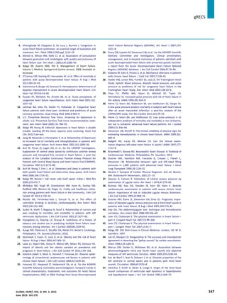
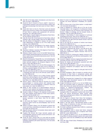
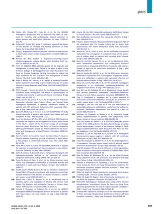
![131. Ronco C, Haapio M, House AA, Anavekar N, Bellomo R. Cardiorenal
syndrome. J Am Coll Cardiol 2008;52:1527–39.
132. Ronco C, McCullough P, Anker SD, et al., for the ADQI Consensus
Group. Cardio-renal syndromes: report from the consensus
conference of the acute dialysis quality initiative. Eur Heart J 2010;
31:703–11.
133. Felker GM, Lee KL, Bull DA, et al., for the NHLBI Heart Failure
Clinical Research Network Diuretic strategies in patients with
acute decompensated heart failure. N Engl J Med 2011;364:
797–805.
134. Packer M, O’Connor CM, Ghali JK, et al., for the Prospective
Randomized Amlodipine Survival Evaluation Study Group Effect of
amlodipine on morbidity and mortality in severe chronic heart
failure. N Engl J Med 1996;335:1107–14.
135. Bradley TD, Logan AG, Kimoff RJ, et al., for the CANPAP Investigators
Continuous positive airway pressure for central sleep apnea and
heart failure. N Engl J Med 2005;353:2025–33.
136. Kaneko Y, Floras JS, Usui K, et al. Cardiovascular effects of contin-
uous positive airway pressure in patients with heart failure and
obstructive sleep apnea. N Engl J Med 2003;348:1233–41.
137. Nichol KL, Margolis KL,Wuorenma J,Von Sternberg T.The efficacy and
cost effectiveness of vaccination against influenza among elderly
persons living in the community. N Engl J Med 1994;331:778–84.
138. Opasich C, Febo O, Riccardi PG, et al. Concomitant factors of
decompensation in chronic heart failure. Am J Cardiol 1996;78:354–7.
139. Ghali JK, Kadakia S, Cooper R, Ferlinz J. Precipitating factors leading
to decompensation of heart failure: traits among urban blacks. Arch
Intern Med 1988;148:2013–6.
We have started to publish a series of white papers with the primary intent of translating currently available evidence to fit our
global audience in the low and middle income countries. This article is the first of the series of many to follow. Major Society
Guidelines (like those from the AHA, ACC, and ESC) have been written with a focus on developed nations with superior health care
delivery systems, adequate infrastructure, and monetary sufficiency. Our journal, Global Heart, targets audiences with few of
these resources, and many of those recommendations may seem impractical in their health care environment. Our journal has
taken it upon itself to digest these outlines and present them in a form more palatable to users in developing countries. A white
paper is an authoritative report or guide helping readers to understand an issue, solve a problem, or make a decision [1], and,
while being an expert consensus statement, it is not an unalterable policy statement [1,2]. The expert end users and their societies
may modify its recommendations based on the needs of their individual health care environment. We intend this expert
consensus statement to provoke a thoughtful debate, just as all good white papers should [3].
REFERENCES
1. White paper. Available at: http://en.wikipedia.org/wiki/White_paper. Accessed May 24, 2013.
2. Doerr, Audrey D. The role of white papers. In: Doern GB, Aucoin P. The Structures of Policy-making in Canada. Toronto:
MacMillan; 1971:179e203.
3. Pemberton JE. Government green papers. Library World 1969;71:49.
jgRECS
170 GLOBAL HEART, VOL. 8, NO. 2, 2013
June 2013: 141-170](https://image.slidesharecdn.com/lmicwhitepaperforsystolichf-160210084559/85/Lmic-white-paperforsystolichf-30-320.jpg)


![APPROACH TO FEVER IN PEDIATRICS[1].pptTT](https://cdn.slidesharecdn.com/ss_thumbnails/approachtofeverinpediatrics1-260125081456-d559e079-thumbnail.jpg?width=640&height=640&fit=bounds)


