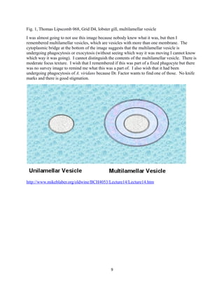
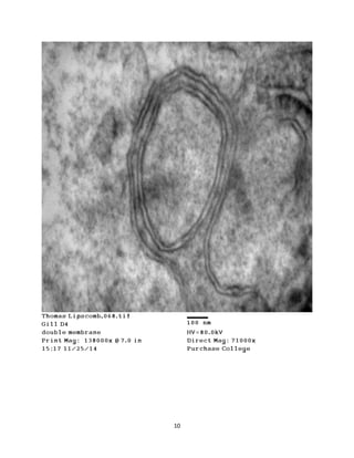
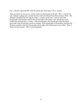
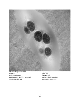
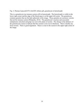
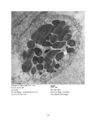
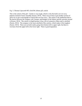
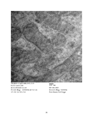
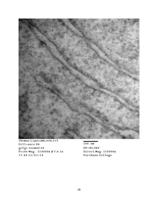
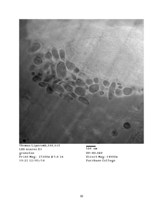
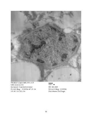
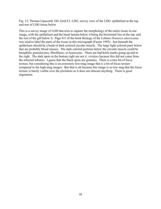
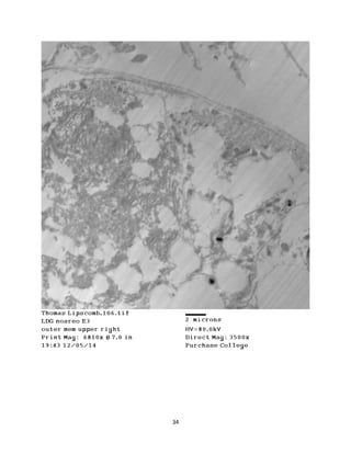
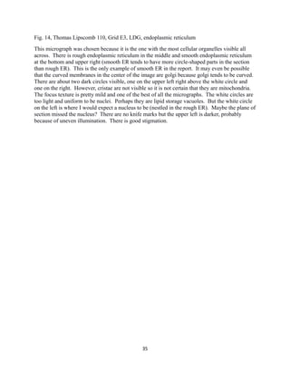
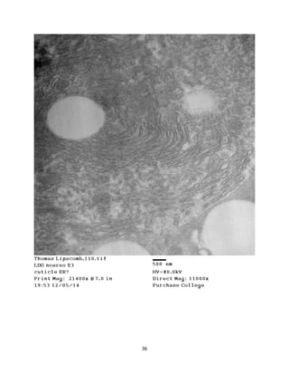
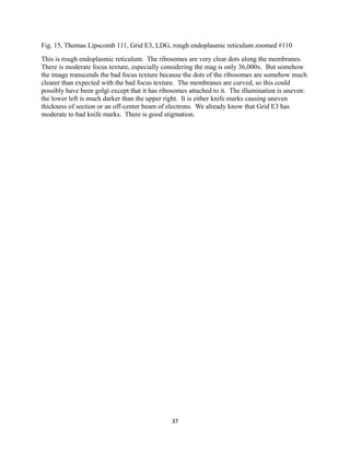
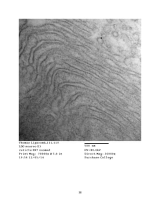
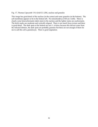
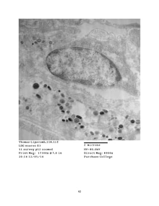
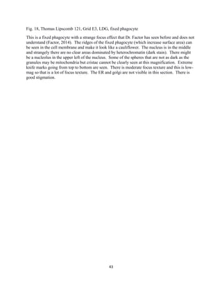
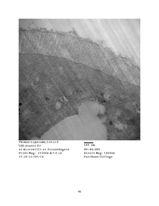
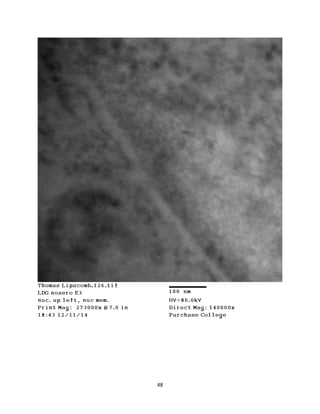
Thomas Lipscomb
Transmission Electron Microscopy and Cell Ultrastructure (BIO.4650)
Fall 2014
TEM Micrographs of Homarus americanus:
Lobster Digestive Gland and Aerococcus
viridans-Infected Gill
1
Introduction
This series of transmission electron microscope micrographs images two organs in
Homarus americanus: lobster digestive gland and Aerococcus viridans-infected gill. Aerococcus
viridans-infected gill is being investigated because the lab is trying to understand the pathology
of Aerococcus viridans, especially how they survive uptake into the lobster by vacuoles and then
how the filaments that Aerococcus viridans releases might be the cause of the vacuoles
pathologically growing too large. Non-infected lobster digestive gland is being investigated by
me because it is the control group. Aerococcus viridans-infected lobster digestive gland is an
experimental group and others are comparing it to the non-infected lobster digestive gland
healthy tissue.
I hope to see the gill tissue (and even the blood spaces feeding it) containing
pathologically large vacuoles containing Aerococcus viridans. I hope to see the cuticle of the
gill, granulocytes, mitochondria, smooth and rough endoplasmic reticulum, and nuclei. I hope to
see the lobster digestive gland containing microvilli, fixed phagocytes, more granulocytes, more
endoplasmic reticulum, more nuclei, and more mitochondria.
One point of comparison between the tissues of the gill and LDG is that gill is an
Aerococcus viridans-infected tissue and the LDG is not. The blood spaces in the gill and the
blood spaces in the LDG can be compared. The gill filaments are similar in structure to the
LDG’s microvilli, but the gill filaments are much larger. The cell components of the cells in the
gill and the cells in the LDG can be compared: endoplasmic reticulum, mitochondria, nucleus,
and vesicles. I did not find golgi anywhere even though it should be present (Factor, 2014).
The lobster digestive gland is located in the midgut (Factor, 1995). It is primarily
responsible for the synthesis and secretion of digestive enzymes, the final digestion of food, and
absorption of nutrients (Factor, 1995). It is also involved in excretion, lipid and carbohydrate
metabolism, storage of lipids, and storage of inorganic reserves (Factor, 1995). The lobster
digestive gland is a pair of large, complex glands, each with its own duct, lying primarily in the
cephalothorax (Factor, 1995). Each gland is divided into three lobes (Factor, 1995). Each lobe
has a series of blindly ending digestive tubules that connect to the duct (Factor, 1995). The
tubules are approximately 100 µm in diameter (Factor, 1995). Circular and longitudinal
contractile bundles of myofilaments lie between the basement membrane and the tubules (Factor,
1995). Microvilli increase the surface area of the LDG tubules (Factor, 2014). The tubules are
interspersed with hepatic arterioles that are covered in fixed phagocytes (Factor, 1995). The
arterioles supply the tubules with blood (Factor, 1995). The fixed phagocytes contain electron-
dense granules and are ridged to increase surface area (Factor, 1995). The fixed phagocytes are
part of the lobster’s immune system by filtering foreign material from the bloodstream and being
a barrier between any germs that might enter from the ingested food in the LDG, into the
bloodstream (Factor, 1995). The fixed phagocytes engulf germs like Aerococcus viridans, but
Aerococcus viridans somehow survives in the phagocytic vacuoles and over time the phagocytic
vacuoles become pathologically large, perhaps because of the filaments that Aerococcus viridans
secretes (Factor, 2014).
The lobster gill contains two blood vessels: the branchial stem afferent vessel that carries
blood from the gill to the heart, and the branchial stem efferent vessel that carries blood away
from the heart to the gill (Factor, 2014). Those two blood vessels are next to each other and
2
separated by a septum and covered by a circular vessel (Factor, 2014). The circular vessel is
covered by gill filaments, that are long tubes hanging from the circular vessel (Factor, 2014).
Blood circulates through the gill filaments (Factor, 2014).
Our transmission electron microscope uses an electron beam generated from an electron
cloud around a heated tungsten filament, focused through several apertures and electron lenses,
to image a thin section (Factor, 2014). The section is stained with electron-dense materials so
when the electron beam passes through the section the stained parts absorb more electrons than
the non-stained parts, which creates enough contrast to show detail in the image (Factor, 2014).
Materials and Methods
Lobster Digestive Gland (LDG) (No author, Appendix 3)
On the first day, the samples went through primary fixation: >6 hours-overnight with 3%
glutaraldehyde in 0.2 M NaCac buffer at 4ºC. On the second day, the sample was rinsed with
buffer four times, each for >15 minutes. Then six hours of secondary fixation of the samples
with 1% OsO4 in buffer, occurred. Then >6 hours-overnight secondary fixation of the samples
with another 1% OsO4 in buffer, occurred. On the third day, the sample was rinsed with buffer
for >15 minutes, twice and rinsed with distilled water for >15 minutes, four times. Then the
sample was en bloc stained >6 hours-overnight, with 1% aq. uranyl acetate at room temperature.
On the fourth day, the sample was dehydrated for >15 minutes (each) with 30% ethanol, then
50% ethanol, then 70% ethanol, then 90% ethanol, then >1 hour with 100% ethanol over CuSO4,
then >2-3 hours with 100% ethanol over CuSO4. Then the sample was dehydrated for >15
minutes (each) with 50/50 ethanol and propylene oxide, 100% propylene oxide, and again with
100% propylene oxide. Then the sample was infiltrated for >2 hours with 50/50 propylene oxide
and embedding medium and infiltrated again, for >2-3 hours, with 100% embedding medium.
Then the sample was infiltrated for >6 hours-overnight with 100% embedding medium. On the
fifth day, the sample was embedded for the last time (in a plastic block), cured at room
temperature for >1 hour, and polymerized in a 60ºC oven for 12-24 hours.
The block was trimmed down with a razor blade until tissue was reached. Then thick
sections were made with the LKB/Cambridge Huxley Microtome to smooth out the block face.
Then thin sections (100 nm) were made with the LKB/Cambridge Huxley Microtome and put on
copper grids for viewing in the TEM.
The TEM was stigmated with reference to a holey grid (Factor, 2012). A background
image with nothing in the TEM’s stage was taken to calibrate the AMT Digital Camera (Factor,
2012). The copper grid was inserted into the specimen rod, sent through the airlock, and placed
on the TEM’s stage (Factor, 2012). When the copper grid with the sample was viewed in the
TEM, the first condenser lens aperture (300 µm) and second objective aperture (50 µm) were
used with the filament at a tension of 80 kV (Factor, 2012). But the LDG staining was very faint,
so micrographs 123-126 used the third objective aperture (30 µm) to improve contrast. The
micrographs of the samples were recorded using the AMT Digital Camera in quality image mode
with autogain on (Factor, 2012). Micrographs 1-73 were not well focused. Starting with image
74, before each image was taken the AMT Digital Camera was removed and the image was
focused with reference to the focusing screen, then the beam was spread until it was dim enough
for the camera and the camera was reinserted (Factor, 2012).
3
Aerococcus viridans-Infected Gill (Surace, 2012)
Six live Homarus americanus lobsters were purchased from a local commercial vendor.
Five lobsters were kept in a 30 gallon tank containing Instant Ocean artificial sea water at 15ºC
and 28 ppt salinity. A 50 gallon capacity filter with a foam and charcoal insert filtered the sea
water to keep organic debris and chlorine levels to a minimum. The tank was in a temperature-
controlled room and the lobsters were monitored closely. The lobsters were fed a diet of catfish
fillets.
Dr. James Daly of Purchase College provided a suspension of culture grown Aerococcus
viridans bacterium, diluted to 5·106
bacteria per 1.0 M of inoculant. This culture originated from
Dr. Richard Nilford, CT Laboratory of the National marine Fisheries Service, who isolated the
bacterium from an infected lobster. Dr. Daly’s culture was injected into the five (experimental)
lobsters but not injected into the sixth (control) lobster. The times of allowed uptake for the five
lobsters were 90 minutes, 24 hours, three days, one week, and finally a natural demise after eight
days. The control and 90 minute uptake specimens were kept cool and moist in the laboratory
before being sacrificed. All specimens were anesthetized in a freezer, humanely euthanized, and
dissected to remove tissue samples.
Digestive gland tissue, gills, and blood samples were obtained from all six lobsters and
fixed with a Lysine Acetate, Ruthenium Red, Osmium Tetroxide fixation method, following the
procedure from the Hammerschmidt et al. (2005) experiment. Specimens were initially fixed
with 2.0% formaldehyde and 2.5% glutaraldehyde in 0.1 M sodium cacodylate buffer. The
buffer contained 0.075% ruthenium red and 0.075 M lysine acetate. The initial fixation was
performed in chilled solutions with a pH of 6.9. Starting April 18th
2012, samples from the
lobsters were placed in this fixative, which was then stored in the refrigerator until all lobsters
were sacrificed (April 26th
2012) and the remaining steps of the procedure could be carried out.
Then a buffer rinse was performed on the specimens in the 0.1 M sodium cacodylate
buffer containing 0.075% ruthenium red; the specimens were rinsed for a total of three hours.
The second fixation was then performed with the same fixative used in the initial fixation, except
without lysine acetate. Samples were then rinsed in the 0.1 M sodium cacodylate buffer
containing 0.075% ruthenium red for one hour at room temperature for a total of three rinses.
The third fixation of the samples was carried out with 1.0% osmium tetroxide solution in 0.1 M
sodium cacodylate buffer containing 0.075% ruthenium red, for one hour at ambient room
temperature, followed by three rinses in 0.1 M sodium cacodylate buffer containing 0.075%
ruthenium red at room temperature for one hour. A second set of specimen samples were fixed
differently for Frank Surace studying the fixed phagocytes, but were not used in my project.
After fixation and rinses, the samples prepared in lysine acetate / ruthenium red were
dehydrated and embedded. The dehydration consisted of ethanol concentrations of 10%, 30%,
50%, 70%, 90% and two 100% dehydrations over CuSO4. Each step in the series was carried out
for a total of 30 minutes each with exceptions to the 100% dehydrations. The first 100%
dehydration was carried out for one hour on ice while the second was performed for 24 hours on
ice.
Specimens were then infiltrated with a solution consisting of a 50/50 mixture of 100%
ethanol and acetonitrile. Two 100% acetonitrile infiltrations, each 15 minutes, followed.
4
Specimen embedding was then performed on tissue with a 50/50 mixture of acetonitrile and
Araldite 502 – Embed 812 plastic resin embedding medium for 15 minutes. Tissues were then
embedded in 100% plastic resin medium, each overnight. Specimens were allowed to cure for
one hour at room temperature and then taken and put into molds with fresh Araldite 502 –
Embed 812 plastic resin medium (from Electron Microscopy Sciences, Inc.) and polymerized in
an oven at 60-70ºC to form blocks for sectioning. The resulting blocks were coded with the
corresponding vial from which they were taken.
The block was trimmed down with a razor blade until tissue was reached. Then thick
sections were made with the LKB/Cambridge Huxley Microtome to smooth out the block face.
Then thin sections (100 nm) were made with the LKB/Cambridge Huxley Microtome and put on
copper grids for viewing in the TEM.
The TEM was stigmated with reference to a holey grid (Factor, 2012). A background
image with nothing in the TEM’s stage was taken to calibrate the AMT Digital Camera (Factor,
2012). The copper grid was inserted into the specimen rod, sent through the airlock, and placed
on the TEM’s stage (Factor, 2012). When the copper grid with the sample was viewed in the
TEM, the first condenser lens aperture (300 µm) and second objective aperture (50 µm) were
used with the filament at a tension of 80 kV (Factor, 2012). The micrographs of the samples
were recorded using the AMT Digital Camera in quality image mode with autogain on (Factor,
2012). Micrographs 1-73 were not well focused. Starting with image 74, before each image was
taken the AMT Digital Camera was removed and the image was focused with reference to the
focusing screen, then the beam was spread until it was dim enough for the camera and the
camera was reinserted (Factor, 2012).
5
Grid Tracking Form
Summary of Micrographs
6
Fig. 1, Thomas Lipscomb 068, 71,000x, lobster gill, multilamellar vesicle
Fig. 2, Thomas Lipscomb 069, 22,000x, lobster gill, blood space with A. viridans
Fig. 3, Thomas Lipscomb 075, 11,000x, lobster gill, granulocyte in hemolymph
Fig. 4, Thomas Lipscomb 081, 71,000x, lobster gill, A. viridans with filaments in vacuole
Fig. 5, Thomas Lipscomb 083, 8,900x, lobster gill, cuticle
Fig. 6, Thomas Lipscomb 084, 14,000x, lobster gill, nucleus and mitochondria (several cells?)
Fig. 7, Thomas Lipscomb 090, 5,600x, lobster gill, cuticle on left epithelium on right nucleus at
bottom left rough ER and mitochondria in middle
Fig. 8, Thomas Lipscomb 091, 14,000x, lobster gill, mitochondria and rough ER
Fig. 9, Thomas Lipscomb 092, 56,000x, lobster gill, mitochondria
Fig. 10, Thomas Lipscomb 094, 110,000x, lobster gill, rough endoplasmic reticulum
Fig. 11, Thomas Lipscomb 100, 14,000x, LDG, granulocyte
Fig. 12, Thomas Lipscomb 105, 11,000x, LDG, nucleus between lipid storage vacuoles
Fig. 13, Thomas Lipscomb 106, 3,500x, LDG, survey view of the LDG: epithelium at the top
and rest of LDG tissue below
Fig. 14, Thomas Lipscomb 110, 11,000x, LDG, endoplasmic reticulum
Fig. 15, Thomas Lipscomb 111, 36,000x, LDG, rough endoplasmic reticulum zoomed #110
Fig. 16, Thomas Lipscomb 116, 4,400x, LDG, lipid storage vacuoles and microvilli on lower left
Fig. 17, Thomas Lipscomb 118, 8,900x, LDG, nucleus and granules
Fig. 18, Thomas Lipscomb 121, 8,900x, LDG, fixed phagocyte
Fig. 19, Thomas Lipscomb 124, 14,000x, LDG, microvilli
Fig. 20, Thomas Lipscomb 126, 140,000x, LDG, nucleus upper right, nuclear membrane
7
Micrographs with Captions and Observations
8
Fig. 1, Thomas Lipscomb 068, Grid D4, lobster gill, multilamellar vesicle
I was almost going to not use this image because nobody knew what it was, but then I
remembered multilamellar vesicles, which are vesicles with more than one membrane. The
cytoplasmic bridge at the bottom of the image suggests that the multilamellar vesicle is
undergoing phagocytosis or exocytosis (without seeing which way it was moving I cannot know
which way it was going). I cannot distinguish the contents of the multilamellar vesicle. There is
moderate focus texture. I wish that I remembered if this was part of a fixed phagocyte but there
was no survey image to remind me what this was a part of. I also wish that it had been
undergoing phagocytosis of A. viridans because Dr. Factor wants to find one of those. No knife
marks and there is good stigmation.
http://www.mikeblaber.org/oldwine/BCH4053/Lecture14/Lecture14.htm
9
10
Fig. 2, Thomas Lipscomb 069, Grid D4, lobster gill, blood space with A. viridans
These are about six Aerococcus viridans inside of a blood space in the gill. The A. viridans not
only multiply in phagocytic vacuoles but also within the blood of the lobster (Factor, 2014). The
filaments extending from the capsule of the A. viridans can be seen. I am not sure if the
hemolymph is the powdery stuff on the left and right of the image or the smooth part in the
middle that surrounds the A. viridans. If the hemolymph is the smooth space then the blood
space ends when the powdery section is reached. If the hemolymph is the powdery stuff then the
filaments somehow repel the hemolymph and the edge of the blood space is not visible. There is
almost no focus texture. There is good stigmation.
11
12
Fig. 3, Thomas Lipscomb 075, Grid D5, lobster gill, granulocyte in hemolymph
This is a granulocyte (an immune system cell) in hemolymph. The hemolymph is visible in the
lower right corner and the edge of the blood space is in the upper left corner. The granulocyte
contains granules that are the dark spheroids in this image. Those granules are secretory vesicles
that aid the immune system (Wikipedia, 2014). The granulocyte’s nucleus and associated
endoplasmic reticulum and golgi was not in this plane of section. The change in the darkness of
the granulocytes seems to indicate that this section is not even in thickness. There is hardly any
focus texture. There is good stigmation. There is a tear in the section in the upper right corner of
the image.
13
14
Fig. 4, Thomas Lipscomb 081, Grid D5, lobster gill, A. viridans with filaments in vacuole
This is the highest-mag micrograph of A. viridans in the report. It was created to best show the
filaments that extend from the A. viridans capsule. This image was of A. viridans in a vacuole,
not another area such as a blood vessel. The working hypothesis is that the filaments cause the
vacuole to pathologically enlarge, but that has not yet been proven (Factor, 2014). There is
almost no focus texture, and that is good for a high mag micrograph. The two A. viridans are
probably together because they can remain a dyad after binary fission (Factor, 2014). They are
called filaments not extracellular matrix, as the caption guesses. There are no knife marks.
There is good stigmation.
15
16
Fig. 5, Thomas Lipscomb 083, Grid D4, lobster gill, cuticle
This is the cuticle of the gill. Cuticle is very tough, which is why the knife cut it in wavy
patterns instead of more smoothly (Factor, 2014). There was no time to get another section of
gill to try to get a micrograph of cuticle that was less wavy. The cuticle is the epithelium that is
the barrier between the carapace, gill, foregut, and hindgut of the lobster and the seawater around
it (Factor, 2014). The cuticle is tough because it has a lot of connective fibers and is calcified
(Factor, 2014). The carapace is the most calcified of the cuticles, which makes it the toughest
(Factor, 2014). There are no knife marks going from bottom left to upper right, but there is
waviness from the upper left to the lower right. There is good stigmation.
17
18
Fig. 6, Thomas Lipscomb 084, Grid D4, lobster gill, nucleus and mitochondria (several cells?)
Thin sections are not always in convenient planes. For example, if the thin section is not
perpendicular enough to the membrane then the bilayer looks like a monolayer. The problem
here appears to be that the section passed through several cells at an awkward angle so only the
nucleus (found by the darkness of its heterochromatin) at the top makes sense. The rest of the
image is a jumble of mitochondria and what may or may not be endoplasmic reticulum or golgi
and may or may not be cell membranes. There is almost no focus texture, but this is a low mag
image so that is less apparent. There is good stigmation.
19
20
Fig. 7, Thomas Lipscomb 090, Grid D4, lobster gill, cuticle on left epithelium on right nucleus at
bottom left rough ER and mitochondria in middle
There is cuticle on the left, epithelium on the right, a nucleus at the bottom left, and rough ER
and mitochondria in the middle. This is the micrograph that has the biggest collection of
organelles in one micrograph. But the clarity of the image is not as good as Figure 14, which
also has a huge field of organelles but is mostly rough ER. There are knife marks going to the
right and slightly down. There is moderate focus texture. There is good stigmation.
21
22
Fig. 8, Thomas Lipscomb 091, Grid D4, lobster gill, mitochondria and rough ER
This is the best survey image of mitochondria in the report. It shows the location of the
mitochondria in relation to the rough endoplasmic reticulum. The mitochondria appear to be
evenly distributed around the cell. The nucleus may be at the bottom right if those dark areas are
heterochromatin. There is almost no focus texture, but this is a low mag image so that is less
apparent. The illumination may be off-center. The rough ER here is slightly curved but not
curved enough to be golgi, as the caption guesses it is. There is good stigmation.
23
24
Fig. 9, Thomas Lipscomb 092, Grid D4, lobster gill, mitochondria
This is the only high-mag image of mitochondria in the report. The inner membrane, cristae
(inner membrane folds), and outer membrane are clearly visible. When taking this image I did
not think that I could see anything more if I went to a higher magnification, but now I think that
if I had I might have seen mitochondrial ribosomes as I had seen ribosomes in the ER. I also had
thought that the mitochondrial DNA is not densely packed enough to be visible at any
magnification, but in the lecture page 12 there is a TEM pictures of DNA or RNA that burst from
a bacteriophage (Factor, 2014), so maybe even unpacked DNA can be seen sometimes. There is
some focus texture but at this high mag that is pretty much unavoidable. The right of the image
is slightly brighter than the left, either because of a misaligned electron beam or different section
thickness. There are no knife marks. There is good stigmation.
25
26
Fig. 10, Thomas Lipscomb 094, Grid D4, lobster gill, rough endoplasmic reticulum
The gill micrographs need one image that is at least 140,000x but all of the 140,000x pictures of
gill were blurry, so this 110,000x image is in its place. This is the only high-mag image of rough
endoplasmic reticulum in the report, however Figure 15 somehow shows the ribosomes in the
rough endoplasmic reticulum better even though it is only mag 36,000x. There are periodic
blurry dots on these membranes, which I assume are ribosomes attached to the membranes. The
membranes here are very clear, but strangely the ribosomes are not, they are much more blurry
than I expected, especially compared to Figure 15. For some reason the right of this image is
lighter than the left, either because of uneven section thickness or a misaligned electron beam
that is not pointing towards the center but towards the right. There is some focus texture but at
this high mag that is pretty much unavoidable. This is rough ER, not golgi as the caption
guesses. There are no knife marks. There is good stigmation.
27
28
Fig. 11, Thomas Lipscomb 100, Grid E3, LDG, granulocyte
This is a granulocyte somewhere in the LDG. This section appears to have uneven thickness,
with a light and thin part at the top and a dark and thick part at the bottom. The nucleus of the
granulocyte is to the right. The granulocyte contains granules that are the dark spheroids in this
image. Those granules are secretory vesicles that aid the immune system (Wikipedia, 2014).
This is the worst micrograph of the twenty because of the serious thickness unevenness. But it is
the only image of a granulocyte in LDG. Bad knife marks going up and to the right are visible,
and unfortunately only one grid (Grid E3) gave me good micrographs of the LDG despite me
trying to get better sections after I took this image. So bad knife marks will plague Figures 11-
20. This image is too low mag for focus texture to appear. There is good stigmation.
29
30
Fig. 12, Thomas Lipscomb 105, Grid E3, LDG, nucleus between lipid storage vacuoles
This was an island of darkness in a giant field of light-colored lipid storage vacuoles. It could be
a blood space, with what looks like hemolymph, feeding the lipid-storage cells around it, but it is
strange that there are no fixed phagocytes visible around the blood space, so it is probably not a
blood space. The dark portion in the center also looks similar to heterochromatin and
euchromatin, so the dark portion is probably a nucleus. The knife marks here are a lot less
pronounced than a lot of the other micrographs of Grid E3 but you can still see them, they are
almost vertical. The focus texture is pretty mild and one of the best of all the micrographs.
There is good stigmation.
31
32
Fig. 13, Thomas Lipscomb 106, Grid E3, LDG, survey view of the LDG: epithelium at the top
and rest of LDG tissue below
This is a survey image of LGD that tries to capture the morphology of the entire tissue in one
image, with the epithelium and the basal lamina below it being the horizontal line at the top, and
the rest of the gill below it. Page 411 of the book Biology of the Lobster Homarys americanus
was used to label the parts of the tissue in this micrograph (Factor 1995). Just beneath the
epithelium should be a band of dark-colored circular muscle. The large light-colored parts below
that are probably blood sinuses. The dark colored portions below the circular muscle could be
basophilic granulocytes, fibroblasts, or hemocytes. There are bad knife marks going up and to
the right. The dark spots at the bottom right are not A. viridans because this did not come from
the infected lobsters. I guess that the black spots are granules. There is a tiny bit of focus
texture, but considering this is an extremely low-mag image that is a lot of focus texture
compared to the high-mag images. But that is ok because this image is so low mag that the focus
texture is barely visible over the pixilation so it does not obscure anything. There is good
stigmation.
33
34
Fig. 14, Thomas Lipscomb 110, Grid E3, LDG, endoplasmic reticulum
This micrograph was chosen because it is the one with the most cellular organelles visible all
across. There is rough endoplasmic reticulum in the middle and smooth endoplasmic reticulum
at the bottom and upper right (smooth ER tends to have more circle-shaped parts in the section
than rough ER). This is the only example of smooth ER in the report. It may even be possible
that the curved membranes in the center of the image are golgi because golgi tends to be curved.
There are about two dark circles visible, one on the upper left right above the white circle and
one on the right. However, cristae are not visible so it is not certain that they are mitochondria.
The focus texture is pretty mild and one of the best of all the micrographs. The white circles are
too light and uniform to be nuclei. Perhaps they are lipid storage vacuoles. But the white circle
on the left is where I would expect a nucleus to be (nestled in the rough ER). Maybe the plane of
section missed the nucleus? There are no knife marks but the upper left is darker, probably
because of uneven illumination. There is good stigmation.
35
36
Fig. 15, Thomas Lipscomb 111, Grid E3, LDG, rough endoplasmic reticulum zoomed #110
This is rough endoplasmic reticulum. The ribosomes are very clear dots along the membranes.
There is moderate focus texture, especially considering the mag is only 36,000x. But somehow
the image transcends the bad focus texture because the dots of the ribosomes are somehow much
clearer than expected with the bad focus texture. The membranes are curved, so this could
possibly have been golgi except that it has ribosomes attached to it. The illumination is uneven:
the lower left is much darker than the upper right. It is either knife marks causing uneven
thickness of section or an off-center beam of electrons. We already know that Grid E3 has
moderate to bad knife marks. There is good stigmation.
37
38
Fig. 16, Thomas Lipscomb 116, Grid E3, LDG, lipid storage vacuoles and microvilli on lower
left
The LDG, like the human liver, stores lipids in lipid storage vacuoles. The lipid storage vacuoles
are the light-colored spheres. The microvilli of the LDG are in the lower left, so these lipid
storage vacuoles are pretty close to the lipids’ point of origin inside of the food in the digestive
tubules of the LDG. Cells like these tend to have the nucleus and ER as far away from the
microvilli as possible (Factor, 2014) so they are safe and do not get in the way. I assume that
these lipid storage vacuoles are travelling from the microvilli in the lower left towards the blood
space past the upper right of the image, to transport the food from the LDG to the rest of the
lobster. There are no nuclei, ER, golgi, or mitochondria in this micrograph. The dark spots at
the top, left, and bottom are not A. viridans because this did not come from the infected lobsters.
I guess that the black spots are contamination because they do not have the regularity in size and
shape that granules have. There are bad knife marks (going up and to the right) in this image.
There is mild focus texture, but considering that this is 4,400x that is actually moderate focus
texture. There is good stigmation.
39
40
Fig. 17, Thomas Lipscomb 118, Grid E3, LDG, nucleus and granules
This image has good detail of the nucleus (in the center) and some granules (at the bottom). The
cell membrane appears to be in the bottom left. No mitochondria or ER are visible. There is
clearly some heterochromatin (dark stain) in the nucleus and the lighter stains are euchromatin.
The knife marks are moderate and vertically aligned. There is not much focus texture and there
is good detail. The dark spots at the bottom are not A. viridans because this did not come from
the infected lobsters, the dark spots are probably granules but there are not enough of them for
me to call this cell a granulocyte. There is good stigmation.
41
42
Fig. 18, Thomas Lipscomb 121, Grid E3, LDG, fixed phagocyte
This is a fixed phagocyte with a strange focus effect that Dr. Factor has seen before and does not
understand (Factor, 2014). The ridges of the fixed phagocyte (which increase surface area) can
be seen in the cell membrane and make it look like a cauliflower. The nucleus is in the middle
and strangely there are no clear areas dominated by heterochromatin (dark stain). There might
be a nucleolus in the upper left of the nucleus. Some of the spheres that are not as dark as the
granules may be mitochondria but cristae cannot be clearly seen at this magnification. Extreme
knife marks going from top to bottom are seen. There is moderate focus texture and this is low-
mag so that is a lot of focus texture. The ER and golgi are not visible in this section. There is
good stigmation.
43
44
Fig. 19, Thomas Lipscomb 124, Grid E3, LDG, microvilli
This is the only high mag image of the microvilli in the LDG in this report. The microvilli are at
the top of the image and are a forest of tubes that increase the surface area of the cell membrane
to increase the rate of food absorption (Factor, 2014). Figures 11-18 had less contrast than what I
thought was optimal, so this figure was made using objective aperture 3 (30 µm) of the TEM, not
the standard objective aperture 2 (50 µm) that were used for Figures 1-18. At the bottom of this
image is what appears to be a lipid storage vacuole. I was surprised at the lack of detail and
structures in the cytoplasm between the microvilli and the lipid storage vacuole. The nucleus
and its associated endoplasmic reticulum and golgi would not be in this portion because it would
be at the basal end of the cell, not the apical end with the microvilli (Factor, 2014). The
mitochondria might be at this apical end but none are visible in this section. There are bad knife
marks going up and to the left, which we have seen before in grid E3. There is moderate focus
texture and this is a moderate-mag image. There is good stigmation.
45
46
Fig. 20, Thomas Lipscomb 126, 140,000x, LDG, nucleus upper right, nuclear membrane
Figures 11-18 had less contrast than what I thought was optimal, so this figure was made using
objective aperture 3 (30 µm) of the TEM, not the standard objective aperture 2 (50 µm) that were
used for Figures 1-18. In the middle of this image is the nuclear membrane. The nucleus is in
the upper right corner (not the upper left as the image caption says, that was a mistake). The
cytoplasm is in the lower left corner. The nuclear membrane in-between can be seen clearly but
unlike the other high-mag micrographs the dark lines are not very visible. There is very bad
focus texture, the worst in the entire report. Maybe the dark parts are not as clear because the
LDG was not stained as heavily as the gill. Without strong dark parts to focus on I had much
more trouble focusing this image because I could only do it based on the worminess of the
background, which may be why the focus is so off. There is good stigmation.
47
48
Discussion
Of course both Gill and LDG have nuclei, rough and smooth endoplasmic reticulum,
golgi, and mitochondria. However, smooth ER was only found in LDG (Figure 14) and that
makes sense because there should be a lot more smooth ER in LDG because there is a lot more
for smooth ER to do in LDG (handling lipid storage vacuoles for example) than in gill. Golgi
was not conclusively found anywhere because I am not good enough at identifying it. I only
look for unusually curved endoplasmic reticulum to say it might be golgi.
I got good images of blood spaces in the gill (Figure 2) and the LDG (Figure 13).
Granulocytes were found in both Gill (Figure 4) and LDG (Figure 12). A fixed phagocyte was
only found in the LDG (Figure 18) not the gill even though both should have them (the fixed
phagocytes filter the blood). Lipid storage vacuoles were found in the LDG (Figures 12, 13, 14,
and 16) not the gill, as expected. Microvilli were found in the LDG (Figures 16 and 19) not the
gill, as expected. The gill also have microvilli-like structures radiating from the circular vessel
(Factor, 2014), but they are much larger than microvilli because they need the space to have their
own blood circulation. Aerococcus viridans were only found in the gill not the LDG, as expected
because only the lobster that the gill came from was infected with A. viridans, not the lobster that
the LDG came from.
49
Literature Cited
1. Factor, J.R. “Transmission Electron Microscopy and Cell Ultrastructure.” BIO 4650 class
lecture. SUNY Purchase, Purchase. August 25, 2014 to December 11, 2014. PowerPoint
lecture.
2. Factor, J.R. 1995. Biology of the Lobster Homarus americanus. (First Edition.) Waltham:
Academic Press, Inc. Chapter 15, The Digestive System, pp. 409-421.
3. Appendix 3. TEM Plastic: Araldite 502/Embed-812TM
Embedding Tracking Form. TEM
Embedding Procedure – For Araldite 502/Embed-812TM
Plastic (Procedure for Tissues
without Polystyrene Microspheres).
4. Surace, F. 2012. Testing for Capsules in Aerococcus viridans: The Disease Agent for
Gaffkemia in American Lobsters. SUNY Purchase Biology BA thesis.
5. Factor, J.R. 2012. Operating Instructions for FEI/Philips Morgagni (M268) Transmission
Electron Microscope.
6. Granulocyte - Wikipedia, the Free Encyclopedia. (2014, November 16). Retrieved
December 14, 2014, from http://en.wikipedia.org/wiki/Granulocyte
50

Lipscomb, Thomas - TEM'14 - Report

  • 1.
    Thomas Lipscomb Transmission ElectronMicroscopy and Cell Ultrastructure (BIO.4650) Fall 2014 TEM Micrographs of Homarus americanus: Lobster Digestive Gland and Aerococcus viridans-Infected Gill 1
  • 2.
    Introduction This series oftransmission electron microscope micrographs images two organs in Homarus americanus: lobster digestive gland and Aerococcus viridans-infected gill. Aerococcus viridans-infected gill is being investigated because the lab is trying to understand the pathology of Aerococcus viridans, especially how they survive uptake into the lobster by vacuoles and then how the filaments that Aerococcus viridans releases might be the cause of the vacuoles pathologically growing too large. Non-infected lobster digestive gland is being investigated by me because it is the control group. Aerococcus viridans-infected lobster digestive gland is an experimental group and others are comparing it to the non-infected lobster digestive gland healthy tissue. I hope to see the gill tissue (and even the blood spaces feeding it) containing pathologically large vacuoles containing Aerococcus viridans. I hope to see the cuticle of the gill, granulocytes, mitochondria, smooth and rough endoplasmic reticulum, and nuclei. I hope to see the lobster digestive gland containing microvilli, fixed phagocytes, more granulocytes, more endoplasmic reticulum, more nuclei, and more mitochondria. One point of comparison between the tissues of the gill and LDG is that gill is an Aerococcus viridans-infected tissue and the LDG is not. The blood spaces in the gill and the blood spaces in the LDG can be compared. The gill filaments are similar in structure to the LDG’s microvilli, but the gill filaments are much larger. The cell components of the cells in the gill and the cells in the LDG can be compared: endoplasmic reticulum, mitochondria, nucleus, and vesicles. I did not find golgi anywhere even though it should be present (Factor, 2014). The lobster digestive gland is located in the midgut (Factor, 1995). It is primarily responsible for the synthesis and secretion of digestive enzymes, the final digestion of food, and absorption of nutrients (Factor, 1995). It is also involved in excretion, lipid and carbohydrate metabolism, storage of lipids, and storage of inorganic reserves (Factor, 1995). The lobster digestive gland is a pair of large, complex glands, each with its own duct, lying primarily in the cephalothorax (Factor, 1995). Each gland is divided into three lobes (Factor, 1995). Each lobe has a series of blindly ending digestive tubules that connect to the duct (Factor, 1995). The tubules are approximately 100 µm in diameter (Factor, 1995). Circular and longitudinal contractile bundles of myofilaments lie between the basement membrane and the tubules (Factor, 1995). Microvilli increase the surface area of the LDG tubules (Factor, 2014). The tubules are interspersed with hepatic arterioles that are covered in fixed phagocytes (Factor, 1995). The arterioles supply the tubules with blood (Factor, 1995). The fixed phagocytes contain electron- dense granules and are ridged to increase surface area (Factor, 1995). The fixed phagocytes are part of the lobster’s immune system by filtering foreign material from the bloodstream and being a barrier between any germs that might enter from the ingested food in the LDG, into the bloodstream (Factor, 1995). The fixed phagocytes engulf germs like Aerococcus viridans, but Aerococcus viridans somehow survives in the phagocytic vacuoles and over time the phagocytic vacuoles become pathologically large, perhaps because of the filaments that Aerococcus viridans secretes (Factor, 2014). The lobster gill contains two blood vessels: the branchial stem afferent vessel that carries blood from the gill to the heart, and the branchial stem efferent vessel that carries blood away from the heart to the gill (Factor, 2014). Those two blood vessels are next to each other and 2
  • 3.
    separated by aseptum and covered by a circular vessel (Factor, 2014). The circular vessel is covered by gill filaments, that are long tubes hanging from the circular vessel (Factor, 2014). Blood circulates through the gill filaments (Factor, 2014). Our transmission electron microscope uses an electron beam generated from an electron cloud around a heated tungsten filament, focused through several apertures and electron lenses, to image a thin section (Factor, 2014). The section is stained with electron-dense materials so when the electron beam passes through the section the stained parts absorb more electrons than the non-stained parts, which creates enough contrast to show detail in the image (Factor, 2014). Materials and Methods Lobster Digestive Gland (LDG) (No author, Appendix 3) On the first day, the samples went through primary fixation: >6 hours-overnight with 3% glutaraldehyde in 0.2 M NaCac buffer at 4ºC. On the second day, the sample was rinsed with buffer four times, each for >15 minutes. Then six hours of secondary fixation of the samples with 1% OsO4 in buffer, occurred. Then >6 hours-overnight secondary fixation of the samples with another 1% OsO4 in buffer, occurred. On the third day, the sample was rinsed with buffer for >15 minutes, twice and rinsed with distilled water for >15 minutes, four times. Then the sample was en bloc stained >6 hours-overnight, with 1% aq. uranyl acetate at room temperature. On the fourth day, the sample was dehydrated for >15 minutes (each) with 30% ethanol, then 50% ethanol, then 70% ethanol, then 90% ethanol, then >1 hour with 100% ethanol over CuSO4, then >2-3 hours with 100% ethanol over CuSO4. Then the sample was dehydrated for >15 minutes (each) with 50/50 ethanol and propylene oxide, 100% propylene oxide, and again with 100% propylene oxide. Then the sample was infiltrated for >2 hours with 50/50 propylene oxide and embedding medium and infiltrated again, for >2-3 hours, with 100% embedding medium. Then the sample was infiltrated for >6 hours-overnight with 100% embedding medium. On the fifth day, the sample was embedded for the last time (in a plastic block), cured at room temperature for >1 hour, and polymerized in a 60ºC oven for 12-24 hours. The block was trimmed down with a razor blade until tissue was reached. Then thick sections were made with the LKB/Cambridge Huxley Microtome to smooth out the block face. Then thin sections (100 nm) were made with the LKB/Cambridge Huxley Microtome and put on copper grids for viewing in the TEM. The TEM was stigmated with reference to a holey grid (Factor, 2012). A background image with nothing in the TEM’s stage was taken to calibrate the AMT Digital Camera (Factor, 2012). The copper grid was inserted into the specimen rod, sent through the airlock, and placed on the TEM’s stage (Factor, 2012). When the copper grid with the sample was viewed in the TEM, the first condenser lens aperture (300 µm) and second objective aperture (50 µm) were used with the filament at a tension of 80 kV (Factor, 2012). But the LDG staining was very faint, so micrographs 123-126 used the third objective aperture (30 µm) to improve contrast. The micrographs of the samples were recorded using the AMT Digital Camera in quality image mode with autogain on (Factor, 2012). Micrographs 1-73 were not well focused. Starting with image 74, before each image was taken the AMT Digital Camera was removed and the image was focused with reference to the focusing screen, then the beam was spread until it was dim enough for the camera and the camera was reinserted (Factor, 2012). 3
  • 4.
    Aerococcus viridans-Infected Gill(Surace, 2012) Six live Homarus americanus lobsters were purchased from a local commercial vendor. Five lobsters were kept in a 30 gallon tank containing Instant Ocean artificial sea water at 15ºC and 28 ppt salinity. A 50 gallon capacity filter with a foam and charcoal insert filtered the sea water to keep organic debris and chlorine levels to a minimum. The tank was in a temperature- controlled room and the lobsters were monitored closely. The lobsters were fed a diet of catfish fillets. Dr. James Daly of Purchase College provided a suspension of culture grown Aerococcus viridans bacterium, diluted to 5·106 bacteria per 1.0 M of inoculant. This culture originated from Dr. Richard Nilford, CT Laboratory of the National marine Fisheries Service, who isolated the bacterium from an infected lobster. Dr. Daly’s culture was injected into the five (experimental) lobsters but not injected into the sixth (control) lobster. The times of allowed uptake for the five lobsters were 90 minutes, 24 hours, three days, one week, and finally a natural demise after eight days. The control and 90 minute uptake specimens were kept cool and moist in the laboratory before being sacrificed. All specimens were anesthetized in a freezer, humanely euthanized, and dissected to remove tissue samples. Digestive gland tissue, gills, and blood samples were obtained from all six lobsters and fixed with a Lysine Acetate, Ruthenium Red, Osmium Tetroxide fixation method, following the procedure from the Hammerschmidt et al. (2005) experiment. Specimens were initially fixed with 2.0% formaldehyde and 2.5% glutaraldehyde in 0.1 M sodium cacodylate buffer. The buffer contained 0.075% ruthenium red and 0.075 M lysine acetate. The initial fixation was performed in chilled solutions with a pH of 6.9. Starting April 18th 2012, samples from the lobsters were placed in this fixative, which was then stored in the refrigerator until all lobsters were sacrificed (April 26th 2012) and the remaining steps of the procedure could be carried out. Then a buffer rinse was performed on the specimens in the 0.1 M sodium cacodylate buffer containing 0.075% ruthenium red; the specimens were rinsed for a total of three hours. The second fixation was then performed with the same fixative used in the initial fixation, except without lysine acetate. Samples were then rinsed in the 0.1 M sodium cacodylate buffer containing 0.075% ruthenium red for one hour at room temperature for a total of three rinses. The third fixation of the samples was carried out with 1.0% osmium tetroxide solution in 0.1 M sodium cacodylate buffer containing 0.075% ruthenium red, for one hour at ambient room temperature, followed by three rinses in 0.1 M sodium cacodylate buffer containing 0.075% ruthenium red at room temperature for one hour. A second set of specimen samples were fixed differently for Frank Surace studying the fixed phagocytes, but were not used in my project. After fixation and rinses, the samples prepared in lysine acetate / ruthenium red were dehydrated and embedded. The dehydration consisted of ethanol concentrations of 10%, 30%, 50%, 70%, 90% and two 100% dehydrations over CuSO4. Each step in the series was carried out for a total of 30 minutes each with exceptions to the 100% dehydrations. The first 100% dehydration was carried out for one hour on ice while the second was performed for 24 hours on ice. Specimens were then infiltrated with a solution consisting of a 50/50 mixture of 100% ethanol and acetonitrile. Two 100% acetonitrile infiltrations, each 15 minutes, followed. 4
  • 5.
    Specimen embedding wasthen performed on tissue with a 50/50 mixture of acetonitrile and Araldite 502 – Embed 812 plastic resin embedding medium for 15 minutes. Tissues were then embedded in 100% plastic resin medium, each overnight. Specimens were allowed to cure for one hour at room temperature and then taken and put into molds with fresh Araldite 502 – Embed 812 plastic resin medium (from Electron Microscopy Sciences, Inc.) and polymerized in an oven at 60-70ºC to form blocks for sectioning. The resulting blocks were coded with the corresponding vial from which they were taken. The block was trimmed down with a razor blade until tissue was reached. Then thick sections were made with the LKB/Cambridge Huxley Microtome to smooth out the block face. Then thin sections (100 nm) were made with the LKB/Cambridge Huxley Microtome and put on copper grids for viewing in the TEM. The TEM was stigmated with reference to a holey grid (Factor, 2012). A background image with nothing in the TEM’s stage was taken to calibrate the AMT Digital Camera (Factor, 2012). The copper grid was inserted into the specimen rod, sent through the airlock, and placed on the TEM’s stage (Factor, 2012). When the copper grid with the sample was viewed in the TEM, the first condenser lens aperture (300 µm) and second objective aperture (50 µm) were used with the filament at a tension of 80 kV (Factor, 2012). The micrographs of the samples were recorded using the AMT Digital Camera in quality image mode with autogain on (Factor, 2012). Micrographs 1-73 were not well focused. Starting with image 74, before each image was taken the AMT Digital Camera was removed and the image was focused with reference to the focusing screen, then the beam was spread until it was dim enough for the camera and the camera was reinserted (Factor, 2012). 5
  • 6.
  • 7.
    Fig. 1, ThomasLipscomb 068, 71,000x, lobster gill, multilamellar vesicle Fig. 2, Thomas Lipscomb 069, 22,000x, lobster gill, blood space with A. viridans Fig. 3, Thomas Lipscomb 075, 11,000x, lobster gill, granulocyte in hemolymph Fig. 4, Thomas Lipscomb 081, 71,000x, lobster gill, A. viridans with filaments in vacuole Fig. 5, Thomas Lipscomb 083, 8,900x, lobster gill, cuticle Fig. 6, Thomas Lipscomb 084, 14,000x, lobster gill, nucleus and mitochondria (several cells?) Fig. 7, Thomas Lipscomb 090, 5,600x, lobster gill, cuticle on left epithelium on right nucleus at bottom left rough ER and mitochondria in middle Fig. 8, Thomas Lipscomb 091, 14,000x, lobster gill, mitochondria and rough ER Fig. 9, Thomas Lipscomb 092, 56,000x, lobster gill, mitochondria Fig. 10, Thomas Lipscomb 094, 110,000x, lobster gill, rough endoplasmic reticulum Fig. 11, Thomas Lipscomb 100, 14,000x, LDG, granulocyte Fig. 12, Thomas Lipscomb 105, 11,000x, LDG, nucleus between lipid storage vacuoles Fig. 13, Thomas Lipscomb 106, 3,500x, LDG, survey view of the LDG: epithelium at the top and rest of LDG tissue below Fig. 14, Thomas Lipscomb 110, 11,000x, LDG, endoplasmic reticulum Fig. 15, Thomas Lipscomb 111, 36,000x, LDG, rough endoplasmic reticulum zoomed #110 Fig. 16, Thomas Lipscomb 116, 4,400x, LDG, lipid storage vacuoles and microvilli on lower left Fig. 17, Thomas Lipscomb 118, 8,900x, LDG, nucleus and granules Fig. 18, Thomas Lipscomb 121, 8,900x, LDG, fixed phagocyte Fig. 19, Thomas Lipscomb 124, 14,000x, LDG, microvilli Fig. 20, Thomas Lipscomb 126, 140,000x, LDG, nucleus upper right, nuclear membrane 7
  • 8.
    Micrographs with Captionsand Observations 8
  • 9.
    Fig. 1, ThomasLipscomb 068, Grid D4, lobster gill, multilamellar vesicle I was almost going to not use this image because nobody knew what it was, but then I remembered multilamellar vesicles, which are vesicles with more than one membrane. The cytoplasmic bridge at the bottom of the image suggests that the multilamellar vesicle is undergoing phagocytosis or exocytosis (without seeing which way it was moving I cannot know which way it was going). I cannot distinguish the contents of the multilamellar vesicle. There is moderate focus texture. I wish that I remembered if this was part of a fixed phagocyte but there was no survey image to remind me what this was a part of. I also wish that it had been undergoing phagocytosis of A. viridans because Dr. Factor wants to find one of those. No knife marks and there is good stigmation. http://www.mikeblaber.org/oldwine/BCH4053/Lecture14/Lecture14.htm 9
  • 10.
  • 11.
    Fig. 2, ThomasLipscomb 069, Grid D4, lobster gill, blood space with A. viridans These are about six Aerococcus viridans inside of a blood space in the gill. The A. viridans not only multiply in phagocytic vacuoles but also within the blood of the lobster (Factor, 2014). The filaments extending from the capsule of the A. viridans can be seen. I am not sure if the hemolymph is the powdery stuff on the left and right of the image or the smooth part in the middle that surrounds the A. viridans. If the hemolymph is the smooth space then the blood space ends when the powdery section is reached. If the hemolymph is the powdery stuff then the filaments somehow repel the hemolymph and the edge of the blood space is not visible. There is almost no focus texture. There is good stigmation. 11
  • 12.
  • 13.
    Fig. 3, ThomasLipscomb 075, Grid D5, lobster gill, granulocyte in hemolymph This is a granulocyte (an immune system cell) in hemolymph. The hemolymph is visible in the lower right corner and the edge of the blood space is in the upper left corner. The granulocyte contains granules that are the dark spheroids in this image. Those granules are secretory vesicles that aid the immune system (Wikipedia, 2014). The granulocyte’s nucleus and associated endoplasmic reticulum and golgi was not in this plane of section. The change in the darkness of the granulocytes seems to indicate that this section is not even in thickness. There is hardly any focus texture. There is good stigmation. There is a tear in the section in the upper right corner of the image. 13
  • 14.
  • 15.
    Fig. 4, ThomasLipscomb 081, Grid D5, lobster gill, A. viridans with filaments in vacuole This is the highest-mag micrograph of A. viridans in the report. It was created to best show the filaments that extend from the A. viridans capsule. This image was of A. viridans in a vacuole, not another area such as a blood vessel. The working hypothesis is that the filaments cause the vacuole to pathologically enlarge, but that has not yet been proven (Factor, 2014). There is almost no focus texture, and that is good for a high mag micrograph. The two A. viridans are probably together because they can remain a dyad after binary fission (Factor, 2014). They are called filaments not extracellular matrix, as the caption guesses. There are no knife marks. There is good stigmation. 15
  • 16.
  • 17.
    Fig. 5, ThomasLipscomb 083, Grid D4, lobster gill, cuticle This is the cuticle of the gill. Cuticle is very tough, which is why the knife cut it in wavy patterns instead of more smoothly (Factor, 2014). There was no time to get another section of gill to try to get a micrograph of cuticle that was less wavy. The cuticle is the epithelium that is the barrier between the carapace, gill, foregut, and hindgut of the lobster and the seawater around it (Factor, 2014). The cuticle is tough because it has a lot of connective fibers and is calcified (Factor, 2014). The carapace is the most calcified of the cuticles, which makes it the toughest (Factor, 2014). There are no knife marks going from bottom left to upper right, but there is waviness from the upper left to the lower right. There is good stigmation. 17
  • 18.
  • 19.
    Fig. 6, ThomasLipscomb 084, Grid D4, lobster gill, nucleus and mitochondria (several cells?) Thin sections are not always in convenient planes. For example, if the thin section is not perpendicular enough to the membrane then the bilayer looks like a monolayer. The problem here appears to be that the section passed through several cells at an awkward angle so only the nucleus (found by the darkness of its heterochromatin) at the top makes sense. The rest of the image is a jumble of mitochondria and what may or may not be endoplasmic reticulum or golgi and may or may not be cell membranes. There is almost no focus texture, but this is a low mag image so that is less apparent. There is good stigmation. 19
  • 20.
  • 21.
    Fig. 7, ThomasLipscomb 090, Grid D4, lobster gill, cuticle on left epithelium on right nucleus at bottom left rough ER and mitochondria in middle There is cuticle on the left, epithelium on the right, a nucleus at the bottom left, and rough ER and mitochondria in the middle. This is the micrograph that has the biggest collection of organelles in one micrograph. But the clarity of the image is not as good as Figure 14, which also has a huge field of organelles but is mostly rough ER. There are knife marks going to the right and slightly down. There is moderate focus texture. There is good stigmation. 21
  • 22.
  • 23.
    Fig. 8, ThomasLipscomb 091, Grid D4, lobster gill, mitochondria and rough ER This is the best survey image of mitochondria in the report. It shows the location of the mitochondria in relation to the rough endoplasmic reticulum. The mitochondria appear to be evenly distributed around the cell. The nucleus may be at the bottom right if those dark areas are heterochromatin. There is almost no focus texture, but this is a low mag image so that is less apparent. The illumination may be off-center. The rough ER here is slightly curved but not curved enough to be golgi, as the caption guesses it is. There is good stigmation. 23
  • 24.
  • 25.
    Fig. 9, ThomasLipscomb 092, Grid D4, lobster gill, mitochondria This is the only high-mag image of mitochondria in the report. The inner membrane, cristae (inner membrane folds), and outer membrane are clearly visible. When taking this image I did not think that I could see anything more if I went to a higher magnification, but now I think that if I had I might have seen mitochondrial ribosomes as I had seen ribosomes in the ER. I also had thought that the mitochondrial DNA is not densely packed enough to be visible at any magnification, but in the lecture page 12 there is a TEM pictures of DNA or RNA that burst from a bacteriophage (Factor, 2014), so maybe even unpacked DNA can be seen sometimes. There is some focus texture but at this high mag that is pretty much unavoidable. The right of the image is slightly brighter than the left, either because of a misaligned electron beam or different section thickness. There are no knife marks. There is good stigmation. 25
  • 26.
  • 27.
    Fig. 10, ThomasLipscomb 094, Grid D4, lobster gill, rough endoplasmic reticulum The gill micrographs need one image that is at least 140,000x but all of the 140,000x pictures of gill were blurry, so this 110,000x image is in its place. This is the only high-mag image of rough endoplasmic reticulum in the report, however Figure 15 somehow shows the ribosomes in the rough endoplasmic reticulum better even though it is only mag 36,000x. There are periodic blurry dots on these membranes, which I assume are ribosomes attached to the membranes. The membranes here are very clear, but strangely the ribosomes are not, they are much more blurry than I expected, especially compared to Figure 15. For some reason the right of this image is lighter than the left, either because of uneven section thickness or a misaligned electron beam that is not pointing towards the center but towards the right. There is some focus texture but at this high mag that is pretty much unavoidable. This is rough ER, not golgi as the caption guesses. There are no knife marks. There is good stigmation. 27
  • 28.
  • 29.
    Fig. 11, ThomasLipscomb 100, Grid E3, LDG, granulocyte This is a granulocyte somewhere in the LDG. This section appears to have uneven thickness, with a light and thin part at the top and a dark and thick part at the bottom. The nucleus of the granulocyte is to the right. The granulocyte contains granules that are the dark spheroids in this image. Those granules are secretory vesicles that aid the immune system (Wikipedia, 2014). This is the worst micrograph of the twenty because of the serious thickness unevenness. But it is the only image of a granulocyte in LDG. Bad knife marks going up and to the right are visible, and unfortunately only one grid (Grid E3) gave me good micrographs of the LDG despite me trying to get better sections after I took this image. So bad knife marks will plague Figures 11- 20. This image is too low mag for focus texture to appear. There is good stigmation. 29
  • 30.
  • 31.
    Fig. 12, ThomasLipscomb 105, Grid E3, LDG, nucleus between lipid storage vacuoles This was an island of darkness in a giant field of light-colored lipid storage vacuoles. It could be a blood space, with what looks like hemolymph, feeding the lipid-storage cells around it, but it is strange that there are no fixed phagocytes visible around the blood space, so it is probably not a blood space. The dark portion in the center also looks similar to heterochromatin and euchromatin, so the dark portion is probably a nucleus. The knife marks here are a lot less pronounced than a lot of the other micrographs of Grid E3 but you can still see them, they are almost vertical. The focus texture is pretty mild and one of the best of all the micrographs. There is good stigmation. 31
  • 32.
  • 33.
    Fig. 13, ThomasLipscomb 106, Grid E3, LDG, survey view of the LDG: epithelium at the top and rest of LDG tissue below This is a survey image of LGD that tries to capture the morphology of the entire tissue in one image, with the epithelium and the basal lamina below it being the horizontal line at the top, and the rest of the gill below it. Page 411 of the book Biology of the Lobster Homarys americanus was used to label the parts of the tissue in this micrograph (Factor 1995). Just beneath the epithelium should be a band of dark-colored circular muscle. The large light-colored parts below that are probably blood sinuses. The dark colored portions below the circular muscle could be basophilic granulocytes, fibroblasts, or hemocytes. There are bad knife marks going up and to the right. The dark spots at the bottom right are not A. viridans because this did not come from the infected lobsters. I guess that the black spots are granules. There is a tiny bit of focus texture, but considering this is an extremely low-mag image that is a lot of focus texture compared to the high-mag images. But that is ok because this image is so low mag that the focus texture is barely visible over the pixilation so it does not obscure anything. There is good stigmation. 33
  • 34.
  • 35.
    Fig. 14, ThomasLipscomb 110, Grid E3, LDG, endoplasmic reticulum This micrograph was chosen because it is the one with the most cellular organelles visible all across. There is rough endoplasmic reticulum in the middle and smooth endoplasmic reticulum at the bottom and upper right (smooth ER tends to have more circle-shaped parts in the section than rough ER). This is the only example of smooth ER in the report. It may even be possible that the curved membranes in the center of the image are golgi because golgi tends to be curved. There are about two dark circles visible, one on the upper left right above the white circle and one on the right. However, cristae are not visible so it is not certain that they are mitochondria. The focus texture is pretty mild and one of the best of all the micrographs. The white circles are too light and uniform to be nuclei. Perhaps they are lipid storage vacuoles. But the white circle on the left is where I would expect a nucleus to be (nestled in the rough ER). Maybe the plane of section missed the nucleus? There are no knife marks but the upper left is darker, probably because of uneven illumination. There is good stigmation. 35
  • 36.
  • 37.
    Fig. 15, ThomasLipscomb 111, Grid E3, LDG, rough endoplasmic reticulum zoomed #110 This is rough endoplasmic reticulum. The ribosomes are very clear dots along the membranes. There is moderate focus texture, especially considering the mag is only 36,000x. But somehow the image transcends the bad focus texture because the dots of the ribosomes are somehow much clearer than expected with the bad focus texture. The membranes are curved, so this could possibly have been golgi except that it has ribosomes attached to it. The illumination is uneven: the lower left is much darker than the upper right. It is either knife marks causing uneven thickness of section or an off-center beam of electrons. We already know that Grid E3 has moderate to bad knife marks. There is good stigmation. 37
  • 38.
  • 39.
    Fig. 16, ThomasLipscomb 116, Grid E3, LDG, lipid storage vacuoles and microvilli on lower left The LDG, like the human liver, stores lipids in lipid storage vacuoles. The lipid storage vacuoles are the light-colored spheres. The microvilli of the LDG are in the lower left, so these lipid storage vacuoles are pretty close to the lipids’ point of origin inside of the food in the digestive tubules of the LDG. Cells like these tend to have the nucleus and ER as far away from the microvilli as possible (Factor, 2014) so they are safe and do not get in the way. I assume that these lipid storage vacuoles are travelling from the microvilli in the lower left towards the blood space past the upper right of the image, to transport the food from the LDG to the rest of the lobster. There are no nuclei, ER, golgi, or mitochondria in this micrograph. The dark spots at the top, left, and bottom are not A. viridans because this did not come from the infected lobsters. I guess that the black spots are contamination because they do not have the regularity in size and shape that granules have. There are bad knife marks (going up and to the right) in this image. There is mild focus texture, but considering that this is 4,400x that is actually moderate focus texture. There is good stigmation. 39
  • 40.
  • 41.
    Fig. 17, ThomasLipscomb 118, Grid E3, LDG, nucleus and granules This image has good detail of the nucleus (in the center) and some granules (at the bottom). The cell membrane appears to be in the bottom left. No mitochondria or ER are visible. There is clearly some heterochromatin (dark stain) in the nucleus and the lighter stains are euchromatin. The knife marks are moderate and vertically aligned. There is not much focus texture and there is good detail. The dark spots at the bottom are not A. viridans because this did not come from the infected lobsters, the dark spots are probably granules but there are not enough of them for me to call this cell a granulocyte. There is good stigmation. 41
  • 42.
  • 43.
    Fig. 18, ThomasLipscomb 121, Grid E3, LDG, fixed phagocyte This is a fixed phagocyte with a strange focus effect that Dr. Factor has seen before and does not understand (Factor, 2014). The ridges of the fixed phagocyte (which increase surface area) can be seen in the cell membrane and make it look like a cauliflower. The nucleus is in the middle and strangely there are no clear areas dominated by heterochromatin (dark stain). There might be a nucleolus in the upper left of the nucleus. Some of the spheres that are not as dark as the granules may be mitochondria but cristae cannot be clearly seen at this magnification. Extreme knife marks going from top to bottom are seen. There is moderate focus texture and this is low- mag so that is a lot of focus texture. The ER and golgi are not visible in this section. There is good stigmation. 43
  • 44.
  • 45.
    Fig. 19, ThomasLipscomb 124, Grid E3, LDG, microvilli This is the only high mag image of the microvilli in the LDG in this report. The microvilli are at the top of the image and are a forest of tubes that increase the surface area of the cell membrane to increase the rate of food absorption (Factor, 2014). Figures 11-18 had less contrast than what I thought was optimal, so this figure was made using objective aperture 3 (30 µm) of the TEM, not the standard objective aperture 2 (50 µm) that were used for Figures 1-18. At the bottom of this image is what appears to be a lipid storage vacuole. I was surprised at the lack of detail and structures in the cytoplasm between the microvilli and the lipid storage vacuole. The nucleus and its associated endoplasmic reticulum and golgi would not be in this portion because it would be at the basal end of the cell, not the apical end with the microvilli (Factor, 2014). The mitochondria might be at this apical end but none are visible in this section. There are bad knife marks going up and to the left, which we have seen before in grid E3. There is moderate focus texture and this is a moderate-mag image. There is good stigmation. 45
  • 46.
  • 47.
    Fig. 20, ThomasLipscomb 126, 140,000x, LDG, nucleus upper right, nuclear membrane Figures 11-18 had less contrast than what I thought was optimal, so this figure was made using objective aperture 3 (30 µm) of the TEM, not the standard objective aperture 2 (50 µm) that were used for Figures 1-18. In the middle of this image is the nuclear membrane. The nucleus is in the upper right corner (not the upper left as the image caption says, that was a mistake). The cytoplasm is in the lower left corner. The nuclear membrane in-between can be seen clearly but unlike the other high-mag micrographs the dark lines are not very visible. There is very bad focus texture, the worst in the entire report. Maybe the dark parts are not as clear because the LDG was not stained as heavily as the gill. Without strong dark parts to focus on I had much more trouble focusing this image because I could only do it based on the worminess of the background, which may be why the focus is so off. There is good stigmation. 47
  • 48.
  • 49.
    Discussion Of course bothGill and LDG have nuclei, rough and smooth endoplasmic reticulum, golgi, and mitochondria. However, smooth ER was only found in LDG (Figure 14) and that makes sense because there should be a lot more smooth ER in LDG because there is a lot more for smooth ER to do in LDG (handling lipid storage vacuoles for example) than in gill. Golgi was not conclusively found anywhere because I am not good enough at identifying it. I only look for unusually curved endoplasmic reticulum to say it might be golgi. I got good images of blood spaces in the gill (Figure 2) and the LDG (Figure 13). Granulocytes were found in both Gill (Figure 4) and LDG (Figure 12). A fixed phagocyte was only found in the LDG (Figure 18) not the gill even though both should have them (the fixed phagocytes filter the blood). Lipid storage vacuoles were found in the LDG (Figures 12, 13, 14, and 16) not the gill, as expected. Microvilli were found in the LDG (Figures 16 and 19) not the gill, as expected. The gill also have microvilli-like structures radiating from the circular vessel (Factor, 2014), but they are much larger than microvilli because they need the space to have their own blood circulation. Aerococcus viridans were only found in the gill not the LDG, as expected because only the lobster that the gill came from was infected with A. viridans, not the lobster that the LDG came from. 49
  • 50.
    Literature Cited 1. Factor,J.R. “Transmission Electron Microscopy and Cell Ultrastructure.” BIO 4650 class lecture. SUNY Purchase, Purchase. August 25, 2014 to December 11, 2014. PowerPoint lecture. 2. Factor, J.R. 1995. Biology of the Lobster Homarus americanus. (First Edition.) Waltham: Academic Press, Inc. Chapter 15, The Digestive System, pp. 409-421. 3. Appendix 3. TEM Plastic: Araldite 502/Embed-812TM Embedding Tracking Form. TEM Embedding Procedure – For Araldite 502/Embed-812TM Plastic (Procedure for Tissues without Polystyrene Microspheres). 4. Surace, F. 2012. Testing for Capsules in Aerococcus viridans: The Disease Agent for Gaffkemia in American Lobsters. SUNY Purchase Biology BA thesis. 5. Factor, J.R. 2012. Operating Instructions for FEI/Philips Morgagni (M268) Transmission Electron Microscope. 6. Granulocyte - Wikipedia, the Free Encyclopedia. (2014, November 16). Retrieved December 14, 2014, from http://en.wikipedia.org/wiki/Granulocyte 50