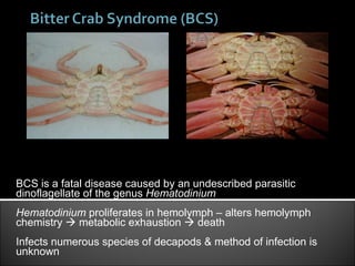
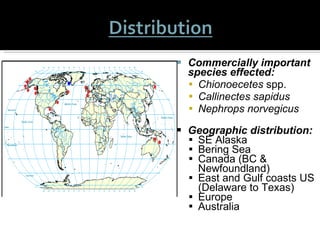
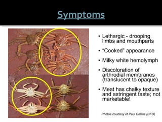
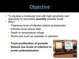
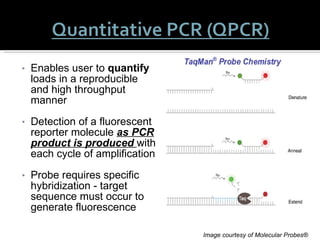
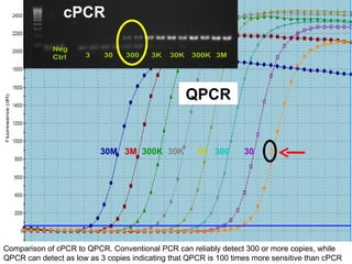
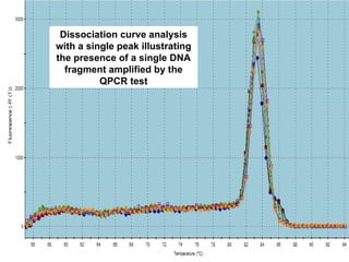
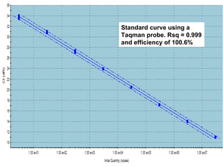
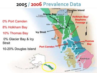
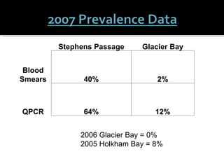
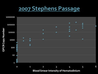
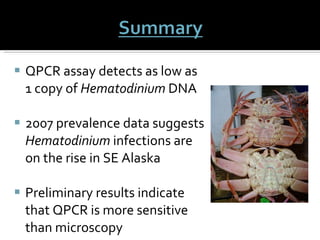
L.M. CROSSON*, C.S. FRIEDMAN. School of Aquatic and Fishery Sciences, University of Washington, Seattle, WA 98195. J.F. MORADO. National Oceanic & Atmospheric Administration, AFSC, 7600 Sand Point Way NE, Seattle, WA 98115.
BCS is a fatal disease caused by an undescribed parasitic dinoflagellate of the genus  Hematodinium  Hematodinium  proliferates in hemolymph – alters hemolymph chemistry    metabolic exhaustion    death Infects numerous species of decapods & method of infection is unknown Left : Heavily infected Chionoecetes bairdi Right : Hematodinium infected (top) versus healthy (bottom) C. bairdi
Commercially important species effected:   Chionoecetes  spp. Callinectes sapidus Nephrops norvegicus Geographic distribution: SE Alaska  Bering Sea  Canada (BC & Newfoundland) East and Gulf coasts US (Delaware to Texas) Europe Australia Figure 1.  World-wide reports of  Hematodinium  related disease.  Red dots = historical  disease; Blue dot = recent report.
Lethargic - drooping limbs and mouthparts “ Cooked” appearance Milky white hemolymph Discoloration of arthrodial membranes (translucent to opaque)  Meat has chalky texture and astringent taste; not marketable! Photos courtesy of Paul Collins (DFO) Symptoms
Prior to 1985 – 7 decapod species reported Currently reported in over 20 species  (Morado et al. 2005; Stentiford & Shields 2005) Areas in SE Alaska have shown prevalence of BCS as high as  95%   (Meyers et al. 1990) Severe economic losses ($3 million US) have been attributed to this parasite on the  C. bairdi  fishery  (Meyers et al. 1987)
Higher in juvenile decapods - die before they recruit into the fishery  (Messick 1994, Morado et al. 2000)   The parasite is more prevalent in female crabs providing additional potential for negative impacts  (Stentiford & Shields 2005) Highest prevalence of infection - July through October Peak mortalities in August and September Cool winter temps thought to inhibit parasite reproduction
http://www.skio.peachnet.edu/people/lee/images/parasitelifecycle.jpg
Slide courtesy of Dr. Carolyn Friedman
Only 2 species have been fully described H. perezi  (Chatton & Poisson 1931) H. australis   (Hudson & Shields 1994) Other have not because: (1) few differentiating morphological characteristics exist (2) not all parasitic forms are encountered in infected hosts Concern:  Without additional characterization of the established and novel strains, the number of species and the extent of host specificity remains unknown
May occur during post-molt  (Shields et al. 2005) Enhanced by cannibalism or mating behavior  (Meyers et al. 1990)  Infectious stage is not known (dinospore?)  Do prey species or seawater serve as parasite vectors or reservoirs? Dinospores of Hematodinium in C. opilio. Photo courtesy of Dr. Jeff Shields
In vitro  transmission not successful due to loss of infectivity  (Meyers et al. 1987, Appleton & Vickerman 1998, Shields and Squyars 2000)  Partial life histories have been described  from infected tissues   (Meyers et al. 1987, 1990, Eaton et al. 1991, Stentiford & Shields 2005) Concern:  To resolve the mode of transmission  external life history stages  require  further characterization  Hematodinium in C. sapidus hemolymph.  Photo courtesy of Dr. Jeff Shields
Microscopy/Histology : Detects pathogen  and  indicates infection  Semi-quantitative Can be used for downstream analysis Considered “gold standard” Limitations: Expensive - long time frame & skilled eye Can  not  be used to look for pathogen vectors  Conventional PCR (cPCR) : More sensitive High throughput Limitations: Only provides presence-absence data  Not quantitative!
To develop a molecular tool with high sensitivity and specificity to accurately  quantify  parasite loads Why? Tolerance level of infection before symptomatic Infection level before fatal Depth or temperature refuge Reservoirs such as seawater or plankton Track proliferation of parasite Detects low levels of infection to  avoid underestimation
Enables user to  quantify  loads in a reproducible and high throughput manner  Detection of a fluorescent reporter molecule  as PCR product is produced   with each cycle of amplification Probe requires specific hybridization - target sequence must occur to generate fluorescence Image courtesy of Molecular Probes®
Align all known 18S rDNA sequence of  Hematodinium  and related genera Design primer/probe combinations Develop a  Hematodinium -plasmid containing the amplified segment of the parasite genome Photos courtesy of Nate Wight 3 M  300K  30K  3K  300  30
Design primers Conventional PCR Insert amplicon into plasmid Clone  multiple copies of plasmid Purify plasmid Quantify plasmid copy number Create dilution set Create standard curve with QPCR Graphic courtesy of Nate Wight
30M   3M   300K   30K   3K   300   30  3   Comparison of cPCR to QPCR. Conventional PCR can reliably detect 300 or more copies, while QPCR can detect as low as 3 copies indicating that QPCR is 100 times more sensitive than cPCR cPCR QPCR
Dissociation curve analysis with a single peak illustrating the presence of a single DNA fragment amplified by the QPCR test
Standard curve using a Taqman probe. Rsq = 0.999 and efficiency of 100.6%
ADF&G/NOAA Oct 2007 Tanner crab survey Sampled 60 Tanner crabs from each location to provide 95% confidence of detecting infections with a prevalence of ≥ 5% Hemolymph was extracted from the arthrodial membrane using a sterile syringe and preserved in EtOH Gross characteristics were recorded and hemolymph smears were taken for microscopy to use as a ‘gold standard’   Seawater from each location sampled, filtered, and preserved in EtOH
Port Camden  Holkham Bay/ Stephens Passage Glacier Bay Icy Strait Douglas Island 0% Port Camden 8% Holkham Bay 10% Thomas Bay 0% Glacier Bay & Icy Strait 10-20% Douglas Island Thomas Bay
2006 Glacier Bay = 0% 2005 Holkham Bay = 8% Stephens Passage Glacier Bay Blood Smears 40% 2% QPCR 64% 12%
 
QPCR assay detects as low as 1 copy of  Hematodinium  DNA 2007 prevalence data suggests  Hematodinium  infections are on the rise in SE Alaska Preliminary results indicate that QPCR is more sensitive than microscopy
Assess the best way to coordinate blood smear and QPCR data taking into consideration the polymorphic nature of  Hematodinium Continuing to process samples in order to validate the diagnostic and analytical sensitivity and specificity of QPCR assay
Implement QPCR assay to monitor the effects of  Hematodinium  on size frequencies and populations trends Identify potential vectors or reservoirs - providing key life history information about the parasite Assess disease dynamics and impacts to provide alternative harvesting strategies to minimize losses
Support for this project  NPRB NOAA SAFS Special thanks to all my collaborators ADF&G Morado Lab Friedman Lab NSA-PCS student award NSA travel award
Any Questions?

2009 NSA

  • 1.
    L.M. CROSSON*, C.S.FRIEDMAN. School of Aquatic and Fishery Sciences, University of Washington, Seattle, WA 98195. J.F. MORADO. National Oceanic & Atmospheric Administration, AFSC, 7600 Sand Point Way NE, Seattle, WA 98115.
  • 2.
    BCS is afatal disease caused by an undescribed parasitic dinoflagellate of the genus Hematodinium Hematodinium proliferates in hemolymph – alters hemolymph chemistry  metabolic exhaustion  death Infects numerous species of decapods & method of infection is unknown Left : Heavily infected Chionoecetes bairdi Right : Hematodinium infected (top) versus healthy (bottom) C. bairdi
  • 3.
    Commercially important specieseffected: Chionoecetes spp. Callinectes sapidus Nephrops norvegicus Geographic distribution: SE Alaska Bering Sea Canada (BC & Newfoundland) East and Gulf coasts US (Delaware to Texas) Europe Australia Figure 1. World-wide reports of Hematodinium related disease. Red dots = historical disease; Blue dot = recent report.
  • 4.
    Lethargic - droopinglimbs and mouthparts “ Cooked” appearance Milky white hemolymph Discoloration of arthrodial membranes (translucent to opaque) Meat has chalky texture and astringent taste; not marketable! Photos courtesy of Paul Collins (DFO) Symptoms
  • 5.
    Prior to 1985– 7 decapod species reported Currently reported in over 20 species (Morado et al. 2005; Stentiford & Shields 2005) Areas in SE Alaska have shown prevalence of BCS as high as 95% (Meyers et al. 1990) Severe economic losses ($3 million US) have been attributed to this parasite on the C. bairdi fishery (Meyers et al. 1987)
  • 6.
    Higher in juveniledecapods - die before they recruit into the fishery (Messick 1994, Morado et al. 2000) The parasite is more prevalent in female crabs providing additional potential for negative impacts (Stentiford & Shields 2005) Highest prevalence of infection - July through October Peak mortalities in August and September Cool winter temps thought to inhibit parasite reproduction
  • 7.
  • 8.
    Slide courtesy ofDr. Carolyn Friedman
  • 9.
    Only 2 specieshave been fully described H. perezi (Chatton & Poisson 1931) H. australis (Hudson & Shields 1994) Other have not because: (1) few differentiating morphological characteristics exist (2) not all parasitic forms are encountered in infected hosts Concern: Without additional characterization of the established and novel strains, the number of species and the extent of host specificity remains unknown
  • 10.
    May occur duringpost-molt (Shields et al. 2005) Enhanced by cannibalism or mating behavior (Meyers et al. 1990) Infectious stage is not known (dinospore?) Do prey species or seawater serve as parasite vectors or reservoirs? Dinospores of Hematodinium in C. opilio. Photo courtesy of Dr. Jeff Shields
  • 11.
    In vitro transmission not successful due to loss of infectivity (Meyers et al. 1987, Appleton & Vickerman 1998, Shields and Squyars 2000) Partial life histories have been described from infected tissues (Meyers et al. 1987, 1990, Eaton et al. 1991, Stentiford & Shields 2005) Concern: To resolve the mode of transmission external life history stages require further characterization Hematodinium in C. sapidus hemolymph. Photo courtesy of Dr. Jeff Shields
  • 12.
    Microscopy/Histology : Detectspathogen and indicates infection Semi-quantitative Can be used for downstream analysis Considered “gold standard” Limitations: Expensive - long time frame & skilled eye Can not be used to look for pathogen vectors Conventional PCR (cPCR) : More sensitive High throughput Limitations: Only provides presence-absence data Not quantitative!
  • 13.
    To develop amolecular tool with high sensitivity and specificity to accurately quantify parasite loads Why? Tolerance level of infection before symptomatic Infection level before fatal Depth or temperature refuge Reservoirs such as seawater or plankton Track proliferation of parasite Detects low levels of infection to avoid underestimation
  • 14.
    Enables user to quantify loads in a reproducible and high throughput manner Detection of a fluorescent reporter molecule as PCR product is produced with each cycle of amplification Probe requires specific hybridization - target sequence must occur to generate fluorescence Image courtesy of Molecular Probes®
  • 15.
    Align all known18S rDNA sequence of Hematodinium and related genera Design primer/probe combinations Develop a Hematodinium -plasmid containing the amplified segment of the parasite genome Photos courtesy of Nate Wight 3 M 300K 30K 3K 300 30
  • 16.
    Design primers ConventionalPCR Insert amplicon into plasmid Clone multiple copies of plasmid Purify plasmid Quantify plasmid copy number Create dilution set Create standard curve with QPCR Graphic courtesy of Nate Wight
  • 17.
    30M 3M 300K 30K 3K 300 30 3 Comparison of cPCR to QPCR. Conventional PCR can reliably detect 300 or more copies, while QPCR can detect as low as 3 copies indicating that QPCR is 100 times more sensitive than cPCR cPCR QPCR
  • 18.
    Dissociation curve analysiswith a single peak illustrating the presence of a single DNA fragment amplified by the QPCR test
  • 19.
    Standard curve usinga Taqman probe. Rsq = 0.999 and efficiency of 100.6%
  • 20.
    ADF&G/NOAA Oct 2007Tanner crab survey Sampled 60 Tanner crabs from each location to provide 95% confidence of detecting infections with a prevalence of ≥ 5% Hemolymph was extracted from the arthrodial membrane using a sterile syringe and preserved in EtOH Gross characteristics were recorded and hemolymph smears were taken for microscopy to use as a ‘gold standard’ Seawater from each location sampled, filtered, and preserved in EtOH
  • 21.
    Port Camden Holkham Bay/ Stephens Passage Glacier Bay Icy Strait Douglas Island 0% Port Camden 8% Holkham Bay 10% Thomas Bay 0% Glacier Bay & Icy Strait 10-20% Douglas Island Thomas Bay
  • 22.
    2006 Glacier Bay= 0% 2005 Holkham Bay = 8% Stephens Passage Glacier Bay Blood Smears 40% 2% QPCR 64% 12%
  • 23.
  • 24.
    QPCR assay detectsas low as 1 copy of Hematodinium DNA 2007 prevalence data suggests Hematodinium infections are on the rise in SE Alaska Preliminary results indicate that QPCR is more sensitive than microscopy
  • 25.
    Assess the bestway to coordinate blood smear and QPCR data taking into consideration the polymorphic nature of Hematodinium Continuing to process samples in order to validate the diagnostic and analytical sensitivity and specificity of QPCR assay
  • 26.
    Implement QPCR assayto monitor the effects of Hematodinium on size frequencies and populations trends Identify potential vectors or reservoirs - providing key life history information about the parasite Assess disease dynamics and impacts to provide alternative harvesting strategies to minimize losses
  • 27.
    Support for thisproject NPRB NOAA SAFS Special thanks to all my collaborators ADF&G Morado Lab Friedman Lab NSA-PCS student award NSA travel award
  • 28.

Editor's Notes

  • #3 BCS is a fatal disease caused by an undescribed parasitic dinoflagellate of the genus hematodinium. The dinoflagellate proliferates in the hemolymph or blood and alters the hemolymph chemistry leading to metabolic exhaustion of the crab and ultimately death. Hematodinium is known to infect over 20 different species of decapods however the method of infection has yet to be determined.
  • #4 Some commercially important species effected by this disease are snow and tanner crabs, blue crabs, and the norwegian lobster. The map shows worldwide reports of hematodinium related disease. The distribution of this disease is global, having severe impacts in AK, Canada, the eastern US, Europe and even Australia. It is also important to note the majority of new infections have occurred in commercially important decapods in northern temperate zones that are experiencing increased warmer temperatures.
  • #5 Some signs that are indicative of hematodinium infection are that the animal becomes lethargic with drooping limbs and mouthparts, its exoskeleton will have a cooked appearance as shown by the infected crabs highlighted in yellow, the animals hemolymph will be milky white in color, and the arthrodial membranes will turn from translucent to opaque. While hematodinium is not known to cause harm to humans, it does however give the animals meat a chalky texture and bitter taste deeming it unmarketable.
  • #6 Prior to 1985 there were 7 decapod species reported to harbor hematodinium infections. Currently the disease has been reported in over 20 species worldwide with some areas in SE alaska, such as lynn canal, show prevalences as high as 95% of the population sampled. Mortalities and fishery impacts associated with hematodinium epidemics and the potential for this pathogen to impact affected commercial decapod abundance and distribution patterns are significant. Economic losses attributed to this parasite in the tanner crab fishery alone are estimated to be at least 3 million dollars.
  • #7 While epidemics and mortalities of commercial size decapods are significant, evidence suggests that hematodinium associated disease is more prevalent in juvenile decapods than in adults and that a high percentage of small infected crustaceans die before they can recruit into the fishery. The parasite is also more prevalent in female than male crab providing additional potential for population and fishery impacts. For example strong bering sea snow crab cohorts in 2000,2001,2002 did not materialize and recruit into the fishery. A potential cause for these failures is Hematodinium. The highest prevalences of infection are found July through October with peak mortalities occurring in August and September. Cooler winter temperatures are thought to inhibit parasite reproduction.
  • #8 This is a simplistic diagram of the life cycle of a parasitic dinoflagellate. For most dinoflagellates, the dinospore is the infectious stage and once entering a host will begin to grow as a trophont, then become a mobile plasmodia, and reproduce to form new spores.
  • #9 Here are some typical parasite morphologies. Note the elongated motile plasmodia stage is very easy to discriminate. However, the trophont stage of the parasite looks very similar to the actual hemocytes of the host which can make diagnosis of this disease by microscopy alone particularly difficult and can lead to underestimation of disease prevalence.
  • #10 To date only 2 species of hematodinium have been fully described from life history stages encountered in infected crabs. Others have not because few differentiating morphological characteristics exist and not all parasitic forms are encountered in infected hosts. Hematodinium from Tanner crab may be a new species however without additional characterization of the established and novel strains, the number of species and the extent of host specificity remains unknown.
  • #11 Disease transmission is thought to occur during the post-molt period and may be enhanced by cannibalism or mating behavior. As I mentioned earlier for many parasitic dinoflagellates the dinospore is the infectious stage, whether this is true for hematodinium awaits further examination. We also do not know if prey species or seawater may possibly be serving as parasite vectors or reservoirs.
  • #12 So far, in vitro transmission of the disease has not been successful due to loss of infectivity and only partial life histories have been described from infected tissues. In order to resolve the mode of transmission external life history stages require further characterization.
  • #13 The traditional technique used for pathogen detection is histology. Histology allows you to detect the pathogen and indicate infection status, it is semi quantitative and can be used for downstream analysis such as ISH however the processes is very expensive, time consuming and takes a well trained eye. It also cannot be used to look for pathogen vectors. A molecular techniques that is more sensitive and allows for higher throughput is cPCR but with cPCR you are limited to only presence-absence data. It is not quantitative.
  • #14 The goal of my project is to develop a molecular tool with high sensitivity and specificity to accurately quantify parasite loads. Being able to quantify parasite loads allows for many new questions to be addressed such as what is the tolerance level of infection before an animal becomes symptomatic? At what infection level is the parasite fatal to its host? Is there a temperature or depth refuge for the host? Do seawater or plankton act as reservoirs for the parasite? Theses are all important questions that we can address using QPCR to help track the proliferation of hematodinium and also to detect very low levels of infection to avoid underestimation of disease prevalence.
  • #15 QPCR enables the user to quantify loads in a reproducible and high throughput manner. The Taqman probe chemistry employed detects and measures fluorescence as PCR product is produced with each cycle of amplification unlike endpoint detection via cPCR. The probe requires specific hybridization therefore the target sequence must be present to generate fluorescence.
  • #16 I designed my QPCR assay by first aligning all known 18S rDNA sequences of Hematodinium and related genera using clustalW. I selected sequence areas unique to only hematodinium where I designed primer and probe combinations using Primer3 software. I was then able to develop a hematodinium plasmid standard containing the amplified segment of the parasite genome amplified by my primers. After quantifying the plasmid copy number, a dilution set of known copy numbers was prepared to create a standard curve.
  • #17 Here is a nice schematic of the steps: I designed my primers, ran a cPCR, took the PCR product or amplicon, inserted it into a plasmid, cloned the plasmid, purified it, quantified it, created a dilution set of known copy numbers, and generated a standard curve with QPCR which I can now use to determine copy numbers of hematodinium DNA in my preserved blood samples.
  • #21 In Oct of 2006, I was fortunate enough to join ADF&G for their Tanner crab survey where I sampled ~ 60 tanner crab from each location to provide 95% confidence of detecting infections with a prevalence of >5%. Hemolymph was extracted from the arthrodial membrane using a sterile syringe and preserved in ethanol. For every crab sampled gross characteristics were recorded and hemolymph smears were taken for microscopy which will be used as a gold standard against our QPCR assay. Seawater was also sampled, filtered, and preserved from each location to look for parasite vectors.
  • #22 The prevalences of crabs with visible signs of infection ranged from 0% in Port Camden to ~8% in Holkham Bay and ~10% in crabs from Thomas Bay. These prevalences are similar to those observed on previous cruises with nearly no Hematodinium observed in crabs from Glacier Bay and Icy Strait and ~10-20% in those collected near Douglas Island.
  • #24 A lot of difference in the zero ……..realtive concordinance in a log linear relationship X blood smear intensity of hemat Y qpcr copy number
  • #26 In the process of assessing the best way to coordinate data taking into consideration the polymorphic nature of parasite Validating diagnostic and analysitcal sensitivity and specificity.