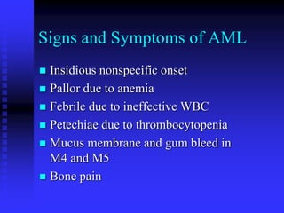
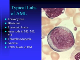
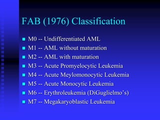
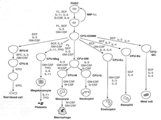
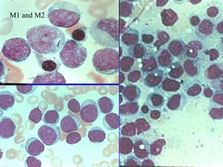
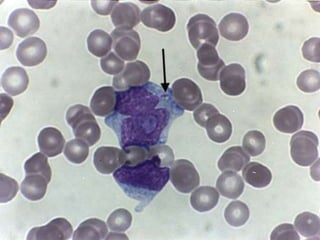
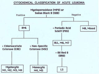
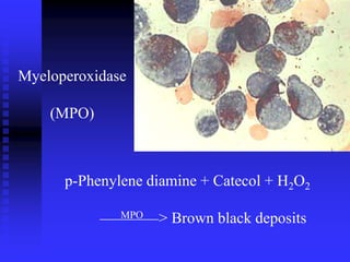
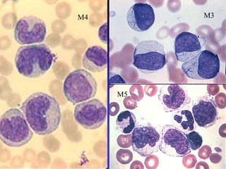
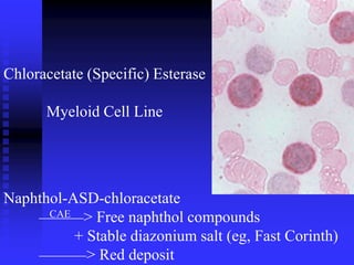
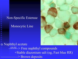
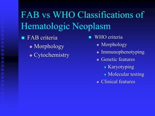
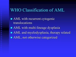
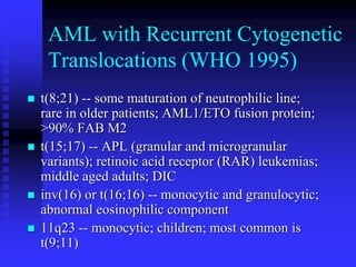
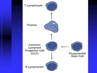
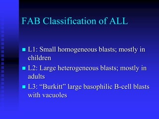
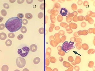
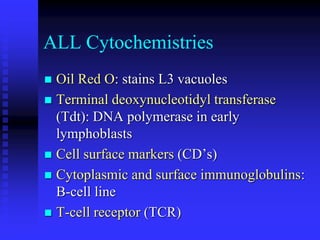
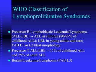
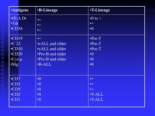
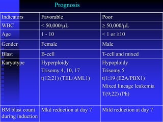
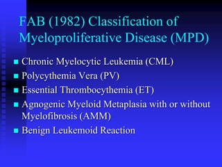
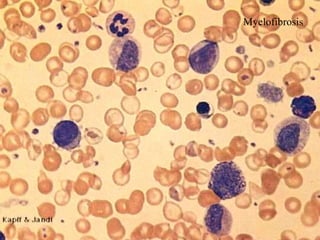
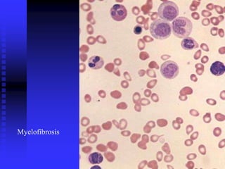
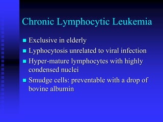
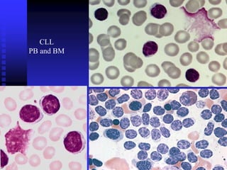
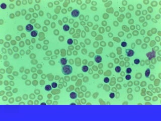
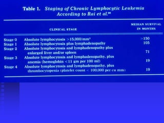
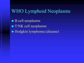
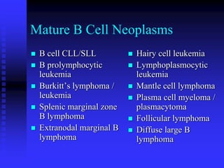
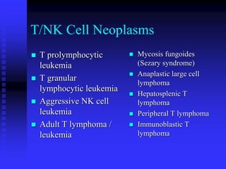
Week 10: Leukemia
 AML
 ALL
 FAB classification
 WHO classification
 Cytochemistries
 MPO
 SBB
 PAS
 ORO
 CD markers
 Flowcytometry
 CML
 CLL
 Karyotype
 Ph chromosome
 LAP
Signs and Symptoms of AML
 Insidious nonspecific onset
 Pallor due to anemia
 Febrile due to ineffective WBC
 Petechiae due to thrombocytopenia
 Mucus membrane and gum bleed in
M4 and M5
 Bone pain
Typical Labs
of AML
 Leukocytosis
 Blastemia
 Leukemic hiatus
 Auer rods in M2, M3,
M4
 Thrombocytopenia
 Anemia
 >20% blasts in BM
Other Findings
 CD 13 and CD 33 in flowcytometry
 Cytochemistries
 Myeloperoxidase
 Sudan black B
 Choloroacetate esterase (specific)
 Nonspecific esterase
FAB (1976) Classification
 M0 -- Undifferentiated AML
 M1 -- AML without maturation
 M2 -- AML with maturation
 M3 -- Acute Promyelocytic Leukemia
 M4 -- Acute Meylomonocytic Leukemia
 M5 -- Acute Monocytic Leukemia
 M6 -- Erythroleukemia (DiGuglielmo’s)
 M7 -- Megakaryoblastic Leukemia
M1 and M2
Myeloperoxidase
(MPO)
p-Phenylene diamine + Catecol + H2O2
MPO > Brown black deposits
M3
M5
M4
Chloracetate (Specific) Esterase
Myeloid Cell Line
Naphthol-ASD-chloracetate
CAE > Free naphthol compounds
+ Stable diazonium salt (eg, Fast Corinth)
> Red deposit
Non-Specific Esterase
Monocytic Line
 Naphthyl acetate
ANAE > Free naphthyl compounds
+Stable diazonium salt (eg, Fast blue RR)
> Brown deposits
Double Esterase in M4
NSE with Fl inhibition
Histiocyte
FAB vs WHO Classifications of
Hematologic Neoplasm
 FAB criteria
 Morphology
 Cytochemistry
 WHO criteria
 Morphology
 Immunophenotyping
 Genetic features
 Karyotyping
 Molecular testing
 Clinical features
WHO Classification of AML
 AML with recurrent cytogenic
translocations
 AML with multi-lineage dysplasia
 AML and myelodysplasia, therapy related
 AML, not otherwise categorized
AML with Recurrent Cytogenetic
Translocations (WHO 1995)
 t(8;21) -- some maturation of neutrophilic line;
rare in older patients; AML1/ETO fusion protein;
>90% FAB M2
 t(15;17) -- APL (granular and microgranular
variants); retinoic acid receptor (RAR) leukemias;
middle aged adults; DIC
 inv(16) or t(16;16) -- monocytic and granulocytic;
abnormal eosinophilic component
 11q23 -- monocytic; children; most common is
t(9;11)
FAB Classification of ALL
 L1: Small homogeneous blasts; mostly in
children
 L2: Large heterogeneous blasts; mostly in
adults
 L3: “Burkitt” large basophilic B-cell blasts
with vacuoles
L3
L2
Periodic Acid Schiff
Periodic acid + Glycogen
oxidation > Aldehyde + Schiff reagent
(para-rosaniline, Na metabisulfite)
> Red deposit
ALL Cytochemistries
 Oil Red O: stains L3 vacuoles
 Terminal deoxynucleotidyl transferase
(Tdt): DNA polymerase in early
lymphoblasts
 Cell surface markers (CD’s)
 Cytoplasmic and surface immunoglobulins:
B-cell line
 T-cell receptor (TCR)
WHO Classification of
Lymphoproliferative Syndromes
 Precursor B Lymphoblastic Leukemia/Lymphoma
(ALL/LBL) -- ALL in children (80-85% of
childhood ALL); LBL in young adults and rare;
FAB L1 or L2 blast morphology
 Precursor T ALL/LBL -- 15% of childhood ALL
and 25% of adult ALL
 Burkitt Leukemia/Lymphoma (FAB L3)
•Antigens •B-Lineage •T-Lineage
•HLA Dr
•Tdt
•CD34
•+
•+
•+
•0 to +
•+
•0
•CD19
•C 22
•CD10
•CD20
•Cyt-
•SIg
•+
•cALL and older
•cALL and older
•Pre-B and older
•Pre-B and older
•B-ALL
•Pre-T
•Pre-T
•Pre-T
•0
•0
•0
•CD7
•CD3
•CD5
•CD2
•CD1
•0
•0
•0
•0
•0
•+
•+
•+
•T-ALL
•T-ALL
Indicators Favorable Poor
WBC < 50,000/L  50,000/L
Age 1 - 10 < 1 or 10
Gender Female Male
Blast B-cell T-cell and mixed
Karyotype Hyperploidy
Trisomy 4, 10, 17
t(12;21) (TEL/AML1)
Hypoploidy
Trisomy 5
t(1;19 (E2A/PBX1)
Mixed lineage leukemia
T(9;22) (Ph)
BM blast count
during induction
Mkd reduction at day 7 Mild reduction at day 7
Prognosis
FAB (1982) Classification of
Myeloproliferative Disease (MPD)
 Chronic Myelocytic Leukemia (CML)
 Polycythemia Vera (PV)
 Essential Thrombocythemia (ET)
 Agnogenic Myeloid Metaplasia with or without
Myelofibrosis (AMM)
 Benign Leukemoid Reaction
WHO Classification of MPS
(1997)
 CML becomes
 CML, Ph + t(9;22) BCR/ABL
 Chronic Neutrophilic Leukemia (CNL)
 Chronic Eosinophilic Leukemia and Hyper-
eosinophilic Syndrome (CEL/HES)
 PV remains PV
 ET remains ET
 AMM becomes
 Chronic Idiopathic Myelofibrosis
Myelofibrosis
Myelofibrosis
Chronic Lymphocytic Leukemia
 Exclusive in elderly
 Lyphocytosis unrelated to viral infection
 Hyper-mature lymphocytes with highly
condensed nuclei
 Smudge cells: preventable with a drop of
bovine albumin
CLL
PB and BM
WHO Lymphoid Neoplasms
 B cell neoplasms
 T/NK cell neoplasms
 Hodgkin lymphoma (disease)
Mature B Cell Neoplasms
 B cell CLL/SLL
 B prolymphocytic
leukemia
 Burkitt’s lymphoma /
leukemia
 Splenic marginal zone
B lymphoma
 Extranodal marginal B
lymphoma
 Hairy cell leukemia
 Lymphoplasmocytic
leukemia
 Mantle cell lymphoma
 Plasma cell myeloma /
plasmacytoma
 Follicular lymphoma
 Diffuse large B
lymphoma
T/NK Cell Neoplasms
 T prolymphocytic
leukemia
 T granular
lymphocytic leukemia
 Aggressive NK cell
leukemia
 Adult T lymphoma /
leukemia
 Mycosis fungoides
(Sezary syndrome)
 Anaplastic large cell
lymphoma
 Hepatosplenic T
lymphoma
 Peripheral T lymphoma
 Immunoblastic T
lymphoma

Leukemia (1).ppt and bio marker and cbc

  • 1.
    Week 10: Leukemia AML  ALL  FAB classification  WHO classification  Cytochemistries  MPO  SBB  PAS  ORO  CD markers  Flowcytometry  CML  CLL  Karyotype  Ph chromosome  LAP
  • 2.
    Signs and Symptomsof AML  Insidious nonspecific onset  Pallor due to anemia  Febrile due to ineffective WBC  Petechiae due to thrombocytopenia  Mucus membrane and gum bleed in M4 and M5  Bone pain
  • 3.
    Typical Labs of AML Leukocytosis  Blastemia  Leukemic hiatus  Auer rods in M2, M3, M4  Thrombocytopenia  Anemia  >20% blasts in BM
  • 4.
    Other Findings  CD13 and CD 33 in flowcytometry  Cytochemistries  Myeloperoxidase  Sudan black B  Choloroacetate esterase (specific)  Nonspecific esterase
  • 5.
    FAB (1976) Classification M0 -- Undifferentiated AML  M1 -- AML without maturation  M2 -- AML with maturation  M3 -- Acute Promyelocytic Leukemia  M4 -- Acute Meylomonocytic Leukemia  M5 -- Acute Monocytic Leukemia  M6 -- Erythroleukemia (DiGuglielmo’s)  M7 -- Megakaryoblastic Leukemia
  • 7.
  • 10.
    Myeloperoxidase (MPO) p-Phenylene diamine +Catecol + H2O2 MPO > Brown black deposits
  • 11.
  • 12.
    Chloracetate (Specific) Esterase MyeloidCell Line Naphthol-ASD-chloracetate CAE > Free naphthol compounds + Stable diazonium salt (eg, Fast Corinth) > Red deposit
  • 13.
    Non-Specific Esterase Monocytic Line Naphthyl acetate ANAE > Free naphthyl compounds +Stable diazonium salt (eg, Fast blue RR) > Brown deposits
  • 14.
    Double Esterase inM4 NSE with Fl inhibition Histiocyte
  • 15.
    FAB vs WHOClassifications of Hematologic Neoplasm  FAB criteria  Morphology  Cytochemistry  WHO criteria  Morphology  Immunophenotyping  Genetic features  Karyotyping  Molecular testing  Clinical features
  • 16.
    WHO Classification ofAML  AML with recurrent cytogenic translocations  AML with multi-lineage dysplasia  AML and myelodysplasia, therapy related  AML, not otherwise categorized
  • 17.
    AML with RecurrentCytogenetic Translocations (WHO 1995)  t(8;21) -- some maturation of neutrophilic line; rare in older patients; AML1/ETO fusion protein; >90% FAB M2  t(15;17) -- APL (granular and microgranular variants); retinoic acid receptor (RAR) leukemias; middle aged adults; DIC  inv(16) or t(16;16) -- monocytic and granulocytic; abnormal eosinophilic component  11q23 -- monocytic; children; most common is t(9;11)
  • 19.
    FAB Classification ofALL  L1: Small homogeneous blasts; mostly in children  L2: Large heterogeneous blasts; mostly in adults  L3: “Burkitt” large basophilic B-cell blasts with vacuoles
  • 20.
  • 21.
    Periodic Acid Schiff Periodicacid + Glycogen oxidation > Aldehyde + Schiff reagent (para-rosaniline, Na metabisulfite) > Red deposit
  • 22.
    ALL Cytochemistries  OilRed O: stains L3 vacuoles  Terminal deoxynucleotidyl transferase (Tdt): DNA polymerase in early lymphoblasts  Cell surface markers (CD’s)  Cytoplasmic and surface immunoglobulins: B-cell line  T-cell receptor (TCR)
  • 23.
    WHO Classification of LymphoproliferativeSyndromes  Precursor B Lymphoblastic Leukemia/Lymphoma (ALL/LBL) -- ALL in children (80-85% of childhood ALL); LBL in young adults and rare; FAB L1 or L2 blast morphology  Precursor T ALL/LBL -- 15% of childhood ALL and 25% of adult ALL  Burkitt Leukemia/Lymphoma (FAB L3)
  • 24.
    •Antigens •B-Lineage •T-Lineage •HLADr •Tdt •CD34 •+ •+ •+ •0 to + •+ •0 •CD19 •C 22 •CD10 •CD20 •Cyt- •SIg •+ •cALL and older •cALL and older •Pre-B and older •Pre-B and older •B-ALL •Pre-T •Pre-T •Pre-T •0 •0 •0 •CD7 •CD3 •CD5 •CD2 •CD1 •0 •0 •0 •0 •0 •+ •+ •+ •T-ALL •T-ALL
  • 25.
    Indicators Favorable Poor WBC< 50,000/L  50,000/L Age 1 - 10 < 1 or 10 Gender Female Male Blast B-cell T-cell and mixed Karyotype Hyperploidy Trisomy 4, 10, 17 t(12;21) (TEL/AML1) Hypoploidy Trisomy 5 t(1;19 (E2A/PBX1) Mixed lineage leukemia T(9;22) (Ph) BM blast count during induction Mkd reduction at day 7 Mild reduction at day 7 Prognosis
  • 26.
    FAB (1982) Classificationof Myeloproliferative Disease (MPD)  Chronic Myelocytic Leukemia (CML)  Polycythemia Vera (PV)  Essential Thrombocythemia (ET)  Agnogenic Myeloid Metaplasia with or without Myelofibrosis (AMM)  Benign Leukemoid Reaction
  • 27.
    WHO Classification ofMPS (1997)  CML becomes  CML, Ph + t(9;22) BCR/ABL  Chronic Neutrophilic Leukemia (CNL)  Chronic Eosinophilic Leukemia and Hyper- eosinophilic Syndrome (CEL/HES)  PV remains PV  ET remains ET  AMM becomes  Chronic Idiopathic Myelofibrosis
  • 28.
  • 29.
  • 31.
    Chronic Lymphocytic Leukemia Exclusive in elderly  Lyphocytosis unrelated to viral infection  Hyper-mature lymphocytes with highly condensed nuclei  Smudge cells: preventable with a drop of bovine albumin
  • 32.
  • 36.
    WHO Lymphoid Neoplasms B cell neoplasms  T/NK cell neoplasms  Hodgkin lymphoma (disease)
  • 37.
    Mature B CellNeoplasms  B cell CLL/SLL  B prolymphocytic leukemia  Burkitt’s lymphoma / leukemia  Splenic marginal zone B lymphoma  Extranodal marginal B lymphoma  Hairy cell leukemia  Lymphoplasmocytic leukemia  Mantle cell lymphoma  Plasma cell myeloma / plasmacytoma  Follicular lymphoma  Diffuse large B lymphoma
  • 38.
    T/NK Cell Neoplasms T prolymphocytic leukemia  T granular lymphocytic leukemia  Aggressive NK cell leukemia  Adult T lymphoma / leukemia  Mycosis fungoides (Sezary syndrome)  Anaplastic large cell lymphoma  Hepatosplenic T lymphoma  Peripheral T lymphoma  Immunoblastic T lymphoma