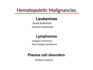
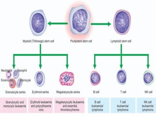
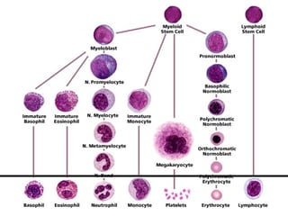
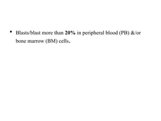
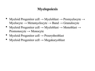
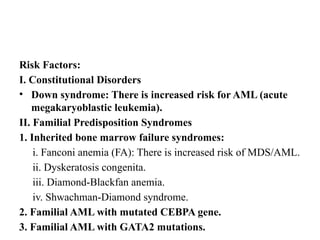
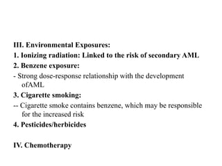
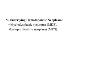
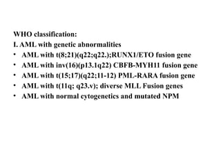
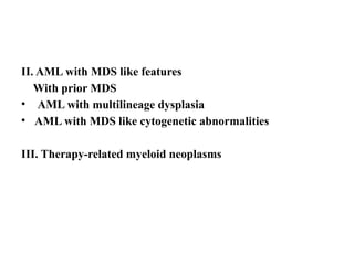
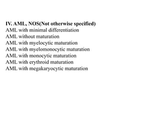
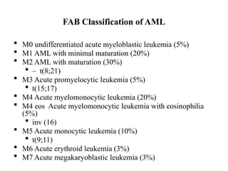
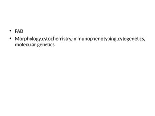
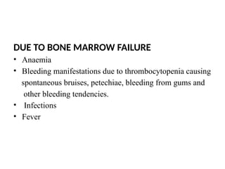
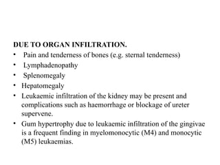
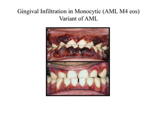
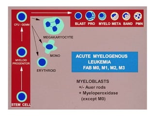
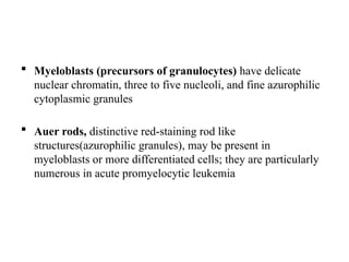
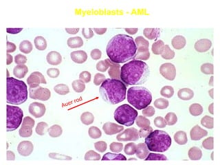
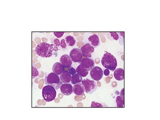
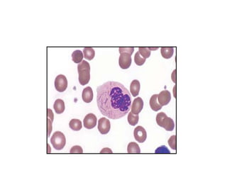
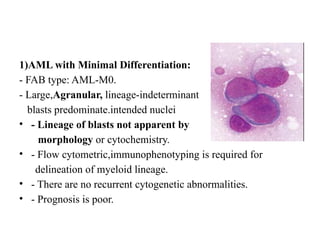
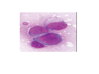
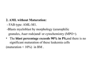
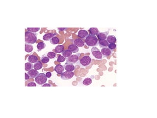
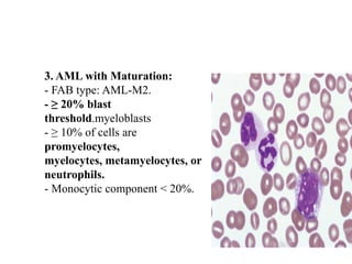
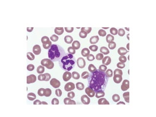
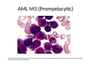
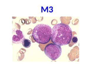
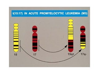
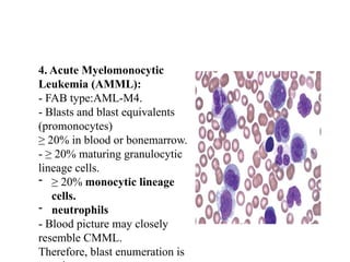
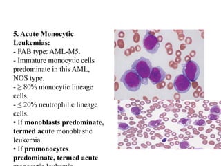
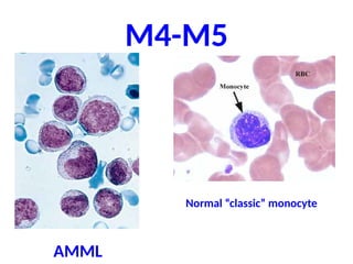
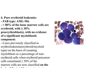
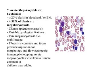
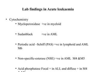
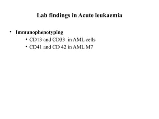
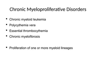
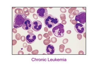
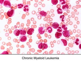
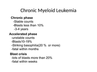
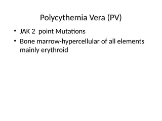
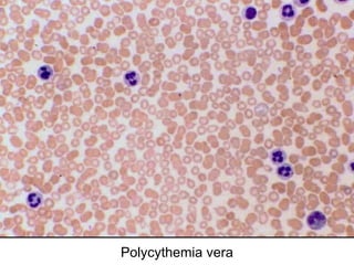
AML CLASSIFICATION
Leukemias
Acute leukemias
Chronic leukemias
Lymphomas
Hodgkin lymphoma
Non-Hodgkin lymphoma
Hematopoietic Malignancies
Plasma cell disorders
Multiple myeloma
 Neoplastic proliferations of white blood cells
- leukaemias and lymphomas, are the most important group of
leucocyte disorders.
leukaemias classified
-on the basis of cell types –
myeloid and lymphoid
-on the basis of natural history of the disease –
acute and chronic
• Acute Myeloid leukaemia
• Acute Lymphoid leukemia (AML and ALL)
• Chronic Myeloid leukaemia
• Chronic Lymphocytic leukaemias (CML and CLL)
Lymphomas which are malignant
• Hodgkin lymphoma or Hodgkin’s disease (HD)
• Non-Hodgkin’s lymphomas (NHL)
Myeloid neoplasms
• Myelodysplastic syndromes (MDS)
• Acute myeloid leukaemia (AML)
• Chronic myeloproliferative disorders
Lymphoid neoplasms
• Neoplasms of lymphoid lineage include leukaemias and
lymphomas of B, T or NK cell origin.
• This group thus includes B cell neoplasms (including plasma
cell disorders), T cell neoplasms, NK cell neoplasms and
• Hodgkin’s disease.
Classification of leukaemias
 FAB (French,American &British) classification—
morphology & cytochemical features
 REAL(Revised European-American classification of
lymphoid neoplasms) –immunophenotypic features
 WHO classification-cytogenetic and molecular features
AML
 Definition
-Acute myeloid leukaemia (AML) is a heterogeneous disease
characterised by infiltration of malignant myeloid cells into
the blood, bone marrow and other tissues.
 Blasts/blast more than 20% in peripheral blood (PB) &/or
bone marrow (BM) cells.
Myelopoiesis
 Myeloid Progenitor cell → Myeloblast → Promyelocyte →
Myelocyte → Metamyelocyte → Band → Granulocyte
 Myeloid Progenitor cell → Myeloblast → Monoblast →
Promonocyte → Monocyte
 Myeloid Progenitor cell → Proerythroblast
 Myeloid Progenitor cell → Megakaryoblast
Risk Factors:
I. Constitutional Disorders
• Down syndrome: There is increased risk for AML (acute
megakaryoblastic leukemia).
II. Familial Predisposition Syndromes
1. Inherited bone marrow failure syndromes:
i. Fanconi anemia (FA): There is increased risk of MDS/AML.
ii. Dyskeratosis congenita.
iii. Diamond-Blackfan anemia.
iv. Shwachman-Diamond syndrome.
2. Familial AML with mutated CEBPA gene.
3. Familial AML with GATA2 mutations.
III. Environmental Exposures:
1. Ionizing radiation: Linked to the risk of secondary AML
2. Benzene exposure:
- Strong dose-response relationship with the development
ofAML
3. Cigarette smoking:
-- Cigarette smoke contains benzene, which may be responsible
for the increased risk
4. Pesticides/herbicides
IV. Chemotherapy
V. Underlying Hematopoietic Neoplasm:
• Myelodysplastic syndrome (MDS),
Myeloproliferative neoplasm (MPN).
WHO classification:
I. AML with genetic abnormalities
• AML with t(8;21)(q22;q22.);RUNX1/ETO fusion gene
• AML with inv(16)(p13.1q22) CBFB-MYH11 fusion gene
• AML with t(15;17)(q22;11-12) PML-RARA fusion gene
• AML with t(11q; q23.v); diverse MLL Fusion genes
• AML with normal cytogenetics and mutated NPM
II. AML with MDS like features
With prior MDS
• AML with multilineage dysplasia
• AML with MDS like cytogenetic abnormalities
III. Therapy-related myeloid neoplasms
IV. AML, NOS(Not otherwise specified)
AML with minimal differentiation
AML without maturation
AML with myelocytic maturation
AML with myelomonocytic maturation
AML with monocytic maturation
AML with erythroid maturation
AML with megakaryocytic maturation
FAB Classification of AML
 M0 undifferentiated acute myeloblastic leukemia (5%)
 M1 AML with minimal maturation (20%)
 M2 AML with maturation (30%)
 – t(8;21)
 M3 Acute promyelocytic leukemia (5%)
 t(15;17)
 M4 Acute myelomonocytic leukemia (20%)
 M4 eos Acute myelomonocytic leukemia with eosinophilia
(5%)
 inv (16)
 M5 Acute monocytic leukemia (10%)
 t(9;11)
 M6 Acute erythroid leukemia (3%)
 M7 Acute megakaryoblastic leukemia (3%)
• FAB
• Morphology,cytochemistry,immunophenotyping,cytogenetics,
molecular genetics
DUE TO BONE MARROW FAILURE
• Anaemia
• Bleeding manifestations due to thrombocytopenia causing
spontaneous bruises, petechiae, bleeding from gums and
other bleeding tendencies.
• Infections
• Fever
DUE TO ORGAN INFILTRATION.
• Pain and tenderness of bones (e.g. sternal tenderness)
• Lymphadenopathy
• Splenomegaly
• Hepatomegaly
• Leukaemic infiltration of the kidney may be present and
complications such as haemorrhage or blockage of ureter
supervene.
• Gum hypertrophy due to leukaemic infiltration of the gingivae
is a frequent finding in myelomonocytic (M4) and monocytic
(M5) leukaemias.
Gingival Infiltration in Monocytic (AML M4 eos)
Variant of AML
 AML mimics a lymphoma by manifesting as a discrete tissue
mass (a so-called granulocytic sarcoma)
Clinical symptoms/Physical Findings
 Extramedullary disease (ie, myeloid sarcoma/chloroma)
– Can also have involvement of lymph nodes, intestine,
mediastinum, ovaries, uterus
Pathogenesis
 Most AMLs harbor mutations in genes encoding transcription
factors that are required for normal myeloid cell differentiation
 t(8;21),inv(16)-RUNX1 and CBFB genes
 Epigenetic alterations
-DNA methylation
-posttranslational modifications of histones
BLAST
• Larger
• High N/C ratio
• Nucleus is large, open chromatin,1-5 nucleoli
• Thin rim to moderate amount of cytoplasm
Myeloblast
4Ms
• More in size
• More nucleoli
• Moderate cytoplasm
• Myeloperoxidase
• Auer rod
 Myeloblasts (precursors of granulocytes) have delicate
nuclear chromatin, three to five nucleoli, and fine azurophilic
cytoplasmic granules
 Auer rods, distinctive red-staining rod like
structures(azurophilic granules), may be present in
myeloblasts or more differentiated cells; they are particularly
numerous in acute promyelocytic leukemia
Myeloblasts - AML
Auer rod
Clinical features:
-Young adults.
- Myeloid sarcomas (tissue infiltration – chloromas).
CBC/Blood morphology:
-Anemia.
-neutropenia
-Thrombocytopenia.
-Circulating blasts; possibly with Auer rods.
- Generally leukocytosis dominated by blasts.
- Evidence of neutrophilic maturation.
-
BMA:
 Increased myeloid blasts.
 Some cases less than the required 20% (low blast count
AML).
 Large blasts, abundant basophilic cytoplasm, numerous
azurophilic granules.
 Characteristic Auer rods: Thin with tapered ends and usually
single within a cell.
 They are seen in cytoplasm of blasts and maturing/mature
granulocytes.
 Abnormal neutrophilic precursors.
.
1)AML with Minimal Differentiation:
- FAB type: AML-M0.
- Large,Agranular, lineage-indeterminant
blasts predominate.intended nuclei
• - Lineage of blasts not apparent by
morphology or cytochemistry.
• - Flow cytometric,immunophenotyping is required for
delineation of myeloid lineage.
• - There are no recurrent cytogenetic abnormalities.
• - Prognosis is poor.
2. AML without Maturation:
- FAB type: AML-M1.
-Blasts myeloblast by morphology (azurophilic
granules, Auer rods)and/ or cytochemistry (MPO+).
• The blast percentage exceeds 90% in PS,and there is no
significant maturation of these leukemia cells
(maturation < 10%) in BM .
3. AML with Maturation:
- FAB type: AML-M2.
- ≥ 20% blast
threshold.myeloblasts
- ≥ 10% of cells are
promyelocytes,
myelocytes, metamyelocytes, or
neutrophils.
- Monocytic component < 20%.
M0M2
FAB - M3
Acute Promyelocytic Leukemia
 Hypergranular promyelocytes
 Increased Auer rods
 15;17 chromosomal translocation
 (retinoic acid receptor gene)
 RAR- Ch17
 PML-Ch 15
Acute promyelocytic leukemia - AML M3
AML M3 (Promyelocytic)
Brunning, RD, McKenna, RW. Tumors of the bone marrow. Atlas of tumor pathology (electronic fascicle), Third series, fascicle 9, 1994, Washington, DC.
Armed Forces Institute of Pathology.
AML M3 (Promyelocytic)
Brunning, RD, McKenna, RW. Tumors of the bone marrow. Atlas of tumor pathology (electronic fascicle), Third series, fascicle 9, 1994, Washington,
DC. Armed Forces Institute of Pathology.
M3
4. Acute Myelomonocytic
Leukemia (AMML):
- FAB type:AML-M4.
- Blasts and blast equivalents
(promonocytes)
≥ 20% in blood or bonemarrow.
- ≥ 20% maturing granulocytic
lineage cells.
- ≥ 20% monocytic lineage
cells.
- neutrophils
- Blood picture may closely
resemble CMML.
Therefore, blast enumeration is
5. Acute Monocytic
Leukemias:
- FAB type: AML-M5.
- Immature monocytic cells
predominate in this AML,
NOS type.
- ≥ 80% monocytic lineage
cells.
- ≤ 20% neutrophilic lineage
cells.
• If monoblasts predominate,
termed acute monoblastic
leukemia.
• If promonocytes
predominate, termed acute
M4-M5
AMML
Normal “classic” monocyte
6. Pure erythroid leukemia:
- FAB type: AML-M6.
- > 80% of the bone marrow cells are
erythroid, with ≥ 30%
proerythroblasts), with no evidence
of a significant myeloblastic
component.
- Cases previously classified as
erythroleukemia(erythroid/myeloid
type) on the basis of counting
myeloblasts as a percentage of non-
erythroid cells when erythroid precursor
cells constituted ≥ 50% of the
marrow cells are now classified on the
7. Acute Megakaryoblastic
Leukemia:
- ≥ 20% blasts in blood and / or BM.
- > 50% of blasts are
megakaryoblasts.
- Clumps (pseudometastasis).
- Variable cytological features.
- Pure megakaryoblastic vs
multilineage.
- Fibrosis is common and it can
preclude aspiration for
morphology and flow cytometric
immunophenotyping. Acute
megakaryoblastic leukemia is more
common in
children than adults.
M6-M7
ERYTHROLEUKEMIA MEGAKARYOCYTIC LEUKEMIA
Lab findings in Acute leukaemia
• Cytochemistry
• Myeloperoxidase +ve in myeloid
• Sudanblack +ve in AML
• Periodic acid –Schiff (PAS) +ve in lymphoid and AML
M6
• Non-specific-esterase (NSE) +ve in AML M4 &M5
• Acid phosphatase Focal + in ALL and diffuse + in M4
Lab findings in Acute leukaemia
• Immunophenotyping
• CD13 and CD33 in AML cells
• CD41 and CD 42 in AML M7
CHRONIC MYELOID LEUKEMIA
Chronic Myeloproliferative Disorders
 Chronic myeloid leukemia
 Polycythemia vera
 Essential thrombocythemia
 Chronic myelofibrosis
 Proliferation of one or more myeloid lineages
Chronic Leukemia
Chronic Myeloid Leukemia
Normal
chromosomes
Chromosomes
in CML
• t(9;22)(q34;q11.2)
• BCR-ABL Fusion-activation of tyrosine kinase-
RAS/JAK/STAT/AKT pathway-cell division,
• Inhibition of apoptosis
• ABL-Abelson murine leukemia virus
• BCR-breakpoint cluster region
Clinical features
• Anemia
• Bleeding
• Massive splenomegaly
• lymphadenopathy
Chronic Myeloid Leukemia
Chronic phase
-Stable counts
-Blasts less than 10%
-3-4 years
Accelerated phase
-unstable counts
-Blasts10-19%
-Striking basophilia(20 % or more)
-fatal within months
Blast crisis
-lots of blasts more than 20%
-fatal within weeks
Polycythemia Vera (PV)
• JAK 2 point Mutations
• Bone marrow-hypercellular of all elements
mainly erythroid
Polycythemia vera
ESSENTIAL THROMBOCYTOPENIA
• JAK 2 MUTATION
• Megakaryocytic lineage
THANK YOU

Acute Myeloid Leukemia Classification.pptx

  • 1.
  • 2.
    Leukemias Acute leukemias Chronic leukemias Lymphomas Hodgkinlymphoma Non-Hodgkin lymphoma Hematopoietic Malignancies Plasma cell disorders Multiple myeloma
  • 3.
     Neoplastic proliferationsof white blood cells - leukaemias and lymphomas, are the most important group of leucocyte disorders.
  • 4.
    leukaemias classified -on thebasis of cell types – myeloid and lymphoid -on the basis of natural history of the disease – acute and chronic
  • 5.
    • Acute Myeloidleukaemia • Acute Lymphoid leukemia (AML and ALL) • Chronic Myeloid leukaemia • Chronic Lymphocytic leukaemias (CML and CLL)
  • 6.
    Lymphomas which aremalignant • Hodgkin lymphoma or Hodgkin’s disease (HD) • Non-Hodgkin’s lymphomas (NHL)
  • 7.
    Myeloid neoplasms • Myelodysplasticsyndromes (MDS) • Acute myeloid leukaemia (AML) • Chronic myeloproliferative disorders
  • 8.
    Lymphoid neoplasms • Neoplasmsof lymphoid lineage include leukaemias and lymphomas of B, T or NK cell origin. • This group thus includes B cell neoplasms (including plasma cell disorders), T cell neoplasms, NK cell neoplasms and • Hodgkin’s disease.
  • 9.
    Classification of leukaemias FAB (French,American &British) classification— morphology & cytochemical features  REAL(Revised European-American classification of lymphoid neoplasms) –immunophenotypic features  WHO classification-cytogenetic and molecular features
  • 12.
    AML  Definition -Acute myeloidleukaemia (AML) is a heterogeneous disease characterised by infiltration of malignant myeloid cells into the blood, bone marrow and other tissues.
  • 13.
     Blasts/blast morethan 20% in peripheral blood (PB) &/or bone marrow (BM) cells.
  • 14.
    Myelopoiesis  Myeloid Progenitorcell → Myeloblast → Promyelocyte → Myelocyte → Metamyelocyte → Band → Granulocyte  Myeloid Progenitor cell → Myeloblast → Monoblast → Promonocyte → Monocyte  Myeloid Progenitor cell → Proerythroblast  Myeloid Progenitor cell → Megakaryoblast
  • 15.
    Risk Factors: I. ConstitutionalDisorders • Down syndrome: There is increased risk for AML (acute megakaryoblastic leukemia). II. Familial Predisposition Syndromes 1. Inherited bone marrow failure syndromes: i. Fanconi anemia (FA): There is increased risk of MDS/AML. ii. Dyskeratosis congenita. iii. Diamond-Blackfan anemia. iv. Shwachman-Diamond syndrome. 2. Familial AML with mutated CEBPA gene. 3. Familial AML with GATA2 mutations.
  • 16.
    III. Environmental Exposures: 1.Ionizing radiation: Linked to the risk of secondary AML 2. Benzene exposure: - Strong dose-response relationship with the development ofAML 3. Cigarette smoking: -- Cigarette smoke contains benzene, which may be responsible for the increased risk 4. Pesticides/herbicides IV. Chemotherapy
  • 17.
    V. Underlying HematopoieticNeoplasm: • Myelodysplastic syndrome (MDS), Myeloproliferative neoplasm (MPN).
  • 18.
    WHO classification: I. AMLwith genetic abnormalities • AML with t(8;21)(q22;q22.);RUNX1/ETO fusion gene • AML with inv(16)(p13.1q22) CBFB-MYH11 fusion gene • AML with t(15;17)(q22;11-12) PML-RARA fusion gene • AML with t(11q; q23.v); diverse MLL Fusion genes • AML with normal cytogenetics and mutated NPM
  • 19.
    II. AML withMDS like features With prior MDS • AML with multilineage dysplasia • AML with MDS like cytogenetic abnormalities III. Therapy-related myeloid neoplasms
  • 20.
    IV. AML, NOS(Nototherwise specified) AML with minimal differentiation AML without maturation AML with myelocytic maturation AML with myelomonocytic maturation AML with monocytic maturation AML with erythroid maturation AML with megakaryocytic maturation
  • 21.
    FAB Classification ofAML  M0 undifferentiated acute myeloblastic leukemia (5%)  M1 AML with minimal maturation (20%)  M2 AML with maturation (30%)  – t(8;21)  M3 Acute promyelocytic leukemia (5%)  t(15;17)  M4 Acute myelomonocytic leukemia (20%)  M4 eos Acute myelomonocytic leukemia with eosinophilia (5%)  inv (16)  M5 Acute monocytic leukemia (10%)  t(9;11)  M6 Acute erythroid leukemia (3%)  M7 Acute megakaryoblastic leukemia (3%)
  • 22.
  • 23.
    DUE TO BONEMARROW FAILURE • Anaemia • Bleeding manifestations due to thrombocytopenia causing spontaneous bruises, petechiae, bleeding from gums and other bleeding tendencies. • Infections • Fever
  • 24.
    DUE TO ORGANINFILTRATION. • Pain and tenderness of bones (e.g. sternal tenderness) • Lymphadenopathy • Splenomegaly • Hepatomegaly • Leukaemic infiltration of the kidney may be present and complications such as haemorrhage or blockage of ureter supervene. • Gum hypertrophy due to leukaemic infiltration of the gingivae is a frequent finding in myelomonocytic (M4) and monocytic (M5) leukaemias.
  • 25.
    Gingival Infiltration inMonocytic (AML M4 eos) Variant of AML
  • 26.
     AML mimicsa lymphoma by manifesting as a discrete tissue mass (a so-called granulocytic sarcoma)
  • 27.
    Clinical symptoms/Physical Findings Extramedullary disease (ie, myeloid sarcoma/chloroma) – Can also have involvement of lymph nodes, intestine, mediastinum, ovaries, uterus
  • 28.
    Pathogenesis  Most AMLsharbor mutations in genes encoding transcription factors that are required for normal myeloid cell differentiation  t(8;21),inv(16)-RUNX1 and CBFB genes  Epigenetic alterations -DNA methylation -posttranslational modifications of histones
  • 29.
    BLAST • Larger • HighN/C ratio • Nucleus is large, open chromatin,1-5 nucleoli • Thin rim to moderate amount of cytoplasm
  • 30.
    Myeloblast 4Ms • More insize • More nucleoli • Moderate cytoplasm • Myeloperoxidase • Auer rod
  • 33.
     Myeloblasts (precursorsof granulocytes) have delicate nuclear chromatin, three to five nucleoli, and fine azurophilic cytoplasmic granules  Auer rods, distinctive red-staining rod like structures(azurophilic granules), may be present in myeloblasts or more differentiated cells; they are particularly numerous in acute promyelocytic leukemia
  • 34.
  • 35.
    Clinical features: -Young adults. -Myeloid sarcomas (tissue infiltration – chloromas). CBC/Blood morphology: -Anemia. -neutropenia -Thrombocytopenia. -Circulating blasts; possibly with Auer rods. - Generally leukocytosis dominated by blasts. - Evidence of neutrophilic maturation. -
  • 36.
    BMA:  Increased myeloidblasts.  Some cases less than the required 20% (low blast count AML).  Large blasts, abundant basophilic cytoplasm, numerous azurophilic granules.  Characteristic Auer rods: Thin with tapered ends and usually single within a cell.  They are seen in cytoplasm of blasts and maturing/mature granulocytes.  Abnormal neutrophilic precursors. .
  • 39.
    1)AML with MinimalDifferentiation: - FAB type: AML-M0. - Large,Agranular, lineage-indeterminant blasts predominate.intended nuclei • - Lineage of blasts not apparent by morphology or cytochemistry. • - Flow cytometric,immunophenotyping is required for delineation of myeloid lineage. • - There are no recurrent cytogenetic abnormalities. • - Prognosis is poor.
  • 41.
    2. AML withoutMaturation: - FAB type: AML-M1. -Blasts myeloblast by morphology (azurophilic granules, Auer rods)and/ or cytochemistry (MPO+). • The blast percentage exceeds 90% in PS,and there is no significant maturation of these leukemia cells (maturation < 10%) in BM .
  • 43.
    3. AML withMaturation: - FAB type: AML-M2. - ≥ 20% blast threshold.myeloblasts - ≥ 10% of cells are promyelocytes, myelocytes, metamyelocytes, or neutrophils. - Monocytic component < 20%.
  • 45.
  • 46.
    FAB - M3 AcutePromyelocytic Leukemia  Hypergranular promyelocytes  Increased Auer rods  15;17 chromosomal translocation  (retinoic acid receptor gene)  RAR- Ch17  PML-Ch 15
  • 47.
  • 48.
    AML M3 (Promyelocytic) Brunning,RD, McKenna, RW. Tumors of the bone marrow. Atlas of tumor pathology (electronic fascicle), Third series, fascicle 9, 1994, Washington, DC. Armed Forces Institute of Pathology.
  • 49.
    AML M3 (Promyelocytic) Brunning,RD, McKenna, RW. Tumors of the bone marrow. Atlas of tumor pathology (electronic fascicle), Third series, fascicle 9, 1994, Washington, DC. Armed Forces Institute of Pathology.
  • 50.
  • 52.
    4. Acute Myelomonocytic Leukemia(AMML): - FAB type:AML-M4. - Blasts and blast equivalents (promonocytes) ≥ 20% in blood or bonemarrow. - ≥ 20% maturing granulocytic lineage cells. - ≥ 20% monocytic lineage cells. - neutrophils - Blood picture may closely resemble CMML. Therefore, blast enumeration is
  • 53.
    5. Acute Monocytic Leukemias: -FAB type: AML-M5. - Immature monocytic cells predominate in this AML, NOS type. - ≥ 80% monocytic lineage cells. - ≤ 20% neutrophilic lineage cells. • If monoblasts predominate, termed acute monoblastic leukemia. • If promonocytes predominate, termed acute
  • 54.
  • 55.
    6. Pure erythroidleukemia: - FAB type: AML-M6. - > 80% of the bone marrow cells are erythroid, with ≥ 30% proerythroblasts), with no evidence of a significant myeloblastic component. - Cases previously classified as erythroleukemia(erythroid/myeloid type) on the basis of counting myeloblasts as a percentage of non- erythroid cells when erythroid precursor cells constituted ≥ 50% of the marrow cells are now classified on the
  • 56.
    7. Acute Megakaryoblastic Leukemia: -≥ 20% blasts in blood and / or BM. - > 50% of blasts are megakaryoblasts. - Clumps (pseudometastasis). - Variable cytological features. - Pure megakaryoblastic vs multilineage. - Fibrosis is common and it can preclude aspiration for morphology and flow cytometric immunophenotyping. Acute megakaryoblastic leukemia is more common in children than adults.
  • 57.
  • 58.
    Lab findings inAcute leukaemia • Cytochemistry • Myeloperoxidase +ve in myeloid • Sudanblack +ve in AML • Periodic acid –Schiff (PAS) +ve in lymphoid and AML M6 • Non-specific-esterase (NSE) +ve in AML M4 &M5 • Acid phosphatase Focal + in ALL and diffuse + in M4
  • 59.
    Lab findings inAcute leukaemia • Immunophenotyping • CD13 and CD33 in AML cells • CD41 and CD 42 in AML M7
  • 60.
  • 61.
    Chronic Myeloproliferative Disorders Chronic myeloid leukemia  Polycythemia vera  Essential thrombocythemia  Chronic myelofibrosis  Proliferation of one or more myeloid lineages
  • 62.
  • 63.
  • 64.
  • 65.
    • t(9;22)(q34;q11.2) • BCR-ABLFusion-activation of tyrosine kinase- RAS/JAK/STAT/AKT pathway-cell division, • Inhibition of apoptosis • ABL-Abelson murine leukemia virus • BCR-breakpoint cluster region
  • 66.
    Clinical features • Anemia •Bleeding • Massive splenomegaly • lymphadenopathy
  • 67.
    Chronic Myeloid Leukemia Chronicphase -Stable counts -Blasts less than 10% -3-4 years Accelerated phase -unstable counts -Blasts10-19% -Striking basophilia(20 % or more) -fatal within months Blast crisis -lots of blasts more than 20% -fatal within weeks
  • 68.
    Polycythemia Vera (PV) •JAK 2 point Mutations • Bone marrow-hypercellular of all elements mainly erythroid
  • 69.
  • 70.
    ESSENTIAL THROMBOCYTOPENIA • JAK2 MUTATION • Megakaryocytic lineage
  • 71.

Editor's Notes

  • #45 Blasts, blasts with AUER rods. Auer rods are clumps of azurophilic granular material that form elongated needles seen in the cytoplasm of leukemic blasts. They are composed of fused lysosomes and contain peroxidase, lysosomal enzymes, and large crystalline inclusions.
  • #50 Acute promyelocytic leukemia, remember promyelocytes have BOTH nucleoli AND nonspecific granules,
  • #54 In AMML, M4, many of the peripheral leukemic cells look like monocytes, while in M5, Acute Monocytic Leukemia, MOST of them look like monocytes. M5 has also been called “Schilling”-type leukemia. (NOT the same Schilling of the B12 Schilling test)
  • #57 In M6, many of the cells may resemble erythroid cells, in M7, many of the cells may resemble megakaryocytes. But in reality you would probably never think the blasts of M7 are related to megakaryocytes.