ACUTE PANCREATITIS- EPIGASTRIC PAIN
#surgicaleducator #epigastricabdominalpain #acutepancreatitis #usmle #babysurgeon #surgicaltutor
• Dear Viewers,
• Greetings from “Surgical Educator”
• Today I have uploaded a video on Acute Pancreatitis- a didactic lecture. I have already uploaded 2 more videos on the same topic, one in case based learning of abdominal pain and another one in image based questions for Hepato-biliary- pancreatic pathologies.
• It is one of the common surgical problems you see in surgical wards.
• I have discussed the various causes for Epigastric pain, etiology, pathology, clinical features, investigations, scoring systems, complications and treatment of Acute Pancreatitis.
• I have also included a mind map and a treatment algorithm for Acute Pancreatitis.
• I hope the video will be very useful and you will enjoy it.
• You can watch all my surgical teaching videos in the following link:
• youtube.com/c/surgicaleducator
• Thank you for watching the video.
This is based on approach to a patient presenting to emergency department complaining of right hypochondriac pain. It includes anatomy, pathophysiology, epidemiology, clinical assessment, investigation, management, complication and disposition of a biliary infection.
This talk was delivered by Dr. Shashikiran Umakanth on 10 Jan, 2016 as part of CME on Infectious Diseases conducted by the Indian Medical Association, Udupi-Karavali branch, Karnataka, India.
Parkinson Plus Syndrome - Multiple System Atrophy: Case Report.
Poster used in CMC MAC 2021.
ABSTRACT
A 61yr/Male, K/C/O T2DM & Parkinson’s disease(PD) on T.Metformin and T.Syndopa for 3 years, presented to us with complaints of unsteadiness of gait, dysarthria, bilateral upper limb tremor. These symptoms started gradually and has been there for last 3 years and it is progressive. Initially, it started as inability to write and difficulty in mixing food due to tremulousness of both hands which worsens with activity. Then it progressed to slurred speech and then to gait unsteadiness. Patient also has urinary incontinence for last 2 years. Patient’s symptoms are more in severity for last 6 months. On examination, patient was having orthostatic hypotension, cogwheel rigidity on bilateral wrist movement and pendular knee jerks. All cerebellar signs were present bilaterally including finger nose test abnormality, past pointing, dysdiadochokinesia, heel shin test abnormality, gross truncal ataxia, wide based gait, impaired tandem walking, gaze evoked nystagmus, scanning speech.
Presence of cerebellar signs, autonomic disturbances, poor response to syndopa, rapid progression, lack of resting tremor at presentation, symmetrical involvement, early speech and gait involvement - are usually NOT seen in PD. So, evaluated further. MRI Brain T2 showed classical cruciform hyperintensity in pons (Hot cross bun sign) & diffuse cerebellar atrophy. Hence the diagnosis MULTIPLE SYSTEM ATROPHY (MSA-C) - Shy Drager Syndrome. This shows the importance of identifying atypical features in PD.
More Related Content
Similar to Leptospirosis Protocol for NABH accredition
This is based on approach to a patient presenting to emergency department complaining of right hypochondriac pain. It includes anatomy, pathophysiology, epidemiology, clinical assessment, investigation, management, complication and disposition of a biliary infection.
This talk was delivered by Dr. Shashikiran Umakanth on 10 Jan, 2016 as part of CME on Infectious Diseases conducted by the Indian Medical Association, Udupi-Karavali branch, Karnataka, India.
Parkinson Plus Syndrome - Multiple System Atrophy: Case Report.
Poster used in CMC MAC 2021.
ABSTRACT
A 61yr/Male, K/C/O T2DM & Parkinson’s disease(PD) on T.Metformin and T.Syndopa for 3 years, presented to us with complaints of unsteadiness of gait, dysarthria, bilateral upper limb tremor. These symptoms started gradually and has been there for last 3 years and it is progressive. Initially, it started as inability to write and difficulty in mixing food due to tremulousness of both hands which worsens with activity. Then it progressed to slurred speech and then to gait unsteadiness. Patient also has urinary incontinence for last 2 years. Patient’s symptoms are more in severity for last 6 months. On examination, patient was having orthostatic hypotension, cogwheel rigidity on bilateral wrist movement and pendular knee jerks. All cerebellar signs were present bilaterally including finger nose test abnormality, past pointing, dysdiadochokinesia, heel shin test abnormality, gross truncal ataxia, wide based gait, impaired tandem walking, gaze evoked nystagmus, scanning speech.
Presence of cerebellar signs, autonomic disturbances, poor response to syndopa, rapid progression, lack of resting tremor at presentation, symmetrical involvement, early speech and gait involvement - are usually NOT seen in PD. So, evaluated further. MRI Brain T2 showed classical cruciform hyperintensity in pons (Hot cross bun sign) & diffuse cerebellar atrophy. Hence the diagnosis MULTIPLE SYSTEM ATROPHY (MSA-C) - Shy Drager Syndrome. This shows the importance of identifying atypical features in PD.
Allergy Induced Acute Coronary Syndrome - Kounis Syndrome: Case Report.
Poster used in CMC MAC 2021.
OBJECTIVE: To discuss a rare occurrence of allergic reaction to NSAID causing Myocardial Infarction.
BACKGROUND: A 21-year-old obese female with no other comorbidities was referred to us with history of chest pain, generalized urticarial rashes and itch suddenly following Inj.IM Diclofenac, which was given for heel pain relief. She was hemodynamically stable, but tachypneic, orthopneic and was having bilateral basal crepitations. ECG revealed significant ST depression & T inversion in II,III,aVF and V2-V6 and ST elevation in aVR. CXR showed pulmonary edema. Diagnosed as ACS following anaphylaxis and loading dose was given along with IM adrenaline, antihistamines, and steroids. Echo revealed global hypokinesia of LV. Cardiac enzymes were elevated. Meanwhile, she had a prompt relief of chestpain, but dyspnea worsened and warranted NIV support. Repeat ECG revealed regression of ST changes correlating with chest pain relief. After 2 days of NIV, patient’s dyspnea improved and weaned from NIV. CAG revealed normal epicardial coronaries. Serial cardiac enzyme levels showed falling trend and ECG was completely normal with no significant ST-T changes. Pre-discharge, repeat echo showed persistence of global hypokinesia. 2weeks later, repeat echo showed dramatic improvement with normal LV systolic function suggesting recovery from myocardial stunning.
RESULTS: This qualifies for the diagnosis of MINOCA (Myocardial Infarction with No Obstructive Coronary Arteries). In the setting of allergic trigger, vasospasm or coronary hypersensitivity is the underlying mechanism- described as KOUNIS SYNDROME.
CONCLUSION: ECG changes and chest discomforts that occur in allergic reactions are not always secondary to distributive/anaphylactic shock. Sometimes heart could be the primarily affected organ by the allergic reaction as in this case. Although <200cases reported globally until 2017, it’s suspected to be frequently overlooked, hence likely to be more prevalent.
Case Report: Brugada Syndrome - A Cardiac Channelopathy.
Poster used for presentation in CMC MAC 2021.
OBJECTIVE: To discuss an interesting case of Brugada syndrome presenting as seizures.
BACKGROUND: A 25-year-old well-informed male presented to us with complaints of seizure on day 3 of an acute febrile illness. He was conscious, oriented, GCS15/15 and system examinations were unremarkable. He had a similar history of seizure during fever 1 year back and was started on anti-epileptics since then and was treated with empirical antibiotics and CSF analysis, MRI brain with seizure protocol and EEG were completely normal during that episode. As described by patient, both episodes were very similar and was like darkening of visual field followed by LOC and bystanders witnessed few jerks involving both sides of body followed by regaining consciousness. This raised suspicion for syncope and ECG revealed RBBB-rSR’ pattern and saddleback STE in V1-V3(type2-brugada pattern-not diagnostic on its own). But on probing, patient revealed SCD in his father at age 42.
RESULTS: Echo revealed structurally normal heart. Expert opinion sought and flecainide challenge test revealed the classical type1 brugada pattern (diagnostic) with coved STE and T inversion in V1-V3 clinching the diagnosis of BRUGADA SYNDROME. Genetic testing for channelopathy was unremarkable. Type 1 Brugada pattern (on provocative testing) along with syncopal event and family history strongly warranted AICD insertion and patient opted for subcutaneous ICD. 6 months later, ICD interrogation revealed occurrence of 1 episode of NSVT, which fell below the ICD intervention threshold.
CONCLUSION: Brugada syndrome is a rare cardiac channelopathy with high risk of SCD in the absence of intervention. Events during fever and family history are very classical. It has male preponderance and more seen in Southeast Asia. All cases of suspected syncopal attacks warrant a thorough search for ECG markers of SCD.
Case Presentation PPT - For TAPICON 2021
Case Report: Allergy Induced Myocardial Infarction - Kounis Syndrome / Coronary Hypersensitivity Disorder / Vasospastic Angina.
Abstract
A young female with no coronary risk factors presented to us with history of chest pain, generalized urticarial rashes and itch suddenly following Inj.IM Diclofenac, which was given for heel pain relief. She was hemodynamically stable, but tachypneic, orthopneic with bilateral basal crepitations.
ECG showed significant ST depression & T inversion in II,III,aVF and V2-V6 and ST elevation in aVR. CXR showed pulmonary edema. Echo revealed global hypokinesia of LV. Cardiac enzymes were elevated. Treated for acute coronary syndrome (ACS) and her pain got relieved. CAG showed normal epicardial coronaries. Repeat ECG showed regression of ST changes correlating with chest pain relief and enzymes were also falling.
Pre-discharge, ECG normalised but echo showed persistence of global hypokinesia. Two weeks later, repeat echo showed dramatic improvement with normal LV systolic function suggesting recovery from myocardial stunning.
This qualifies for the diagnosis of MINOCA (Myocardial Infarction with No Obstructive Coronary Arteries). In the setting of allergic trigger, vasospasm or coronary hypersensitivity is the underlying mechanism - described as KOUNIS SYNDROME.
ECG changes and chest discomforts that occur in allergic reactions are NOT ALWAYS SECONDARY to distributive/anaphylactic shock. Sometimes heart could be the primarily affected organ by the allergic reaction. It is frequently overlooked and its timely recognition and appropriate intervention will improve the outcome.
NVBDCP.pptx Nation vector borne disease control programSapna Thakur
NVBDCP was launched in 2003-2004 . Vector-Borne Disease: Disease that results from an infection transmitted to humans and other animals by blood-feeding arthropods, such as mosquitoes, ticks, and fleas. Examples of vector-borne diseases include Dengue fever, West Nile Virus, Lyme disease, and malaria.
These lecture slides, by Dr Sidra Arshad, offer a quick overview of the physiological basis of a normal electrocardiogram.
Learning objectives:
1. Define an electrocardiogram (ECG) and electrocardiography
2. Describe how dipoles generated by the heart produce the waveforms of the ECG
3. Describe the components of a normal electrocardiogram of a typical bipolar lead (limb II)
4. Differentiate between intervals and segments
5. Enlist some common indications for obtaining an ECG
6. Describe the flow of current around the heart during the cardiac cycle
7. Discuss the placement and polarity of the leads of electrocardiograph
8. Describe the normal electrocardiograms recorded from the limb leads and explain the physiological basis of the different records that are obtained
9. Define mean electrical vector (axis) of the heart and give the normal range
10. Define the mean QRS vector
11. Describe the axes of leads (hexagonal reference system)
12. Comprehend the vectorial analysis of the normal ECG
13. Determine the mean electrical axis of the ventricular QRS and appreciate the mean axis deviation
14. Explain the concepts of current of injury, J point, and their significance
Study Resources:
1. Chapter 11, Guyton and Hall Textbook of Medical Physiology, 14th edition
2. Chapter 9, Human Physiology - From Cells to Systems, Lauralee Sherwood, 9th edition
3. Chapter 29, Ganong’s Review of Medical Physiology, 26th edition
4. Electrocardiogram, StatPearls - https://www.ncbi.nlm.nih.gov/books/NBK549803/
5. ECG in Medical Practice by ABM Abdullah, 4th edition
6. Chapter 3, Cardiology Explained, https://www.ncbi.nlm.nih.gov/books/NBK2214/
7. ECG Basics, http://www.nataliescasebook.com/tag/e-c-g-basics
Title: Sense of Smell
Presenter: Dr. Faiza, Assistant Professor of Physiology
Qualifications:
MBBS (Best Graduate, AIMC Lahore)
FCPS Physiology
ICMT, CHPE, DHPE (STMU)
MPH (GC University, Faisalabad)
MBA (Virtual University of Pakistan)
Learning Objectives:
Describe the primary categories of smells and the concept of odor blindness.
Explain the structure and location of the olfactory membrane and mucosa, including the types and roles of cells involved in olfaction.
Describe the pathway and mechanisms of olfactory signal transmission from the olfactory receptors to the brain.
Illustrate the biochemical cascade triggered by odorant binding to olfactory receptors, including the role of G-proteins and second messengers in generating an action potential.
Identify different types of olfactory disorders such as anosmia, hyposmia, hyperosmia, and dysosmia, including their potential causes.
Key Topics:
Olfactory Genes:
3% of the human genome accounts for olfactory genes.
400 genes for odorant receptors.
Olfactory Membrane:
Located in the superior part of the nasal cavity.
Medially: Folds downward along the superior septum.
Laterally: Folds over the superior turbinate and upper surface of the middle turbinate.
Total surface area: 5-10 square centimeters.
Olfactory Mucosa:
Olfactory Cells: Bipolar nerve cells derived from the CNS (100 million), with 4-25 olfactory cilia per cell.
Sustentacular Cells: Produce mucus and maintain ionic and molecular environment.
Basal Cells: Replace worn-out olfactory cells with an average lifespan of 1-2 months.
Bowman’s Gland: Secretes mucus.
Stimulation of Olfactory Cells:
Odorant dissolves in mucus and attaches to receptors on olfactory cilia.
Involves a cascade effect through G-proteins and second messengers, leading to depolarization and action potential generation in the olfactory nerve.
Quality of a Good Odorant:
Small (3-20 Carbon atoms), volatile, water-soluble, and lipid-soluble.
Facilitated by odorant-binding proteins in mucus.
Membrane Potential and Action Potential:
Resting membrane potential: -55mV.
Action potential frequency in the olfactory nerve increases with odorant strength.
Adaptation Towards the Sense of Smell:
Rapid adaptation within the first second, with further slow adaptation.
Psychological adaptation greater than receptor adaptation, involving feedback inhibition from the central nervous system.
Primary Sensations of Smell:
Camphoraceous, Musky, Floral, Pepperminty, Ethereal, Pungent, Putrid.
Odor Detection Threshold:
Examples: Hydrogen sulfide (0.0005 ppm), Methyl-mercaptan (0.002 ppm).
Some toxic substances are odorless at lethal concentrations.
Characteristics of Smell:
Odor blindness for single substances due to lack of appropriate receptor protein.
Behavioral and emotional influences of smell.
Transmission of Olfactory Signals:
From olfactory cells to glomeruli in the olfactory bulb, involving lateral inhibition.
Primitive, less old, and new olfactory systems with different path
ABDOMINAL TRAUMA in pediatrics part one.drhasanrajab
Abdominal trauma in pediatrics refers to injuries or damage to the abdominal organs in children. It can occur due to various causes such as falls, motor vehicle accidents, sports-related injuries, and physical abuse. Children are more vulnerable to abdominal trauma due to their unique anatomical and physiological characteristics. Signs and symptoms include abdominal pain, tenderness, distension, vomiting, and signs of shock. Diagnosis involves physical examination, imaging studies, and laboratory tests. Management depends on the severity and may involve conservative treatment or surgical intervention. Prevention is crucial in reducing the incidence of abdominal trauma in children.
Muktapishti is a traditional Ayurvedic preparation made from Shoditha Mukta (Purified Pearl), is believed to help regulate thyroid function and reduce symptoms of hyperthyroidism due to its cooling and balancing properties. Clinical evidence on its efficacy remains limited, necessitating further research to validate its therapeutic benefits.
Recomendações da OMS sobre cuidados maternos e neonatais para uma experiência pós-natal positiva.
Em consonância com os ODS – Objetivos do Desenvolvimento Sustentável e a Estratégia Global para a Saúde das Mulheres, Crianças e Adolescentes, e aplicando uma abordagem baseada nos direitos humanos, os esforços de cuidados pós-natais devem expandir-se para além da cobertura e da simples sobrevivência, de modo a incluir cuidados de qualidade.
Estas diretrizes visam melhorar a qualidade dos cuidados pós-natais essenciais e de rotina prestados às mulheres e aos recém-nascidos, com o objetivo final de melhorar a saúde e o bem-estar materno e neonatal.
Uma “experiência pós-natal positiva” é um resultado importante para todas as mulheres que dão à luz e para os seus recém-nascidos, estabelecendo as bases para a melhoria da saúde e do bem-estar a curto e longo prazo. Uma experiência pós-natal positiva é definida como aquela em que as mulheres, pessoas que gestam, os recém-nascidos, os casais, os pais, os cuidadores e as famílias recebem informação consistente, garantia e apoio de profissionais de saúde motivados; e onde um sistema de saúde flexível e com recursos reconheça as necessidades das mulheres e dos bebês e respeite o seu contexto cultural.
Estas diretrizes consolidadas apresentam algumas recomendações novas e já bem fundamentadas sobre cuidados pós-natais de rotina para mulheres e neonatos que recebem cuidados no pós-parto em unidades de saúde ou na comunidade, independentemente dos recursos disponíveis.
É fornecido um conjunto abrangente de recomendações para cuidados durante o período puerperal, com ênfase nos cuidados essenciais que todas as mulheres e recém-nascidos devem receber, e com a devida atenção à qualidade dos cuidados; isto é, a entrega e a experiência do cuidado recebido. Estas diretrizes atualizam e ampliam as recomendações da OMS de 2014 sobre cuidados pós-natais da mãe e do recém-nascido e complementam as atuais diretrizes da OMS sobre a gestão de complicações pós-natais.
O estabelecimento da amamentação e o manejo das principais intercorrências é contemplada.
Recomendamos muito.
Vamos discutir essas recomendações no nosso curso de pós-graduação em Aleitamento no Instituto Ciclos.
Esta publicação só está disponível em inglês até o momento.
Prof. Marcus Renato de Carvalho
www.agostodourado.com
micro teaching on communication m.sc nursing.pdfAnurag Sharma
Microteaching is a unique model of practice teaching. It is a viable instrument for the. desired change in the teaching behavior or the behavior potential which, in specified types of real. classroom situations, tends to facilitate the achievement of specified types of objectives.
Integrating Ayurveda into Parkinson’s Management: A Holistic ApproachAyurveda ForAll
Explore the benefits of combining Ayurveda with conventional Parkinson's treatments. Learn how a holistic approach can manage symptoms, enhance well-being, and balance body energies. Discover the steps to safely integrate Ayurvedic practices into your Parkinson’s care plan, including expert guidance on diet, herbal remedies, and lifestyle modifications.
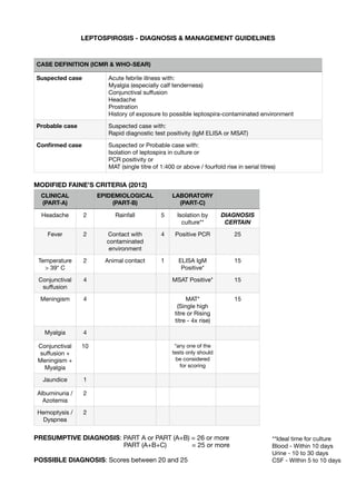
1. LEPTOSPIROSIS - DIAGNOSIS & MANAGEMENT GUIDELINES
MODIFIED FAINE’S CRITERIA (2012)
PRESUMPTIVE DIAGNOSIS: PART A or PART (A+B) = 26 or more
PART (A+B+C) = 25 or more
POSSIBLE DIAGNOSIS: Scores between 20 and 25
CASE DEFINITION (ICMR & WHO-SEAR)
Suspected case Acute febrile illness with:
Myalgia (especially calf tenderness)
Conjunctival su
ff
usion
Headache
Prostration
History of exposure to possible leptospira-contaminated environment
Probable case Suspected case with:
Rapid diagnostic test positivity (IgM ELISA or MSAT)
Con
fi
rmed case Suspected or Probable case with:
Isolation of leptospira in culture or
PCR positivity or
MAT (single titre of 1:400 or above / fourfold rise in serial titres)
CLINICAL
(PART-A)
EPIDEMIOLOGICAL
(PART-B)
LABORATORY
(PART-C)
Headache 2 Rainfall 5 Isolation by
culture**
DIAGNOSIS
CERTAIN
Fever 2 Contact with
contaminated
environment
4 Positive PCR 25
Temperature
> 39* C
2 Animal contact 1 ELISA IgM
Positive*
15
Conjunctival
su
ff
usion
4 MSAT Positive* 15
Meningism 4 MAT*
(Single high
titre or Rising
titre - 4x rise)
15
Myalgia 4
Conjunctival
su
ff
usion +
Meningism +
Myalgia
10 *any one of the
tests only should
be considered
for scoring
Jaundice 1
Albuminuria /
Azotemia
2
Hemoptysis /
Dyspnea
2
**Ideal time for culture
Blood - Within 10 days
Urine - 10 to 30 days
CSF - Within 5 to 10 days
2. Leptospirosis - Suspected / Probable / Con
fi
rmed
90% of cases are
Mild Leptospirosis
(Fever, myalgia,
conjunctival suffusion,
headache BUT NO
JAUNDICE)
Around 10% cases are
Moderate / Severe Leptospirosis
(Fever, myalgia, conjunctival suffusion, headache + JAUNDICE +/-
Multi-organ involvement
Based on clinical
spectrum
OP Treatment
Doxycycline 100mg PO
BD x 7 days or
Amoxicillin 500mg PO
TDS x 7 days or
Ampicillin 500mg
PO TDS x 7days or
Azithromycin 500mg PO
OD x 3 days
Other supportive Rx*
*advise adequate
hydration, bed rest,
antipyretics etc.
RED FLAG SIGNS
(tachypnea, tachycardia
disproportionate to fever,
shock, altered sensorium,
oliguria, bleeding
manifestations etc.)
IP Treatment (Antibiotic Rx +/- Organ speci
fi
c Rx)
Antibiotic therapy:
Penicillin 1.5 million units IV QID x 7 days or
Ceftriaxone 1g IV BD x 7 days or
Doxycycline 200mg IV stat, then 100mg IV BD x 7days
(Doxycycline contraindicated in pregnancy)
Organ speci
fi
c therapy:
RENAL: renal involvement is common.
Mild - only proteinuria and no RFT derangement: No intervention
Severe - AKI: Fluid management +/- diuretics, electrolyte correction,
avoid nephrotoxic drugs, avoid hypotension and hypovolemia +/- RRT (if
indicated by standard RRT guidelines)
HEPATIC: acute liver failure is rare.
Avoid precipitating factors of hepatic encephalopathy - drugs
(hepatotoxic drugs, sedatives etc.), hypovolemia, hypokalemia,
alkalosis, constipation, UGI bleeding.
Jaundice, Hepatomegaly: No intervention
Hepatic encephalopathy: lactulose, rifaximin etc.
LUNG: Most dangerous complication
ARDS / Pulmonary hemorrhage: Continuous O2 therapy, Mechanical
ventilation (if indicated)
HEART:
Myocarditis / Arrhythmia: treatment of speci
fi
c arrhythmia
Shock: treat hypovolemia with
fl
uid replacement. If not responding, add
dopamine or dobutamine.
HEMATOLOGICAL:
Thrombocytopenia: Platelet transfusion (if indicated)
Coagulopathy: Vit.K 5-10mg IV x 3 days +/- Fresh Frozen Plasma
DIC: FFP +/- blood transfusion
NEUROLOGICAL:
Aseptic meningitis: Symptomatic and supportive management.
Hypokalemic paralysis: IV Potassium supplementation
MUSCULOSKELETAL:
Myalgia / Myositis / Rhabdomyolysis : Monitor CPK levels, adequate
hydration, monitor urine output and serum electrolytes.
Arthralgia: No intervention +/- analgesic-antipyretics
All Absent
Any one or more is
present
Always rule out other tropical diseases.
Mixed infections are common.
Important differential diagnosis include:
-Malaria
-Scrub typhus
-Dengue
-Hepatitis
-Enteric fever etc.
3. Leptospirosis
Zoonotic
Pathogenic spirochete – Leptospira interrogans
Rodents and Cattle excrete these organisms in their urine, which contaminates soil and
waterbodies
Mode of transmission: contact of abraded skin or mucous membrane with contaminated
environment
Incubation period: Average 5-14 days with a range 2- 30 days
Risk factors:
o Heavy rainfall and water logging
o Natural disasters like floods
o Seasonal – at the onset of monsoon
o Farmers
o Agricultural field workers
o Fishermen
o Sewer workers
o Livestock handlers
o Mason
o Residence in endemic area
Presentation spectrum:
o Anicteric Leptospirosis: (90%) – Mild form presents like Acute undifferentiated fever
o Icteric Leptospirosis: (5-10%) – Moderate-Severe form
o Weil’s Disease (0-5%) – Severe form
When to suspect Leptospirosis?
o Acute febrile illness + Risk factors + one or more of the following:
Headache
Myalgia
Prostration
Calf muscle tenderness
Conjunctival suffusion
Oliguria / Frothy urine
Jaundice
Haemorrhagic manifestations
Meningeal irritation
Nausea, Vomiting, Abdominal pain, Diarrhoea
Lab investigations to support diagnosis: (Blood, CSF, Urine sample)
o MAT titre of 100/200/400 or above based on endemicity (preferred)
o IgM based immune assays
o Seroconversion or Four-fold rise in MAT titre between acute and convalescent sera
o Direct isolation of organism
o PCR test
4. Lab investigations to assess severity:
o CBC, ESR
o RFT, LFT
o S. Electrolytes
o Urine Routine examination
o CPK
o CXR, ABG
Management:
o IVF and correction of electrolytes
o Mild cases:
Tab. Doxycycline 100mg PO BD X 7 days (preferred) or
Tab. Azithromycin 500mg PO OD X 3 days
o Moderate / Severe cases:
Inj. Penicillin 1.5 million units IV or IM Q6H X 7 days or
Inj. Ceftriaxone 2g IV OD X 7 days
o Chemoprophylaxis:
Tab. Doxycycline 200mg PO once a week or
Tab. Azithromycin 250mg PO once/ twice a week
5. DIAGNOSTIC:
A + B = /> 26
OR
A + B + C = /> 25
POSSIBLE:
A + B = Between 20 to 25
CRITERIA