Leptospirosis diagnosis
•Download as PPTX, PDF•
0 likes•124 views

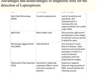
Dark field microscopy can visualize Leptospirosis but lacks sensitivity and specificity, requiring a high concentration of leptospires. The IgM ELISA is widely used but can produce false positives and cannot detect early infection. The microscopic agglutination test is the gold standard but is less sensitive early in disease and is labor intensive, requiring maintained Leptospira strains. PCR can successfully detect Leptospira DNA but requires expensive reagents and large DNA quantities, and cannot identify the infecting serovar.

Recommended
More Related Content
Similar to Leptospirosis diagnosis
Similar to Leptospirosis diagnosis (20)
More from DR RML DELHI
More from DR RML DELHI (20)
Recently uploaded
Recently uploaded (20)
Leptospirosis diagnosis
- 1. Test Advantages Disadvantages Dark Field Microscopy (DFM) Visualize Leptospirosis Lack of sensitivity and specificity. 104 Leptospires/ml is necessary for one organism/field to be visible under DFM. IgM ELISA Most widely used False positive, IgM cannot be detected in early stages of infection and can persist in blood for years. Microscopic Agglutination Test (MAT) Gold Standard Less sensitive in early phase of disease. Labor intensive and complicated procedure as there is a need to maintain Leptospira strain for preparing live antigen. Polymerase Chain Reaction (PCR) Successful in detecting Leptospira DNA in serum and urine samples of patients Expensive reagents, Requires large quantity of DNA. Cannot identify the infecting serovar. Advantages and disadvantages of diagnostic tests for the detection of Leptospirosis