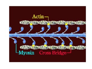
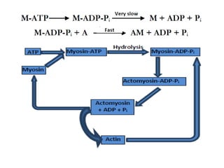
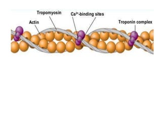
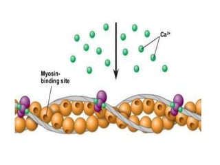
Physiology - HND
Khawaja Taimoor Shahid
Topic-11 Contraction of Muscle:
Sliding Filament Theory
• Sliding Filament Theory
• H. E. Huxley and A. F. Huxley proposed the sliding
filament theory of muscle contraction in 1954.
• This theory states that during muscle contraction the thin
and thick filaments in sarcomeres slide and undergo
shifting.
• When a muscle contracts, the thin actin filaments actively
slide along between the thick myosin filaments and move
closer to the center of the sarcomere. As a result, the
sarcomere becomes shorter.
• When a muscle relaxes or is stretched, the overlap between
thin and thick filaments is reduced, and the sarcomere
elongates.
• The changes in sarcomere length during contraction and
stretch of a muscle, correspond to changes in muscle length.
Explanation
• In a relaxed muscle fiber, the thick and thin filaments
overlap only at the ends of A band. But when muscle fibers
are stimulated by the nervous system, myosin heads are
attached to the myosin-binding sites on actin in the thin
filaments, i.e. cross bridges are formed and the sliding
begins.
• During contraction, the A bands (myosin filaments)
maintain a constant length, whereas the I bands and the H
zone (zones where actin and myosin filaments do not
overlap) become shorter and Z lines get closer.
• When the muscle is stretched, the A band again maintains a
constant length, but the I bands and H zone become longer.
• Neither the myosin thick filaments nor the actin thin
filaments change their lengths when a sarcomere shortens or
is stretched. It is the extent of overlap between actin and
myosin filaments that changes.
Length-Tension Curve
• One of the strongest pieces of evidence in
support of the sliding filament theory comes
from the length-tension relation of a
sarcomere.
• Experimental measurement of the shortening
of length of sarcomere during contraction and
resulting force generates a length-tension
curve. This curve explains the assumptions of
sliding-filament theory.
Explanation of the Curve
• • The tension produced by the muscle is maximal when
the overlap between thick and thin filaments allows the
largest number of cross-bridges to be formed between
actin and myosin.
• • Tension drops off with increased length of sarcomere,
because the thick and thin filaments overlap less and
fewer crossbridges can be formed.
• • It also drops off with decreased length, because thin
filaments begin to collide with one another, preventing
further shortening.
• • The curve also predicts the consequence of sliding
filament theory that no active tension will develop if a
sarcomere is stretched so far that there remains no
overlap between actin and myosin filaments, making it
impossible to develop any crossbidges.
Conclusion
• This curve shows that the tension
produced by a sarcomere is proportional to its
shortening which is due to sliding of thick and
thin filaments and formation of cross bridges
in the sarcomere during contraction. These
were the proposals of sliding filament theory.
So length-tension curve provides a practical
proof of this theory.
Topic-12 Role of ATP in Cross Bridge
Working
Cyclic Attachment and Detachment of Cross-Bridges
• Myosin cross-bridges must attach to binding sites on
actin filaments in order to generate force.
• However, the cross-bridges must also be able to detach
because attached cross-bridges would prevent
filaments from sliding past one another, locking the
muscle at one length.
• In addition, detachment of cross-bridges from actin is
necessary for the muscle to relax.
• So, during contraction, the cross-bridges must attach
and detach from the thin filaments in a cyclic fashion.
Role of ATP in Cross-Bridge
Detachment
• This cyclic attachment and detachment happens
due to the activity of ATP.
• Cross bridges are formed when actin (A) and
myosin (M) bind and form a stable complex called
actomyosin (AM). This happens in the absence of
ATP.
• Cross-bridge detachment occurs in the presence
of ATP. ATP causes the AM complex to rapidly
dissociate into actin and myosin-ATP:
A + M = AM
AM + ATP = A + M-ATP
Cyclic Activity of AM complex
• The Myosin-ATP complex hydrolyzes to form
myosin-ADP-Pi complex.
• However, ADP and Pi unbind from myosin very
slowly.
• The release of ADP and Pi is greatly speeded
up when actin binds to myosin in the myosin-
ADP-Pi complex.
• This binding of actin results in the formation
of another actomyosin complex. This reaction
is kinetically favored as it releases energy.
Rigor Mortis
• After death, human and other animal’s bodies
gradually become rigid. This condition is called
rigor mortis.
• This rigidity happens because ATP’s are not
available in dead body for detachment of actin
and myosin, so muscles cannot relax.
Topic-13 Production of Force for
Sliding of Filaments
Force is Produced by Rotation of Myosin Head
• During muscle contraction, myosin heads make cross-
bridges with actin filaments that pull the thin filament
toward the center of sarcomere. The force for pulling is
produced by the partial rotation of myosin heads.
How Rotation is Produced?
• The rotation is produced when the four sites M1 to M4
of myosin head interact sequentially with the binding
sites of actin filament.
Energy Storage in the Link
• As the myosin head rotates against the actin filament,
the link is stretched elastically and stores mechanical
energy in the link due to tension developed in it.
Transmission of Force to Thick Filament
The tension thus produced is transmitted to the thick filament through the neck
of the myosin molecule. This tension provides force to shorten the sarcomere.
Detachment of Myosin Head
• When the rotation of the head is complete, the myosin
head dissociates from the actin filament and rotates back
to its relaxed position.
• The myosin head detaches from actin when Mg2+ and ATP
bind to the head. The ATP is then hydrolyzed, which is
accompanied by a conformational change in the myosin
head, leaving the head in an energized state, ready to
rebind to a little farther site on the actin filament.
• This cycle is repeated over and over, and the filaments slide
past one another in small incremental steps of attachment,
rotation, and detachment of the many cross-bridges on
each thick filament.
Topic-14 Role of Calcium in
Contraction
Role of Calcium in Cross-bridge Attachment
• Ca2+ plays a crucial role in regulating the
contractile activity of muscles as it helps to
expose the myosin binding sites of actin. This
exposure induces cross bridge formation,
necessary for contraction.
Ca2+ Plays Role by Interacting with
Troponin and Tropomyosin
• Ca2+ induces contraction with the help of two
regulatory proteins troponin and tropomyosin,
associated with actin filaments.
• In a relaxed myofibril, tropomyosin coils around
the actin filament and sterically (physically)
covers the myosin binding sites of actin, thus
preventing actin and myosin from interacting.
• Troponin complex binds to tropomyosin about
every 40 nm along the thin actin filament.
Binding of Ca2+ to Troponin
• Troponin has a high binding affinity for Ca2+ and
each troponin complex binds four Ca2+ ions.
• When Ca2+ binds to troponin, the troponin
molecule undergoes a change in conformation.
• This causes a shifting in the position of
tropomyosin. Tropomyosin movement exposes
the myosin binding sites on the thin actin
filament.
Binding of Ca2+ to Troponin
• Thus, when Ca2+ binds to troponin, it removes the
inhibition of attachment between myosin cross-bridges
and thin filaments. So the thin and thick filaments can
slide past each other, and the muscle fiber contracts.
• The role of Ca2+ in regulating the actin-myosin
interaction via troponin and tropomyosin applies to
vertebrate skeletal and cardiac muscle.
Required Concentration of Ca2+ for Contraction
• The required concentration of Ca2+ ions in cytosol for
binding of cross-bridges to actin is above 10-7 M.
Topic-15 Excitation Contraction
Coupling
Action Potentials Trigger Muscle Contraction
• The skeletal muscles contract in response to an action
potential (nerve impulse) that arrives at the
neuromuscular junction.
Acetylcholine Neurotransmitter
• At the neuromuscular junction, motor neurons release
the neurotransmitter, acetylcholine.
• Acetylcholine binds to the receptors in postsynaptic
muscle fibers. These receptors are ligand-gated ion
channels. Opening of these channels causes movement
of sodium (Na+) and potassium (K+) ions.
End-plate Potential
• The ionic movements cause change of potential
in the muscle cell membrane resulting in
membrane excitation. The potential of excited
muscle fiber membrane is known as end-plate
potential. Membrane excitation results in the
triggering of an all-or-none AP in the fiber
membrane.
• The AP propagates away, exciting the entire
membrane of the muscle fiber and setting in
motion the sequence of events leading to
contraction.
Excitation-Contraction Coupling
• The sequence of events that convert an action potential to
muscle contraction is known as excitation-contraction
coupling.
Latency period
• It takes several milliseconds to begin contraction after the
arrival of an AP.
• This latency is because of the large size of skeletal muscle
fibers which cannot contract unless action potential
spreads deep into the fiber to the vicinity of each myofibril.
• During this latent period, action potential is transmitted
along the transverse tubules (T tubules) deep within the
fiber.
Release of Ca2+ and Contraction
• Transmission of action potential through T
tubules results in the release of calcium ions form
stores of sarcoplasmic reticulum.
• This increases Ca2+ concentration inside the
muscle fiber in the immediate vicinity of the
myofibrils.
• These calcium ions cause the contraction to
begin.
• The net effect of excitation-contraction coupling
is to link an AP in the plasma membrane of the
muscle fiber to the concentration of free Ca2+ in
the cytosol that initiate contraction.
Topic-16 Summary of Muscle
Contraction Mechanism
• Contraction-Relaxation Cycle
• Starting with a relaxed skeletal muscle, the following
sequence of events leads to contraction and then
relaxation of a skeletal muscle fiber:
• 1. The surface membrane of the fiber is depolarized by
an AP due to neuronal input.
• 2. The AP is conducted deep into the muscle fiber
along the T tubules.
• 3. In response to depolarization of the T-tubule
membrane, voltage-sensitive dihydropyridine receptors
in the T-tubule membrane undergo a conformational
change that-through direct mechanical linkage to
ryanodine receptors in the SR membrane-causes
opening of Ca2+ channels in the SR membrane.
• 4. As Ca2+ flow out from the lumen of sarcoplasmic reticulum,
the free Ca2+ concentration of the myoplasm increases from a
resting value of below 10-7 M to an active level of about 10-6
within a few milliseconds.
• 5. Most of the Ca2+ ions that enter the myoplasm bind rapidly
to troponin, inducing a conformational change in the troponin
molecule. This conformational change causes a change in the
position of the tropomyosin, eliminating steric hindrance and
allowing myosin cross-bridges to bind to actin thin filaments.
• 6. Myosin cross-bridges attach to the actin filaments. Myosin
heads rotate against the actin filaments producing force that
pulls the thin filaments toward the center of the sarcomere,
causing the sarcomere to shorten.
• 7. ATP binds to the ATPase site on the myosin head
causing the myosin head to detach from the thin
filament.
• 8. ATP is then hydrolyzed, and the energy of the
hydrolysis is stored as a conformational change in the
myosin molecule, which then reattaches to the next
site along the actin filament and the cycle of binding
and unbinding is repeated.
• 9. Finally, calcium pumps in the SR membrane actively
transport Ca2+ from the myoplasm back into the SR. As
the concentration of free Ca2+ in the myoplasm drops,
Ca2+ bound to troponin is released, allowing
tropomyosin again to inhibit cross-bridge attachment,
so the muscle relaxes. The muscle remains relaxed until
the next depolarization.

lecture 3 physio.pptx

  • 1.
  • 2.
    Topic-11 Contraction ofMuscle: Sliding Filament Theory • Sliding Filament Theory • H. E. Huxley and A. F. Huxley proposed the sliding filament theory of muscle contraction in 1954. • This theory states that during muscle contraction the thin and thick filaments in sarcomeres slide and undergo shifting. • When a muscle contracts, the thin actin filaments actively slide along between the thick myosin filaments and move closer to the center of the sarcomere. As a result, the sarcomere becomes shorter. • When a muscle relaxes or is stretched, the overlap between thin and thick filaments is reduced, and the sarcomere elongates. • The changes in sarcomere length during contraction and stretch of a muscle, correspond to changes in muscle length.
  • 3.
    Explanation • In arelaxed muscle fiber, the thick and thin filaments overlap only at the ends of A band. But when muscle fibers are stimulated by the nervous system, myosin heads are attached to the myosin-binding sites on actin in the thin filaments, i.e. cross bridges are formed and the sliding begins. • During contraction, the A bands (myosin filaments) maintain a constant length, whereas the I bands and the H zone (zones where actin and myosin filaments do not overlap) become shorter and Z lines get closer. • When the muscle is stretched, the A band again maintains a constant length, but the I bands and H zone become longer. • Neither the myosin thick filaments nor the actin thin filaments change their lengths when a sarcomere shortens or is stretched. It is the extent of overlap between actin and myosin filaments that changes.
  • 5.
    Length-Tension Curve • Oneof the strongest pieces of evidence in support of the sliding filament theory comes from the length-tension relation of a sarcomere. • Experimental measurement of the shortening of length of sarcomere during contraction and resulting force generates a length-tension curve. This curve explains the assumptions of sliding-filament theory.
  • 6.
    Explanation of theCurve • • The tension produced by the muscle is maximal when the overlap between thick and thin filaments allows the largest number of cross-bridges to be formed between actin and myosin. • • Tension drops off with increased length of sarcomere, because the thick and thin filaments overlap less and fewer crossbridges can be formed. • • It also drops off with decreased length, because thin filaments begin to collide with one another, preventing further shortening. • • The curve also predicts the consequence of sliding filament theory that no active tension will develop if a sarcomere is stretched so far that there remains no overlap between actin and myosin filaments, making it impossible to develop any crossbidges.
  • 8.
    Conclusion • This curveshows that the tension produced by a sarcomere is proportional to its shortening which is due to sliding of thick and thin filaments and formation of cross bridges in the sarcomere during contraction. These were the proposals of sliding filament theory. So length-tension curve provides a practical proof of this theory.
  • 9.
    Topic-12 Role ofATP in Cross Bridge Working Cyclic Attachment and Detachment of Cross-Bridges • Myosin cross-bridges must attach to binding sites on actin filaments in order to generate force. • However, the cross-bridges must also be able to detach because attached cross-bridges would prevent filaments from sliding past one another, locking the muscle at one length. • In addition, detachment of cross-bridges from actin is necessary for the muscle to relax. • So, during contraction, the cross-bridges must attach and detach from the thin filaments in a cyclic fashion.
  • 10.
    Role of ATPin Cross-Bridge Detachment • This cyclic attachment and detachment happens due to the activity of ATP. • Cross bridges are formed when actin (A) and myosin (M) bind and form a stable complex called actomyosin (AM). This happens in the absence of ATP. • Cross-bridge detachment occurs in the presence of ATP. ATP causes the AM complex to rapidly dissociate into actin and myosin-ATP: A + M = AM AM + ATP = A + M-ATP
  • 11.
    Cyclic Activity ofAM complex • The Myosin-ATP complex hydrolyzes to form myosin-ADP-Pi complex. • However, ADP and Pi unbind from myosin very slowly. • The release of ADP and Pi is greatly speeded up when actin binds to myosin in the myosin- ADP-Pi complex. • This binding of actin results in the formation of another actomyosin complex. This reaction is kinetically favored as it releases energy.
  • 13.
    Rigor Mortis • Afterdeath, human and other animal’s bodies gradually become rigid. This condition is called rigor mortis. • This rigidity happens because ATP’s are not available in dead body for detachment of actin and myosin, so muscles cannot relax.
  • 14.
    Topic-13 Production ofForce for Sliding of Filaments Force is Produced by Rotation of Myosin Head • During muscle contraction, myosin heads make cross- bridges with actin filaments that pull the thin filament toward the center of sarcomere. The force for pulling is produced by the partial rotation of myosin heads. How Rotation is Produced? • The rotation is produced when the four sites M1 to M4 of myosin head interact sequentially with the binding sites of actin filament. Energy Storage in the Link • As the myosin head rotates against the actin filament, the link is stretched elastically and stores mechanical energy in the link due to tension developed in it.
  • 15.
    Transmission of Forceto Thick Filament The tension thus produced is transmitted to the thick filament through the neck of the myosin molecule. This tension provides force to shorten the sarcomere.
  • 16.
    Detachment of MyosinHead • When the rotation of the head is complete, the myosin head dissociates from the actin filament and rotates back to its relaxed position. • The myosin head detaches from actin when Mg2+ and ATP bind to the head. The ATP is then hydrolyzed, which is accompanied by a conformational change in the myosin head, leaving the head in an energized state, ready to rebind to a little farther site on the actin filament. • This cycle is repeated over and over, and the filaments slide past one another in small incremental steps of attachment, rotation, and detachment of the many cross-bridges on each thick filament.
  • 17.
    Topic-14 Role ofCalcium in Contraction Role of Calcium in Cross-bridge Attachment • Ca2+ plays a crucial role in regulating the contractile activity of muscles as it helps to expose the myosin binding sites of actin. This exposure induces cross bridge formation, necessary for contraction.
  • 18.
    Ca2+ Plays Roleby Interacting with Troponin and Tropomyosin • Ca2+ induces contraction with the help of two regulatory proteins troponin and tropomyosin, associated with actin filaments. • In a relaxed myofibril, tropomyosin coils around the actin filament and sterically (physically) covers the myosin binding sites of actin, thus preventing actin and myosin from interacting. • Troponin complex binds to tropomyosin about every 40 nm along the thin actin filament.
  • 20.
    Binding of Ca2+to Troponin • Troponin has a high binding affinity for Ca2+ and each troponin complex binds four Ca2+ ions. • When Ca2+ binds to troponin, the troponin molecule undergoes a change in conformation. • This causes a shifting in the position of tropomyosin. Tropomyosin movement exposes the myosin binding sites on the thin actin filament.
  • 22.
    Binding of Ca2+to Troponin • Thus, when Ca2+ binds to troponin, it removes the inhibition of attachment between myosin cross-bridges and thin filaments. So the thin and thick filaments can slide past each other, and the muscle fiber contracts. • The role of Ca2+ in regulating the actin-myosin interaction via troponin and tropomyosin applies to vertebrate skeletal and cardiac muscle. Required Concentration of Ca2+ for Contraction • The required concentration of Ca2+ ions in cytosol for binding of cross-bridges to actin is above 10-7 M.
  • 23.
    Topic-15 Excitation Contraction Coupling ActionPotentials Trigger Muscle Contraction • The skeletal muscles contract in response to an action potential (nerve impulse) that arrives at the neuromuscular junction. Acetylcholine Neurotransmitter • At the neuromuscular junction, motor neurons release the neurotransmitter, acetylcholine. • Acetylcholine binds to the receptors in postsynaptic muscle fibers. These receptors are ligand-gated ion channels. Opening of these channels causes movement of sodium (Na+) and potassium (K+) ions.
  • 24.
    End-plate Potential • Theionic movements cause change of potential in the muscle cell membrane resulting in membrane excitation. The potential of excited muscle fiber membrane is known as end-plate potential. Membrane excitation results in the triggering of an all-or-none AP in the fiber membrane. • The AP propagates away, exciting the entire membrane of the muscle fiber and setting in motion the sequence of events leading to contraction.
  • 25.
    Excitation-Contraction Coupling • Thesequence of events that convert an action potential to muscle contraction is known as excitation-contraction coupling. Latency period • It takes several milliseconds to begin contraction after the arrival of an AP. • This latency is because of the large size of skeletal muscle fibers which cannot contract unless action potential spreads deep into the fiber to the vicinity of each myofibril. • During this latent period, action potential is transmitted along the transverse tubules (T tubules) deep within the fiber.
  • 26.
    Release of Ca2+and Contraction • Transmission of action potential through T tubules results in the release of calcium ions form stores of sarcoplasmic reticulum. • This increases Ca2+ concentration inside the muscle fiber in the immediate vicinity of the myofibrils. • These calcium ions cause the contraction to begin. • The net effect of excitation-contraction coupling is to link an AP in the plasma membrane of the muscle fiber to the concentration of free Ca2+ in the cytosol that initiate contraction.
  • 27.
    Topic-16 Summary ofMuscle Contraction Mechanism • Contraction-Relaxation Cycle • Starting with a relaxed skeletal muscle, the following sequence of events leads to contraction and then relaxation of a skeletal muscle fiber: • 1. The surface membrane of the fiber is depolarized by an AP due to neuronal input. • 2. The AP is conducted deep into the muscle fiber along the T tubules. • 3. In response to depolarization of the T-tubule membrane, voltage-sensitive dihydropyridine receptors in the T-tubule membrane undergo a conformational change that-through direct mechanical linkage to ryanodine receptors in the SR membrane-causes opening of Ca2+ channels in the SR membrane.
  • 28.
    • 4. AsCa2+ flow out from the lumen of sarcoplasmic reticulum, the free Ca2+ concentration of the myoplasm increases from a resting value of below 10-7 M to an active level of about 10-6 within a few milliseconds. • 5. Most of the Ca2+ ions that enter the myoplasm bind rapidly to troponin, inducing a conformational change in the troponin molecule. This conformational change causes a change in the position of the tropomyosin, eliminating steric hindrance and allowing myosin cross-bridges to bind to actin thin filaments. • 6. Myosin cross-bridges attach to the actin filaments. Myosin heads rotate against the actin filaments producing force that pulls the thin filaments toward the center of the sarcomere, causing the sarcomere to shorten.
  • 29.
    • 7. ATPbinds to the ATPase site on the myosin head causing the myosin head to detach from the thin filament. • 8. ATP is then hydrolyzed, and the energy of the hydrolysis is stored as a conformational change in the myosin molecule, which then reattaches to the next site along the actin filament and the cycle of binding and unbinding is repeated. • 9. Finally, calcium pumps in the SR membrane actively transport Ca2+ from the myoplasm back into the SR. As the concentration of free Ca2+ in the myoplasm drops, Ca2+ bound to troponin is released, allowing tropomyosin again to inhibit cross-bridge attachment, so the muscle relaxes. The muscle remains relaxed until the next depolarization.