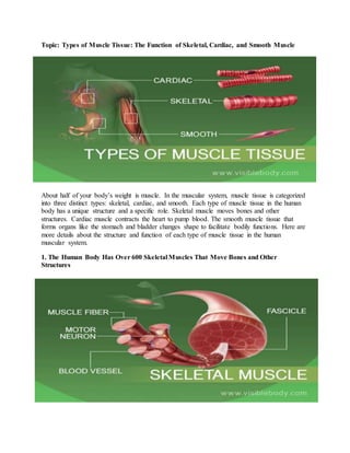
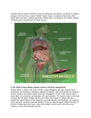
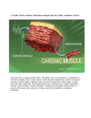
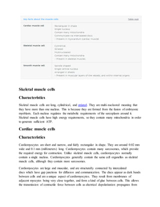
SlideShare a Scribd company logo
1 of 7
Topic: Types of Muscle Tissue: The Function of Skeletal, Cardiac, and Smooth Muscle
About half of your body’s weight is muscle. In the muscular system, muscle tissue is categorized
into three distinct types: skeletal, cardiac, and smooth. Each type of muscle tissue in the human
body has a unique structure and a specific role. Skeletal muscle moves bones and other
structures. Cardiac muscle contracts the heart to pump blood. The smooth muscle tissue that
forms organs like the stomach and bladder changes shape to facilitate bodily functions. Here are
more details about the structure and function of each type of muscle tissue in the human
muscular system.
1. The Human Body Has Over 600 Skeletal Muscles That Move Bones and Other
Structures
sSkeletal muscles attach to and move bones by contracting and relaxing in response to voluntary
messages from the nervous system. Skeletal muscle tissue is composed of long cells called
muscle fibers that have a striated appearance. Muscle fibers are organized into bundles supplied
by blood vessels and innervated by motor neurons.
s
2. The Walls of Many Human Organs Contract and Relax Automatically
Smooth muscle is found in the walls of hollow organs throughout the body. Smooth muscle
contractions are involuntary movements triggered by impulses that travel through the autonomic
nervous system to the smooth muscle tissue. The arrangement of cells within smooth muscle
tissue allows for contraction and relaxation with great elasticity. The smooth muscle in the walls
of organs like the urinary bladder and the uterus allow those organs to expand and relax as
needed. The smooth muscle of the alimentary canal (the digestive tract) facilitates the peristaltic
waves that move swallowed food and nutrients. In the eye smooth muscle changes the shape of
the lens to bring objects into focus. Artery walls include smooth muscle that relaxes and
contracts to move blood through the body
3. Cardiac Muscle Contracts in Response to Signals from the Cardiac Conduction System
The heart wall is composed of three layers. The middle layer, the myocardium, is responsible for
the heart’s pumping action. Cardiac muscle, found only in the myocardium, contracts in response
to signals from the cardiac conduction system to make the heart beat. Cardiac muscle is made
from cells called cardiocytes. Like skeletal muscle cells cardiocytes have a striated appearance,
but their overall structure is shorter and thicker. Cardiocytes are branched, allowing them to
connect with several other cardiocytes, forming a network that facilitates coordinated
contraction.
Skeletal muscle cells
Characteristics
Skeletal muscle cells are long, cylindrical, and striated. They are multi-nucleated meaning that
they have more than one nucleus. This is because they are formed from the fusion of embryonic
myoblasts. Each nucleus regulates the metabolic requirements of the sarcoplasm around it.
Skeletal muscle cells have high energy requirements, so they contain many mitochondria in order
to generate sufficient ATP.
Cardiac muscle cells
Characteristics
Cardiomyocytes are short and narrow, and fairly rectangular in shape. They are around 0.02 mm
wide and 0.1 mm (millimeters) long. Cardiomyocytes contain many sarcosomes, which provide
the required energy for contraction. Unlike skeletal muscle cells, cardiomyocytes normally
contain a single nucleus. Cardiomyocytes generally contain the same cell organelles as skeletal
muscle cells, although they contain more sarcosomes.
Cardiomyocytes are large and muscular, and are structurally connected by intercalated
discs which have gap junctions for diffusion and communication. The discs appear as dark bands
between cells and are a unique aspect of cardiomyocytes. They result from membranes of
adjacent myocytes being very close together, and form a kind of glue between cells. This allows
the transmission of contractile force between cells as electrical depolarization propagates from
cell to cell. The key role of cardiomyocytes is to generate enough contractile force for the heart
to beat effectively. They contract together in unison, causing enough pressure to
force blood around the body.
Smooth muscle cells
Characteristics
Smooth muscle cells are spindle-shaped and contain a single central nucleus. They range from 10
to 600 μm (micrometers) in length, and are the smallest type of muscle cell. They are elastic and
therefore important in the expansion of organs such as the kidneys, lungs, and vagina. The
myofibrils of smooth muscle cells are not aligned like in cardiac and skeletal muscle meaning
that they are not striated, hence, the name smooth.
Smooth muscle cells are arranged together in sheets and this organisation means that they can
contract simultaneously. They have poorly developed sarcoplasmic reticulums and do not
contain T-tubules, due to the restricted size of the cells. However, they do contain other normal
cell organelles such as sarcosomes but in lower numbers.
Topic: Atrphy and hypertrophy
Atrophy and hypertrophy are two opposite conditions that can be found in pathological
or diseased muscles.
Atrophy is characterized by a wasting or loss of the muscle mass (A1) and usually
involves a decrease in the size or cross-sectional area (CSA) (A2) of an individual myofibre or a
number of myofibres. In contrast, hypertrophy is an increase in muscle mass and CSA,
specifically due to an increase in the CSA of individual muscle fibres. As a result, the muscle
strength (A3) and the bone mass are significantly affected.
To maintain homeostasis, the biological response of the human body generates a dynamic
balance between synthetic and degradative processes for both atrophic and hypertrophic muscles.
This dynamic balance occurs in response to any stimuli, due to processes that promote muscle
growth via increased protein content.
Moreover, it can result either from increased protein production, decreased protein breakdown,
or a combination of both of these aspects of protein turnover. The processes that govern the
extent of muscle atrophy are based on the magnitude of the regulated decline in rate of protein
synthesis, increased level of oxidative damage (A4), and subsequent unregulated protein
degradation. For example, inhibitors of the proteosome block increases in protein breakdown
normally seen in atrophy, the level of ubiquitinated conjugates increase during atrophy and genes
that encode various components of the ubiquitin pathway increase during atrophy. An increase in
muscle activity stimulates the expression of a protein growth factor known as insulin-like growth
factor I (IGF-I). IGF-I has been shown to be sufficient to induce hypertrophy through either
autocrine or paracrine mechanisms. IGF-I expression is increased during compensatory
hypertrophy caused experimentally by removing several muscles to force those remaining to take
up the resultant increase in load.
Muscle atrophy
The causes of muscle atrophy are from several sources, such as neuromuscular diseases,
immobilization and denervated conditions. In addition, the muscle atrophy may also take place,
secondary to some devastating injuries or common health problems, such as spinal cord injury
(SCI), ageing and various systemic diseases (A5), respectively. Moreover, the condition may be
exacerbated by starvation, micro-gravity (A6), detraining (A7), reduction in neuromuscular
activity (Fitts et al. 2000), decreased levels of hormones (A8), increases in protein degradation
(A9), decreases in protein synthesis (A10), decreases in protein content, and various forms of
reduced use (A11). Among acute and critically ill patients, the onset of muscle atrophy is rapid
and severe, beginning within 4 h of hospitalization. In the first few weeks during hospitalization,
the antigravity or the extensor group muscles will show greater atrophy than non-antigravity or
flexor group muscles. During extended periods of hospitalization, a prolonged unused limb leads
not only to an impairment of the muscle function (A12), but also to a deleterious alteration in the
muscle morphology (Bloomfield 1997), manifested in symptoms such as a decrease in muscle
mass (A13), a reduction of the muscle fibre diameter, and a reduction in the overall number of
muscle fibres. Moreover, this condition may also have a negative affect on bone health by
decreasing bone mineral density at the lumbar spine, femoral neck and calcaneus. Interestingly,
the duration of immobility has been shown to be positively correlated with the degree of muscle
atrophy (A14). The early signs of muscle atrophy found in these patients are accompanied by
general weakness (A15) and fatigue (A16), especially in the lower limb (A17). In fact, these
clinical signs may be caused either by the medication or pathological condition per se. Therefore,
it is inappropriate to conclude the patient’s condition based only on muscle testing alone. Some
other clinical assessments such as electrodiagnosis, computerized muscle strength analysis and
biochemical analyses are essential for providing verification and further confirmation of the
status of the muscles in question. The clinical assessments for disuse muscle atrophy can be
performed at the bedside, accompanied with the strength assessment by observing muscle
movement, muscle tone, muscle size and muscle strength.
Muscle hypertrophy
Hypertrophy of a muscle is a multidimensional process involving several factors such as
growth factors (GFs), IGFs (A29), clenbuterol, anabolic steroids, hormones (A30), the immune
system, and satellite cells (A31). For example, in a study investigating IGF-I peptide levels in
human muscle following 10 weeks of strength training in old men and women (aged 72–98
years), it was shown that there was a c. 500% increase in the levels of IGF-I within the muscle
fibres of these subjects after the training period, as determined using immunohistochemistry.
This demonstrates that the peptide levels in older muscles may adapt over the longer-term to
exercise training. Indeed, the results of longitudinal strength training studies have confirmed that
the muscles of even very elderly people are able to exhibit a hypertrophy response to resistance
exercise (A32).
IGF-I is also thought to be involved in the activation of satellite cells, satellite cells are
small mononucleate muscle stem cells located between the sarcolemma and basal lamina of
muscle fibres. Recently, the link between satellite cell number and myofibre size has been
demonstrated in both untrained and hypertrophied human muscle fibres. These cells, when
activated, are believed to proliferate and differentiate into myoblasts, which then fuse with
existing fibres, thus providing new nuclei to maintain the ratio of DNA to protein for fibres
undergoing hypertrophy. The link between IGF-I, satellite cells, and hypertrophy has been
shown in studies where localised infusion of IGF-I into the tibialis anterior muscle of adult rats
resulted in an increased total muscle protein and DNA content. More recently, Bamman et al.
(2001) reported a 62% increase in IGF-I mRNA concentration in human muscle 48 h after a
single bout of eccentric resistance type exercise.

More Related Content

Similar to lecture 4.docx

SKELETAL MUSCLE TISSUE: MUSCLE ,TENDON, LIGAMENT
SKELETAL MUSCLE TISSUE: MUSCLE ,TENDON, LIGAMENTSKELETAL MUSCLE TISSUE: MUSCLE ,TENDON, LIGAMENT
SKELETAL MUSCLE TISSUE: MUSCLE ,TENDON, LIGAMENTDawitAlemayehu7
 
Muscular system
Muscular systemMuscular system
Muscular systemKomal Kp
 
Review of Anatomy and Physiology of Musculoskeletal System / Nursing
Review of Anatomy and Physiology of Musculoskeletal System / NursingReview of Anatomy and Physiology of Musculoskeletal System / Nursing
Review of Anatomy and Physiology of Musculoskeletal System / NursingAby Thankachan
 
Introduction to the muscular system
Introduction to the muscular systemIntroduction to the muscular system
Introduction to the muscular systemsania aslam
 
Muscular System.pdf
Muscular System.pdfMuscular System.pdf
Muscular System.pdfPritamjit3
 
Exercise Physiology11. Discuss the relationship between dist.docx
Exercise Physiology11. Discuss the relationship between dist.docxExercise Physiology11. Discuss the relationship between dist.docx
Exercise Physiology11. Discuss the relationship between dist.docxSANSKAR20
 
Chronic Myofascial Pain Syndrome- Final Case Presentation
Chronic Myofascial Pain Syndrome- Final Case PresentationChronic Myofascial Pain Syndrome- Final Case Presentation
Chronic Myofascial Pain Syndrome- Final Case PresentationTeMz Gordonas
 
Muscles and Muscle Tissue
Muscles and Muscle TissueMuscles and Muscle Tissue
Muscles and Muscle TissueSunitaArya17
 
General doctrine about the muscles.
General doctrine about the muscles.General doctrine about the muscles.
General doctrine about the muscles.Tetyana Knyazevych
 
Anatomy and physiology
Anatomy and physiologyAnatomy and physiology
Anatomy and physiologyjazz060493
 
Musculoskeletal System Anatomy and Assessment
Musculoskeletal System Anatomy and AssessmentMusculoskeletal System Anatomy and Assessment
Musculoskeletal System Anatomy and AssessmentJofred Martinez
 
Fundamentals of biomechanics
Fundamentals of biomechanicsFundamentals of biomechanics
Fundamentals of biomechanicsSpringer
 
B asic ap chapter 10 powerpoint 2017
B asic ap chapter 10 powerpoint 2017B asic ap chapter 10 powerpoint 2017
B asic ap chapter 10 powerpoint 2017Kathy Richards
 
Tissue connective and muscule tissue
Tissue connective and muscule tissueTissue connective and muscule tissue
Tissue connective and muscule tissueChetan Sehgal
 
Bio13 .ppt;10 muscle-tissue-and-organization ppt zeny
Bio13 .ppt;10 muscle-tissue-and-organization ppt zenyBio13 .ppt;10 muscle-tissue-and-organization ppt zeny
Bio13 .ppt;10 muscle-tissue-and-organization ppt zenyDevron Jeko
 

Similar to lecture 4.docx (20)

SKELETAL MUSCLE TISSUE: MUSCLE ,TENDON, LIGAMENT
SKELETAL MUSCLE TISSUE: MUSCLE ,TENDON, LIGAMENTSKELETAL MUSCLE TISSUE: MUSCLE ,TENDON, LIGAMENT
SKELETAL MUSCLE TISSUE: MUSCLE ,TENDON, LIGAMENT
 
Muscular system
Muscular systemMuscular system
Muscular system
 
Review of Anatomy and Physiology of Musculoskeletal System / Nursing
Review of Anatomy and Physiology of Musculoskeletal System / NursingReview of Anatomy and Physiology of Musculoskeletal System / Nursing
Review of Anatomy and Physiology of Musculoskeletal System / Nursing
 
muscles anatomy
muscles anatomy muscles anatomy
muscles anatomy
 
Introduction to the muscular system
Introduction to the muscular systemIntroduction to the muscular system
Introduction to the muscular system
 
Muscular System.pdf
Muscular System.pdfMuscular System.pdf
Muscular System.pdf
 
Exercise Physiology11. Discuss the relationship between dist.docx
Exercise Physiology11. Discuss the relationship between dist.docxExercise Physiology11. Discuss the relationship between dist.docx
Exercise Physiology11. Discuss the relationship between dist.docx
 
Chronic Myofascial Pain Syndrome- Final Case Presentation
Chronic Myofascial Pain Syndrome- Final Case PresentationChronic Myofascial Pain Syndrome- Final Case Presentation
Chronic Myofascial Pain Syndrome- Final Case Presentation
 
Muscles and Muscle Tissue
Muscles and Muscle TissueMuscles and Muscle Tissue
Muscles and Muscle Tissue
 
General doctrine about the muscles.
General doctrine about the muscles.General doctrine about the muscles.
General doctrine about the muscles.
 
Anatomy and physiology
Anatomy and physiologyAnatomy and physiology
Anatomy and physiology
 
Muscular system.pptx
Muscular system.pptxMuscular system.pptx
Muscular system.pptx
 
Musculoskeletal System Anatomy and Assessment
Musculoskeletal System Anatomy and AssessmentMusculoskeletal System Anatomy and Assessment
Musculoskeletal System Anatomy and Assessment
 
The Effectors: Muscle
The Effectors: MuscleThe Effectors: Muscle
The Effectors: Muscle
 
Fundamentals of biomechanics
Fundamentals of biomechanicsFundamentals of biomechanics
Fundamentals of biomechanics
 
B asic ap chapter 10 powerpoint 2017
B asic ap chapter 10 powerpoint 2017B asic ap chapter 10 powerpoint 2017
B asic ap chapter 10 powerpoint 2017
 
Tissue connective and muscule tissue
Tissue connective and muscule tissueTissue connective and muscule tissue
Tissue connective and muscule tissue
 
ex.phy. spsc.pptx
ex.phy. spsc.pptxex.phy. spsc.pptx
ex.phy. spsc.pptx
 
Skeletal system
Skeletal systemSkeletal system
Skeletal system
 
Bio13 .ppt;10 muscle-tissue-and-organization ppt zeny
Bio13 .ppt;10 muscle-tissue-and-organization ppt zenyBio13 .ppt;10 muscle-tissue-and-organization ppt zeny
Bio13 .ppt;10 muscle-tissue-and-organization ppt zeny
 

More from Superior college

More from Superior college (20)

15. INFLUENZA.pptx
15. INFLUENZA.pptx15. INFLUENZA.pptx
15. INFLUENZA.pptx
 
14. empyema.pptx
14. empyema.pptx14. empyema.pptx
14. empyema.pptx
 
13. Emphysema.pptx
13. Emphysema.pptx13. Emphysema.pptx
13. Emphysema.pptx
 
12.common cold.pptx
12.common cold.pptx12.common cold.pptx
12.common cold.pptx
 
11a. Bronchitis.pptx
11a. Bronchitis.pptx11a. Bronchitis.pptx
11a. Bronchitis.pptx
 
11.BRONCHIECTASIS.pptx
11.BRONCHIECTASIS.pptx11.BRONCHIECTASIS.pptx
11.BRONCHIECTASIS.pptx
 
10. asthma.pptx
10. asthma.pptx10. asthma.pptx
10. asthma.pptx
 
9. AIRWAY FOREIGN BODY.pptx
9. AIRWAY  FOREIGN BODY.pptx9. AIRWAY  FOREIGN BODY.pptx
9. AIRWAY FOREIGN BODY.pptx
 
8. Acute Pharyngitis.pptx
8. Acute Pharyngitis.pptx8. Acute Pharyngitis.pptx
8. Acute Pharyngitis.pptx
 
7. Lymphatic system 2.pptx
7. Lymphatic system 2.pptx7. Lymphatic system 2.pptx
7. Lymphatic system 2.pptx
 
6. Lymphatic system.pptx
6. Lymphatic system.pptx6. Lymphatic system.pptx
6. Lymphatic system.pptx
 
3. lec 4 integument system PATH-321.pptx
3. lec 4 integument system PATH-321.pptx3. lec 4 integument system PATH-321.pptx
3. lec 4 integument system PATH-321.pptx
 
2. integument system...pptx
2. integument system...pptx2. integument system...pptx
2. integument system...pptx
 
1. introduction to skin PATH-321.pptx
1. introduction to skin PATH-321.pptx1. introduction to skin PATH-321.pptx
1. introduction to skin PATH-321.pptx
 
lecture 6b physio.docx
lecture 6b physio.docxlecture 6b physio.docx
lecture 6b physio.docx
 
lecture 6a physio.docx
lecture 6a physio.docxlecture 6a physio.docx
lecture 6a physio.docx
 
lecture 5 physio.docx
lecture 5 physio.docxlecture 5 physio.docx
lecture 5 physio.docx
 
lecture 3 physio.pptx
lecture 3 physio.pptxlecture 3 physio.pptx
lecture 3 physio.pptx
 
lecture 2 physio ppt.pptx
lecture 2 physio ppt.pptxlecture 2 physio ppt.pptx
lecture 2 physio ppt.pptx
 
lecture 1 physio ppt.pptx
lecture 1 physio ppt.pptxlecture 1 physio ppt.pptx
lecture 1 physio ppt.pptx
 

Recently uploaded

CELL CYCLE Division Science 8 quarter IV.pptx
CELL CYCLE Division Science 8 quarter IV.pptxCELL CYCLE Division Science 8 quarter IV.pptx
CELL CYCLE Division Science 8 quarter IV.pptxJiesonDelaCerna
 
Pharmacognosy Flower 3. Compositae 2023.pdf
Pharmacognosy Flower 3. Compositae 2023.pdfPharmacognosy Flower 3. Compositae 2023.pdf
Pharmacognosy Flower 3. Compositae 2023.pdfMahmoud M. Sallam
 
Computed Fields and api Depends in the Odoo 17
Computed Fields and api Depends in the Odoo 17Computed Fields and api Depends in the Odoo 17
Computed Fields and api Depends in the Odoo 17Celine George
 
18-04-UA_REPORT_MEDIALITERAСY_INDEX-DM_23-1-final-eng.pdf
18-04-UA_REPORT_MEDIALITERAСY_INDEX-DM_23-1-final-eng.pdf18-04-UA_REPORT_MEDIALITERAСY_INDEX-DM_23-1-final-eng.pdf
18-04-UA_REPORT_MEDIALITERAСY_INDEX-DM_23-1-final-eng.pdfssuser54595a
 
Framing an Appropriate Research Question 6b9b26d93da94caf993c038d9efcdedb.pdf
Framing an Appropriate Research Question 6b9b26d93da94caf993c038d9efcdedb.pdfFraming an Appropriate Research Question 6b9b26d93da94caf993c038d9efcdedb.pdf
Framing an Appropriate Research Question 6b9b26d93da94caf993c038d9efcdedb.pdfUjwalaBharambe
 
KSHARA STURA .pptx---KSHARA KARMA THERAPY (CAUSTIC THERAPY)————IMP.OF KSHARA ...
KSHARA STURA .pptx---KSHARA KARMA THERAPY (CAUSTIC THERAPY)————IMP.OF KSHARA ...KSHARA STURA .pptx---KSHARA KARMA THERAPY (CAUSTIC THERAPY)————IMP.OF KSHARA ...
KSHARA STURA .pptx---KSHARA KARMA THERAPY (CAUSTIC THERAPY)————IMP.OF KSHARA ...M56BOOKSTORE PRODUCT/SERVICE
 
ECONOMIC CONTEXT - PAPER 1 Q3: NEWSPAPERS.pptx
ECONOMIC CONTEXT - PAPER 1 Q3: NEWSPAPERS.pptxECONOMIC CONTEXT - PAPER 1 Q3: NEWSPAPERS.pptx
ECONOMIC CONTEXT - PAPER 1 Q3: NEWSPAPERS.pptxiammrhaywood
 
How to Configure Email Server in Odoo 17
How to Configure Email Server in Odoo 17How to Configure Email Server in Odoo 17
How to Configure Email Server in Odoo 17Celine George
 
POINT- BIOCHEMISTRY SEM 2 ENZYMES UNIT 5.pptx
POINT- BIOCHEMISTRY SEM 2 ENZYMES UNIT 5.pptxPOINT- BIOCHEMISTRY SEM 2 ENZYMES UNIT 5.pptx
POINT- BIOCHEMISTRY SEM 2 ENZYMES UNIT 5.pptxSayali Powar
 
internship ppt on smartinternz platform as salesforce developer
internship ppt on smartinternz platform as salesforce developerinternship ppt on smartinternz platform as salesforce developer
internship ppt on smartinternz platform as salesforce developerunnathinaik
 
“Oh GOSH! Reflecting on Hackteria's Collaborative Practices in a Global Do-It...
“Oh GOSH! Reflecting on Hackteria's Collaborative Practices in a Global Do-It...“Oh GOSH! Reflecting on Hackteria's Collaborative Practices in a Global Do-It...
“Oh GOSH! Reflecting on Hackteria's Collaborative Practices in a Global Do-It...Marc Dusseiller Dusjagr
 
Earth Day Presentation wow hello nice great
Earth Day Presentation wow hello nice greatEarth Day Presentation wow hello nice great
Earth Day Presentation wow hello nice greatYousafMalik24
 
Alper Gobel In Media Res Media Component
Alper Gobel In Media Res Media ComponentAlper Gobel In Media Res Media Component
Alper Gobel In Media Res Media ComponentInMediaRes1
 
DATA STRUCTURE AND ALGORITHM for beginners
DATA STRUCTURE AND ALGORITHM for beginnersDATA STRUCTURE AND ALGORITHM for beginners
DATA STRUCTURE AND ALGORITHM for beginnersSabitha Banu
 
Hierarchy of management that covers different levels of management
Hierarchy of management that covers different levels of managementHierarchy of management that covers different levels of management
Hierarchy of management that covers different levels of managementmkooblal
 
EPANDING THE CONTENT OF AN OUTLINE using notes.pptx
EPANDING THE CONTENT OF AN OUTLINE using notes.pptxEPANDING THE CONTENT OF AN OUTLINE using notes.pptx
EPANDING THE CONTENT OF AN OUTLINE using notes.pptxRaymartEstabillo3
 
Biting mechanism of poisonous snakes.pdf
Biting mechanism of poisonous snakes.pdfBiting mechanism of poisonous snakes.pdf
Biting mechanism of poisonous snakes.pdfadityarao40181
 

Recently uploaded (20)

TataKelola dan KamSiber Kecerdasan Buatan v022.pdf
TataKelola dan KamSiber Kecerdasan Buatan v022.pdfTataKelola dan KamSiber Kecerdasan Buatan v022.pdf
TataKelola dan KamSiber Kecerdasan Buatan v022.pdf
 
CELL CYCLE Division Science 8 quarter IV.pptx
CELL CYCLE Division Science 8 quarter IV.pptxCELL CYCLE Division Science 8 quarter IV.pptx
CELL CYCLE Division Science 8 quarter IV.pptx
 
Pharmacognosy Flower 3. Compositae 2023.pdf
Pharmacognosy Flower 3. Compositae 2023.pdfPharmacognosy Flower 3. Compositae 2023.pdf
Pharmacognosy Flower 3. Compositae 2023.pdf
 
Computed Fields and api Depends in the Odoo 17
Computed Fields and api Depends in the Odoo 17Computed Fields and api Depends in the Odoo 17
Computed Fields and api Depends in the Odoo 17
 
18-04-UA_REPORT_MEDIALITERAСY_INDEX-DM_23-1-final-eng.pdf
18-04-UA_REPORT_MEDIALITERAСY_INDEX-DM_23-1-final-eng.pdf18-04-UA_REPORT_MEDIALITERAСY_INDEX-DM_23-1-final-eng.pdf
18-04-UA_REPORT_MEDIALITERAСY_INDEX-DM_23-1-final-eng.pdf
 
Framing an Appropriate Research Question 6b9b26d93da94caf993c038d9efcdedb.pdf
Framing an Appropriate Research Question 6b9b26d93da94caf993c038d9efcdedb.pdfFraming an Appropriate Research Question 6b9b26d93da94caf993c038d9efcdedb.pdf
Framing an Appropriate Research Question 6b9b26d93da94caf993c038d9efcdedb.pdf
 
KSHARA STURA .pptx---KSHARA KARMA THERAPY (CAUSTIC THERAPY)————IMP.OF KSHARA ...
KSHARA STURA .pptx---KSHARA KARMA THERAPY (CAUSTIC THERAPY)————IMP.OF KSHARA ...KSHARA STURA .pptx---KSHARA KARMA THERAPY (CAUSTIC THERAPY)————IMP.OF KSHARA ...
KSHARA STURA .pptx---KSHARA KARMA THERAPY (CAUSTIC THERAPY)————IMP.OF KSHARA ...
 
ECONOMIC CONTEXT - PAPER 1 Q3: NEWSPAPERS.pptx
ECONOMIC CONTEXT - PAPER 1 Q3: NEWSPAPERS.pptxECONOMIC CONTEXT - PAPER 1 Q3: NEWSPAPERS.pptx
ECONOMIC CONTEXT - PAPER 1 Q3: NEWSPAPERS.pptx
 
Model Call Girl in Tilak Nagar Delhi reach out to us at 🔝9953056974🔝
Model Call Girl in Tilak Nagar Delhi reach out to us at 🔝9953056974🔝Model Call Girl in Tilak Nagar Delhi reach out to us at 🔝9953056974🔝
Model Call Girl in Tilak Nagar Delhi reach out to us at 🔝9953056974🔝
 
How to Configure Email Server in Odoo 17
How to Configure Email Server in Odoo 17How to Configure Email Server in Odoo 17
How to Configure Email Server in Odoo 17
 
POINT- BIOCHEMISTRY SEM 2 ENZYMES UNIT 5.pptx
POINT- BIOCHEMISTRY SEM 2 ENZYMES UNIT 5.pptxPOINT- BIOCHEMISTRY SEM 2 ENZYMES UNIT 5.pptx
POINT- BIOCHEMISTRY SEM 2 ENZYMES UNIT 5.pptx
 
internship ppt on smartinternz platform as salesforce developer
internship ppt on smartinternz platform as salesforce developerinternship ppt on smartinternz platform as salesforce developer
internship ppt on smartinternz platform as salesforce developer
 
“Oh GOSH! Reflecting on Hackteria's Collaborative Practices in a Global Do-It...
“Oh GOSH! Reflecting on Hackteria's Collaborative Practices in a Global Do-It...“Oh GOSH! Reflecting on Hackteria's Collaborative Practices in a Global Do-It...
“Oh GOSH! Reflecting on Hackteria's Collaborative Practices in a Global Do-It...
 
Earth Day Presentation wow hello nice great
Earth Day Presentation wow hello nice greatEarth Day Presentation wow hello nice great
Earth Day Presentation wow hello nice great
 
Alper Gobel In Media Res Media Component
Alper Gobel In Media Res Media ComponentAlper Gobel In Media Res Media Component
Alper Gobel In Media Res Media Component
 
DATA STRUCTURE AND ALGORITHM for beginners
DATA STRUCTURE AND ALGORITHM for beginnersDATA STRUCTURE AND ALGORITHM for beginners
DATA STRUCTURE AND ALGORITHM for beginners
 
Hierarchy of management that covers different levels of management
Hierarchy of management that covers different levels of managementHierarchy of management that covers different levels of management
Hierarchy of management that covers different levels of management
 
EPANDING THE CONTENT OF AN OUTLINE using notes.pptx
EPANDING THE CONTENT OF AN OUTLINE using notes.pptxEPANDING THE CONTENT OF AN OUTLINE using notes.pptx
EPANDING THE CONTENT OF AN OUTLINE using notes.pptx
 
Biting mechanism of poisonous snakes.pdf
Biting mechanism of poisonous snakes.pdfBiting mechanism of poisonous snakes.pdf
Biting mechanism of poisonous snakes.pdf
 
9953330565 Low Rate Call Girls In Rohini Delhi NCR
9953330565 Low Rate Call Girls In Rohini  Delhi NCR9953330565 Low Rate Call Girls In Rohini  Delhi NCR
9953330565 Low Rate Call Girls In Rohini Delhi NCR
 

lecture 4.docx

  • 1. Topic: Types of Muscle Tissue: The Function of Skeletal, Cardiac, and Smooth Muscle About half of your body’s weight is muscle. In the muscular system, muscle tissue is categorized into three distinct types: skeletal, cardiac, and smooth. Each type of muscle tissue in the human body has a unique structure and a specific role. Skeletal muscle moves bones and other structures. Cardiac muscle contracts the heart to pump blood. The smooth muscle tissue that forms organs like the stomach and bladder changes shape to facilitate bodily functions. Here are more details about the structure and function of each type of muscle tissue in the human muscular system. 1. The Human Body Has Over 600 Skeletal Muscles That Move Bones and Other Structures
  • 2. sSkeletal muscles attach to and move bones by contracting and relaxing in response to voluntary messages from the nervous system. Skeletal muscle tissue is composed of long cells called muscle fibers that have a striated appearance. Muscle fibers are organized into bundles supplied by blood vessels and innervated by motor neurons. s 2. The Walls of Many Human Organs Contract and Relax Automatically Smooth muscle is found in the walls of hollow organs throughout the body. Smooth muscle contractions are involuntary movements triggered by impulses that travel through the autonomic nervous system to the smooth muscle tissue. The arrangement of cells within smooth muscle tissue allows for contraction and relaxation with great elasticity. The smooth muscle in the walls of organs like the urinary bladder and the uterus allow those organs to expand and relax as needed. The smooth muscle of the alimentary canal (the digestive tract) facilitates the peristaltic waves that move swallowed food and nutrients. In the eye smooth muscle changes the shape of the lens to bring objects into focus. Artery walls include smooth muscle that relaxes and contracts to move blood through the body
  • 3. 3. Cardiac Muscle Contracts in Response to Signals from the Cardiac Conduction System The heart wall is composed of three layers. The middle layer, the myocardium, is responsible for the heart’s pumping action. Cardiac muscle, found only in the myocardium, contracts in response to signals from the cardiac conduction system to make the heart beat. Cardiac muscle is made from cells called cardiocytes. Like skeletal muscle cells cardiocytes have a striated appearance, but their overall structure is shorter and thicker. Cardiocytes are branched, allowing them to connect with several other cardiocytes, forming a network that facilitates coordinated contraction.
  • 4. Skeletal muscle cells Characteristics Skeletal muscle cells are long, cylindrical, and striated. They are multi-nucleated meaning that they have more than one nucleus. This is because they are formed from the fusion of embryonic myoblasts. Each nucleus regulates the metabolic requirements of the sarcoplasm around it. Skeletal muscle cells have high energy requirements, so they contain many mitochondria in order to generate sufficient ATP. Cardiac muscle cells Characteristics Cardiomyocytes are short and narrow, and fairly rectangular in shape. They are around 0.02 mm wide and 0.1 mm (millimeters) long. Cardiomyocytes contain many sarcosomes, which provide the required energy for contraction. Unlike skeletal muscle cells, cardiomyocytes normally contain a single nucleus. Cardiomyocytes generally contain the same cell organelles as skeletal muscle cells, although they contain more sarcosomes. Cardiomyocytes are large and muscular, and are structurally connected by intercalated discs which have gap junctions for diffusion and communication. The discs appear as dark bands between cells and are a unique aspect of cardiomyocytes. They result from membranes of adjacent myocytes being very close together, and form a kind of glue between cells. This allows the transmission of contractile force between cells as electrical depolarization propagates from
  • 5. cell to cell. The key role of cardiomyocytes is to generate enough contractile force for the heart to beat effectively. They contract together in unison, causing enough pressure to force blood around the body. Smooth muscle cells Characteristics Smooth muscle cells are spindle-shaped and contain a single central nucleus. They range from 10 to 600 μm (micrometers) in length, and are the smallest type of muscle cell. They are elastic and therefore important in the expansion of organs such as the kidneys, lungs, and vagina. The myofibrils of smooth muscle cells are not aligned like in cardiac and skeletal muscle meaning that they are not striated, hence, the name smooth. Smooth muscle cells are arranged together in sheets and this organisation means that they can contract simultaneously. They have poorly developed sarcoplasmic reticulums and do not contain T-tubules, due to the restricted size of the cells. However, they do contain other normal cell organelles such as sarcosomes but in lower numbers.
  • 6. Topic: Atrphy and hypertrophy Atrophy and hypertrophy are two opposite conditions that can be found in pathological or diseased muscles. Atrophy is characterized by a wasting or loss of the muscle mass (A1) and usually involves a decrease in the size or cross-sectional area (CSA) (A2) of an individual myofibre or a number of myofibres. In contrast, hypertrophy is an increase in muscle mass and CSA, specifically due to an increase in the CSA of individual muscle fibres. As a result, the muscle strength (A3) and the bone mass are significantly affected. To maintain homeostasis, the biological response of the human body generates a dynamic balance between synthetic and degradative processes for both atrophic and hypertrophic muscles. This dynamic balance occurs in response to any stimuli, due to processes that promote muscle growth via increased protein content. Moreover, it can result either from increased protein production, decreased protein breakdown, or a combination of both of these aspects of protein turnover. The processes that govern the extent of muscle atrophy are based on the magnitude of the regulated decline in rate of protein synthesis, increased level of oxidative damage (A4), and subsequent unregulated protein degradation. For example, inhibitors of the proteosome block increases in protein breakdown normally seen in atrophy, the level of ubiquitinated conjugates increase during atrophy and genes that encode various components of the ubiquitin pathway increase during atrophy. An increase in muscle activity stimulates the expression of a protein growth factor known as insulin-like growth factor I (IGF-I). IGF-I has been shown to be sufficient to induce hypertrophy through either autocrine or paracrine mechanisms. IGF-I expression is increased during compensatory hypertrophy caused experimentally by removing several muscles to force those remaining to take up the resultant increase in load. Muscle atrophy The causes of muscle atrophy are from several sources, such as neuromuscular diseases, immobilization and denervated conditions. In addition, the muscle atrophy may also take place, secondary to some devastating injuries or common health problems, such as spinal cord injury (SCI), ageing and various systemic diseases (A5), respectively. Moreover, the condition may be exacerbated by starvation, micro-gravity (A6), detraining (A7), reduction in neuromuscular activity (Fitts et al. 2000), decreased levels of hormones (A8), increases in protein degradation (A9), decreases in protein synthesis (A10), decreases in protein content, and various forms of reduced use (A11). Among acute and critically ill patients, the onset of muscle atrophy is rapid and severe, beginning within 4 h of hospitalization. In the first few weeks during hospitalization, the antigravity or the extensor group muscles will show greater atrophy than non-antigravity or flexor group muscles. During extended periods of hospitalization, a prolonged unused limb leads not only to an impairment of the muscle function (A12), but also to a deleterious alteration in the
  • 7. muscle morphology (Bloomfield 1997), manifested in symptoms such as a decrease in muscle mass (A13), a reduction of the muscle fibre diameter, and a reduction in the overall number of muscle fibres. Moreover, this condition may also have a negative affect on bone health by decreasing bone mineral density at the lumbar spine, femoral neck and calcaneus. Interestingly, the duration of immobility has been shown to be positively correlated with the degree of muscle atrophy (A14). The early signs of muscle atrophy found in these patients are accompanied by general weakness (A15) and fatigue (A16), especially in the lower limb (A17). In fact, these clinical signs may be caused either by the medication or pathological condition per se. Therefore, it is inappropriate to conclude the patient’s condition based only on muscle testing alone. Some other clinical assessments such as electrodiagnosis, computerized muscle strength analysis and biochemical analyses are essential for providing verification and further confirmation of the status of the muscles in question. The clinical assessments for disuse muscle atrophy can be performed at the bedside, accompanied with the strength assessment by observing muscle movement, muscle tone, muscle size and muscle strength. Muscle hypertrophy Hypertrophy of a muscle is a multidimensional process involving several factors such as growth factors (GFs), IGFs (A29), clenbuterol, anabolic steroids, hormones (A30), the immune system, and satellite cells (A31). For example, in a study investigating IGF-I peptide levels in human muscle following 10 weeks of strength training in old men and women (aged 72–98 years), it was shown that there was a c. 500% increase in the levels of IGF-I within the muscle fibres of these subjects after the training period, as determined using immunohistochemistry. This demonstrates that the peptide levels in older muscles may adapt over the longer-term to exercise training. Indeed, the results of longitudinal strength training studies have confirmed that the muscles of even very elderly people are able to exhibit a hypertrophy response to resistance exercise (A32). IGF-I is also thought to be involved in the activation of satellite cells, satellite cells are small mononucleate muscle stem cells located between the sarcolemma and basal lamina of muscle fibres. Recently, the link between satellite cell number and myofibre size has been demonstrated in both untrained and hypertrophied human muscle fibres. These cells, when activated, are believed to proliferate and differentiate into myoblasts, which then fuse with existing fibres, thus providing new nuclei to maintain the ratio of DNA to protein for fibres undergoing hypertrophy. The link between IGF-I, satellite cells, and hypertrophy has been shown in studies where localised infusion of IGF-I into the tibialis anterior muscle of adult rats resulted in an increased total muscle protein and DNA content. More recently, Bamman et al. (2001) reported a 62% increase in IGF-I mRNA concentration in human muscle 48 h after a single bout of eccentric resistance type exercise.