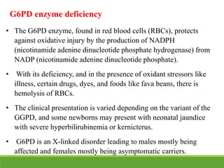
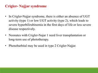
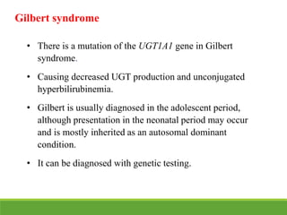
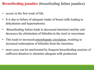
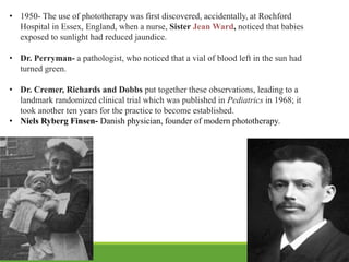
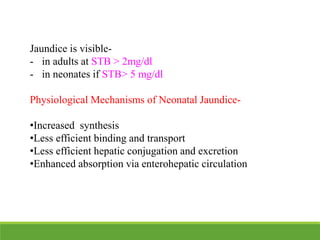
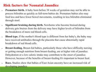
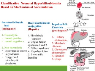
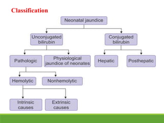
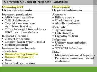
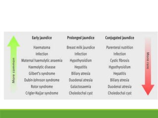
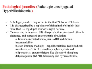
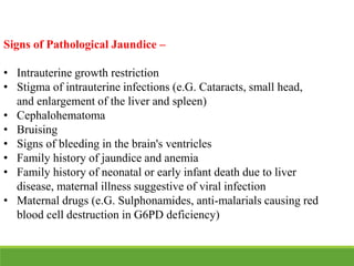
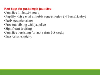
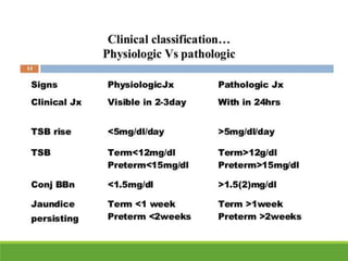
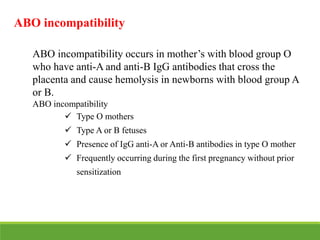
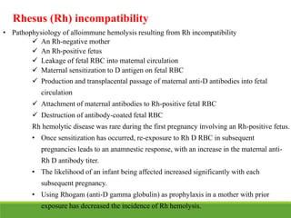
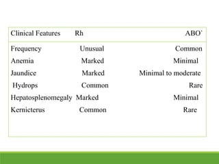
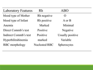
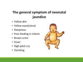
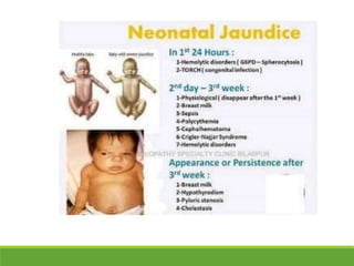
This document discusses jaundice in newborns. It defines jaundice as a yellowing of the skin caused by high bilirubin levels. Jaundice is common in newborns and usually resolves on its own, but sometimes requires treatment. The document covers physiological vs pathological jaundice, clinical assessment of jaundice, risk factors, potential complications like kernicterus, treatment options like phototherapy and exchange transfusion.
This document discusses neonatal jaundice, including its definition, pathophysiology, types, complications, and management. Key points include:
- Jaundice is caused by a buildup of bilirubin, which appears yellow. It is visible in newborns when bilirubin levels reach 5 mg/dL.
- Physiological jaundice is common in newborns and resolves on its own. Pathological jaundice requires treatment to prevent complications like kernicterus.
- Causes of neonatal jaundice include an imbalance between bilirubin production and excretion, as well as breastfeeding issues. Treatment depends on the type and severity of jaundice
This document discusses neonatal jaundice (hyperbilirubinemia) in infants. It covers the definition, causes, types (physiological vs pathological), investigations, management including phototherapy and exchange transfusion, and prevention of kernicterus. Key points include:
- Jaundice is caused by high bilirubin levels which cause yellowing of the skin and eyes. Bilirubin is produced from the breakdown of red blood cells.
- Physiological jaundice appears after 24-72 hours and is common, while pathological jaundice appears within 24 hours and requires treatment.
- Management involves phototherapy to reduce bilirubin levels or exchange transfusion for severe
This document discusses various types of birth injuries that can occur in newborns, including soft tissue injuries, cranial injuries, nerve injuries, fractures, and intra-abdominal injuries. It identifies risk factors for birth injuries such as prematurity, large baby size, breech presentation, and traumatic delivery methods. Each type of injury is defined and examples are provided, along with typical signs, symptoms, and treatment approaches. Nursing management focuses on close physical assessment, monitoring, consultation, and supporting feeding when appropriate.
Management of child with neonatal jaundiceNEHA MALIK
Newborn jaundice is a yellowing of a baby's skin and eyes. Newborn jaundice is very common and can occur when babies have a high level of bilirubin, a yellow pigment produced during normal breakdown of red blood cells.
follow me on my YouTube channel :- medic o mania
Neonatal jaundice is a condition in newborns marked by high levels of bilirubin in the blood, causing yellowing of the skin and whites of the eyes. Bilirubin levels are often higher in neonates due to increased red blood cell breakdown, liver immaturity, and bacterial colonization. Without treatment, hyperbilirubinemia can cause permanent brain damage known as kernicterus. Proper monitoring and treatment is important to prevent dangerous bilirubin levels in newborns.
Neonatal jaundice is one of the most common conditions in newborns. It is caused by high levels of bilirubin in the blood which causes yellowing of the skin. Physiological jaundice is common and appears after 24 hours, peaks by days 3-5, and resolves by 14 days without treatment. Pathological jaundice appears within 24 hours and requires treatment. Proper assessment includes history, physical exam, and lab tests to determine severity and risk of complications like acute bilirubin encephalopathy. Treatment may include phototherapy or exchange transfusion in severe cases. Close monitoring is important to prevent bilirubin neurotoxicity in newborns.
The document discusses neonatal hypoglycemia, including its definition, symptoms, risk factors, treatment, and monitoring. Some key points:
- Neonatal hypoglycemia is defined as a blood glucose level below certain thresholds in the first 24 hours and thereafter. It is a common problem in newborns.
- Babies at higher risk include preterms, those of diabetic mothers, or experiencing other stresses. Symptoms can be nonspecific.
- Treatment involves glucose administration via IV bolus or infusion to raise blood glucose to the normal range. Frequent monitoring is needed until levels stabilize.
- Persistent or resistant hypoglycemia may require additional drugs or referral to a specialist to investigate underlying
This document discusses neonatal jaundice (hyperbilirubinemia). It defines jaundice as an excessive level of bilirubin in the blood characterized by yellowing of the skin. It distinguishes between physiological and pathological jaundice. The causes, pathophysiology, complications, diagnostic evaluation and management including phototherapy, exchange transfusion and drugs are described in detail over multiple pages.
This document discusses neonatal jaundice, also known as hyperbilirubinemia. It defines jaundice as an excessive level of bilirubin in the blood characterized by yellowing of the skin. It distinguishes between physiological jaundice, which occurs in most newborns and resolves on its own, and pathological jaundice, which requires treatment. The causes, diagnostic evaluation, therapeutic management including phototherapy, and nursing considerations for babies with hyperbilirubinemia are described in detail. The goals of treatment are to reduce bilirubin levels and prevent complications like bilirubin toxicity.
This document summarizes information about neonatal jaundice from the Neonatology Department at Hadi Clinic. It discusses the definition of neonatal jaundice as a high bilirubin level in the blood. It describes physiological and pathological jaundice and their causes. It also outlines risk factors for jaundice, evaluation and treatment methods like phototherapy and exchange transfusion, and references guidelines on the treatment of hyperbilirubinemia in infants.
Please find the power point on Phototherapy in jaundice . I tried to present it on understandable way and all the contents are reviewed by experts and from very reliable references. Thank you
This document provides information on neonatal jaundice, including:
- Jaundice is visible bilirubinemia caused by the breakdown of hemoglobin. It becomes apparent on skin at bilirubin levels over 5 mg/dl in neonates.
- Physiological jaundice occurs in 60% of term and 80% of preterm babies in the first week due to immature liver function.
- Pathological jaundice requires treatment if bilirubin rises too quickly, reaches over 15 mg/dl, or persists after 14 days.
- Causes include blood incompatibility, infection, bruising, or breast milk. Evaluation includes history, exam, and lab tests.
-
Neonatal sepsis is a clinical syndrome of systemic illness accompanied by bacteria in the blood occurring in the first month of life. It can be early-onset within the first week of life, usually acquired during birth from the mother, or late-onset between 1 week to 1 month of life, often from the hospital environment. Symptoms are non-specific but can include temperature irregularities, poor feeding, or respiratory distress. Treatment involves blood cultures, antibiotics like ampicillin and gentamicin, and supportive care for complications involving various organ systems. Future treatments may involve immunotherapies and blocking inflammatory responses.
1) Hyperbilirubinemia refers to excessive bilirubin in the blood, seen as yellowing of the skin. It occurs commonly in newborns.
2) Bilirubin is produced from the breakdown of red blood cells. Risk factors for severe hyperbilirubinemia in newborns include blood group incompatibility.
3) Treatment options include phototherapy, which uses light to convert bilirubin to a soluble form excreted in urine, and exchange transfusion, replacing infant blood. Preventive measures promote breastfeeding and monitor high-risk infants.
This document discusses neonatal jaundice, including its causes, assessment, management, and follow up. Key points:
- Neonatal jaundice is common, seen in 60% of term and 80% of preterm infants, and is usually due to increased bilirubin production, defective conjugation, or increased enterohepatic circulation.
- Assessment involves history, physical exam for signs of jaundice, sepsis, or kernicterus, and measuring serum bilirubin levels.
- Management depends on bilirubin levels and underlying conditions, and may include phototherapy, antibiotics for sepsis, hydration, exchange transfusion, or albumin/immunoglobul
Hyperbilirubinemia is an elevated level of bilirubin in the blood that can cause jaundice. Bilirubin is formed from the breakdown of heme from red blood cells and is transported to the liver where it is conjugated and excreted in bile or reabsorbed in the enterohepatic circulation. Elevated bilirubin can be unconjugated or conjugated due to various hepatocellular or cholestatic conditions. Hereditary disorders like Crigler-Najjar syndrome and Gilbert's syndrome involve defects in bilirubin conjugation leading to unconjugated hyperbilirubinemia. Cholestatic diseases impair bile formation and flow causing conjug
This document summarizes the history and mechanisms of jaundice and kernicterus. Some key points include:
- Kernicterus was first described in 1875 and results from bilirubin accumulation in the brain due to hyperbilirubinemia.
- Bilirubin exists in two forms - a water-soluble dianion and insoluble bilirubin acid. The acid form can cross the blood brain barrier.
- Two transporters, MRPs and MDR/PGP, help prevent bilirubin entry into the brain from the blood under normal conditions.
- Kernicterus presents as acute bilirubin encephalopathy with symptoms ranging
This document describes a case of omphalitis, an infection of the umbilical stump, in a 7-day-old male infant. The infant presented with fever, yellowish umbilical discharge, and hypoactivity. Laboratory tests showed elevated white blood cell count and C-reactive protein. The infant was diagnosed with omphalitis and sepsis and started on intravenous antibiotics and supportive care. Omphalitis is a potentially serious infection in neonates that requires prompt treatment with antibiotics and sometimes surgery.
ABO incompatibility occurs when a pregnant woman has a blood type that is incompatible with her fetus's blood type. It can cause hemolytic disease of the newborn (HDN) when the mother's IgG antibodies destroy the fetus's or newborn's red blood cells. The most common form is ABO HDN, which usually results in mild symptoms and does not require treatment. It occurs when an O group mother has an A or B group baby and the mother's anti-A or anti-B antibodies destroy the baby's red blood cells. Investigations of the mother and baby include blood typing and testing the mother's serum for IgG antibodies, while the baby may experience jaundice, anemia and other symptoms. Treatment
1. Neonatal resuscitation may be required for 10% of newborns who need some assistance at birth and 1% who need extensive measures to transition from fetal to neonatal circulation.
2. After birth, clamping of the umbilical cord and expansion of the lungs with air allows oxygen to diffuse across the alveoli as the pulmonary vessels dilate, increasing blood flow to the lungs.
3. If the transition is interrupted, the newborn may be apneic, have low muscle tone, respiratory depression, bradycardia or cyanosis, requiring the steps of resuscitation - assessing airway, providing breathing support and positive pressure ventilation if needed, giving chest compressions if
The document describes a case of a 3-day old baby boy presenting with jaundice. The baby was yellow in color, had difficulty waking and feeding, and cried softly when touched. The most probable causes of jaundice given the baby's age include physiological jaundice, breastfeeding jaundice, or hemolytic diseases like G6PD deficiency. To reach a diagnosis, the physician would need to take a thorough postnatal, perinatal, and family history to determine if the jaundice is pathological or identify any risk factors for underlying conditions.
Breast milk jaundice.
The key features that point to breast milk jaundice in this case are:
- Jaundice onset after the first week of life
- Progressive deepening of jaundice over 1 week
- Exclusively breastfed
- Pale/gray stool color indicating reduced bilirubin conjugation
Breast milk jaundice results from high levels of beta-glucuronidase in breast milk interfering with bilirubin conjugation in the infant's liver and gut. It typically presents after the first week of life and can persist for several weeks if breastfeeding continues.
An exchange transfusion involves slowly removing a patient's blood and replacing it with donor blood in order to remove abnormal components and toxins while maintaining blood volume. It is indicated for hyperbilirubinemia, Rhesus/ABO incompatibility, severe anemia, and other conditions. The procedure requires specialized equipment and staffing in a NICU. Blood is warmed and infused using either a push-pull or isovolumetric method while carefully monitoring the infant. Potential complications include blood clots, changes in blood chemistry, heart/lung problems, and low risk of infection.
This document provides an overview of neonatal jaundice presented by Dr. Binaya Dhakal of Shree Birendra Hospital. It discusses the metabolism of bilirubin, types of neonatal jaundice including physiological, pathological, breastfeeding and breast milk jaundice. It also covers the evaluation, causes, approach and management of hyperbilirubinemia as well as prolonged hyperbilirubinemia. Management options discussed include phototherapy, intravenous immunoglobulins, exchange transfusion and metalloporphyrins. The document concludes with information on kernicterus.
Neonatal jaundice is caused by an accumulation of unconjugated bilirubin in the blood and tissues. Physiological jaundice appears between 2-4 days of life, peaks at 5-6 mg/dL by days 2-4, and resolves by days 5-7. Pathological jaundice appears within 24 hours, bilirubin increases more than 5 mg/dL per day, persists past 14 days, or direct bilirubin is elevated. Causes include hemolytic diseases, breastfeeding, infections, and genetic disorders affecting bilirubin metabolism. Timely diagnosis and treatment are important to prevent neurotoxicity and kernicterus.
Neonatal jaundice, also known as neonatal hyperbilirubinemia, refers to the yellow discoloration of skin and eyes in newborn babies due to high levels of bilirubin. It is common and usually harmless, but in rare cases can lead to kernicterus if bilirubin levels get too high. There are two main types - physiological jaundice which occurs after 24 hours of life and is harmless, and pathological jaundice which occurs earlier and requires treatment. Causes include breast milk jaundice, Rh incompatibility, infections, and liver or blood disorders. Treatment depends on bilirubin levels but may include phototherapy, fluid supplementation, or in severe cases
Neonatal jaundice is the yellowish discoloration of skin due to high bilirubin levels. It can be physiological, appearing 2-3 days after birth and resolving in 10-12 days, or pathological, appearing within 24 hours. Physiological jaundice is caused by the normal breakdown of red blood cells in newborns. Pathological jaundice has underlying causes that interfere with bilirubin production, transport, conjugation or excretion. Mild cases are monitored while moderate-severe cases may require phototherapy or phototherapy with medication to process the bilirubin for excretion.
This document summarizes neonatal jaundice. It notes that a bilirubin level over 5mg/dl causes clinical jaundice in newborns, typically progressing from head to toe. About 50-60% of babies are affected in the first week of life. Physiological jaundice is most common, caused by immature liver pathways and the breakdown of fetal hemoglobin. It describes the typical phases and timelines of physiological jaundice. Pathological jaundice has specific criteria for onset, rise in bilirubin levels, and total bilirubin levels. Causes, clinical assessment, treatment with phototherapy or exchange transfusions, and complications like kernicterus are also outlined
This document provides information on neonatal jaundice including definitions, incidence, pathophysiology, risk factors, clinical presentation, diagnosis, management, and prevention of kernicterus. Some key points include:
- Neonatal jaundice is diagnosed if the total serum bilirubin is >5 mg/dL in a full-term newborn or >7 mg/dL in a preterm newborn.
- Jaundice occurs in 60% of full-term and 80% of preterm infants due to the immature liver's inability to sufficiently conjugate and excrete bilirubin.
- Risk factors for pathological jaundice include jaundice in the first 24 hours of life or
Neonatal Hyperbilirubinemia final I.pptJusticeYegon1
This document discusses neonatal jaundice and hyperbilirubinemia. It begins by defining jaundice as the deposition of bilirubin in the skin and mucous membranes, which is the end product of heme breakdown from red blood cell lysis. It then covers the causes, types, risk factors, investigations, treatments including phototherapy and exchange transfusion, and prevention of neonatal jaundice and hyperbilirubinemia. The key topics are the physiologic and pathologic causes of jaundice, the risks of kernicterus from high bilirubin levels, and the importance of monitoring at-risk infants to prevent severe hyperbilirubinemia.
This document discusses neonatal jaundice and bilirubin metabolism. It covers the following key points:
Physiologic jaundice occurs in 50-65% of newborns as bilirubin levels rise in the first week of life. Breastfeeding jaundice can occur if breastmilk intake is inadequate. Pathologic jaundice has an earlier onset or faster rising bilirubin levels and requires investigation for underlying causes. Treatment options for high bilirubin include phototherapy and exchange transfusions in severe cases to prevent potential brain damage from bilirubin toxicity.
This document discusses jaundice during pregnancy and its management. It defines jaundice and explains the normal physiology of the liver during pregnancy. It then focuses on obstetric cholestasis (OC), defining it as a form of intrahepatic cholestasis associated with pruritus and elevated bile acid levels. The document outlines the causes, signs and symptoms, diagnosis, and maternal and fetal effects of OC. It notes OC can lead to complications like preterm delivery and stillbirth. Prevention measures include a healthy diet, maintaining a healthy weight, and an active lifestyle.
This document discusses neonatal jaundice, providing information on:
- Jaundice is caused by elevated bilirubin levels and presents as a yellowish skin color. It is common in newborns.
- There are various causes of jaundice, including physiological jaundice in the first weeks of life or pathological issues like hemolysis or liver/bile duct problems.
- Clinical assessment of jaundiced newborns should include history, exam, monitoring bilirubin levels, and considering underlying causes if jaundice is early-onset, prolonged, or the baby is unwell. Timely treatment may be required to prevent brain damage in severe cases.
Neonatal jaundice is a common condition that requires medical attention in newborns. It is caused by high levels of bilirubin, which is produced from the breakdown of red blood cells. There are two main types - physiological jaundice, which appears after 24 hours and is harmless, and pathological jaundice, which can be a sign of other health issues and sometimes requires treatment. Pathological jaundice has various potential causes, including blood group incompatibility, infections, or liver problems. Treatment depends on the underlying cause and severity of jaundice, and may involve phototherapy, intravenous immunoglobulins, or exchange transfusions in severe cases to prevent potential brain damage from high bilirub
This document discusses neonatal jaundice, including the physiology of bilirubin, types and causes of jaundice, clinical approach, and management. It covers the sources and properties of bilirubin, defines physiological and non-physiological jaundice, and discusses various causes like blood group incompatibility, red cell membrane defects, enzyme deficiencies, infections, and genetic disorders. The clinical approach section outlines assessment and investigations. Management techniques for unconjugated jaundice include phototherapy and exchange transfusion, while conjugated jaundice may require treatment of the underlying cause.
Neonatal jaundice is yellow discoloration of the skin that occurs in 60-80% of newborns during the first week of life due to high levels of bilirubin in the blood. Bilirubin is produced from the breakdown of red blood cells and is normally processed and excreted by the liver and gallbladder. In newborns, the liver is not fully developed and has difficulty processing bilirubin, leading to jaundice. Jaundice is considered pathological if the bilirubin levels rise too quickly or remain high after two weeks, requiring treatment like phototherapy or blood exchange transfusion to prevent neurological damage from bilirubin toxicity.
This document provides an overview of neonatal cholestasis. It defines cholestasis as diminished bile formation and/or excretion that can result from various disorders. The most common causes of neonatal cholestasis are discussed, including biliary atresia, idiopathic neonatal hepatitis, and choledochal cysts. Signs, symptoms, diagnostic evaluation, and management approaches are described. Liver biopsy may be needed to differentiate between intrahepatic and extrahepatic causes such as biliary atresia. Early surgical intervention via Kasai procedure can improve outcomes for biliary atresia, while supportive care and liver transplantation are options for other etiologies of neonatal cholestasis.
This document discusses neonatal jaundice (hyperbilirubinemia), which refers to an excessive level of bilirubin in the blood that causes jaundice. It notes that physiological jaundice occurs in 50-60% of term and 80% of preterm neonates, but significant jaundice affects only 6% of term babies. The document describes the causes, risk factors, diagnosis, and treatment of pathological jaundice, including phototherapy and exchange transfusion. Nursing considerations for babies receiving phototherapy focus on monitoring temperature and fluid intake to prevent complications during therapy.
This document discusses neonatal jaundice and hyperbilirubinemia. It begins by defining jaundice and hyperbilirubinemia. It then describes the metabolism of bilirubin, including transport to the liver, hepatic uptake, conjugation in the liver, and excretion in bile and stool. Causes of increased bilirubin production are discussed, including increased red blood cell production or breakdown. The document also discusses approaches to evaluating and managing neonatal jaundice and hyperbilirubinemia, including assessing risk factors, monitoring bilirubin levels, and guidelines for initiating phototherapy.
This document discusses neonatal jaundice, including its causes, clinical presentation, evaluation, and management. It describes the normal bilirubin metabolism in newborns and factors that can cause unconjugated or conjugated hyperbilirubinemia. Causes of jaundice in the first 24 hours include ABO incompatibility and G6PD deficiency. Prolonged jaundice beyond 2 weeks requires investigation for possible biliary atresia. Treatment involves phototherapy or exchange transfusion depending on bilirubin levels to prevent kernicterus.
This document outlines the bilirubin pathway, causes of neonatal jaundice, and kernicterus. It discusses physiological versus pathological jaundice and risk factors. The major causes outlined are physiological jaundice, breast milk jaundice, hemolysis, increased enterohepatic circulation, conjugation issues, and hepatocyte uptake defects. Kernicterus signs and prevention are also reviewed.
Neonatal jaundice (hyperbilirubinemia) by Rajiv MavachiRajiv Mavachi
Jaundice is the most common condition that requires medical attention in newborns. The yellow coloration of the skin and sclera in newborns with jaundice is the result of accumulation of unconjugated bilirubin.
Neonatal jaundice occurs in 60% of term and 80% of preterm babies. Despite Neonatal jaundice is one of the commonest neonatal conditions, there are no national practice guidelines for its management in our country. Lack of uniform guidelines and standard practice parameters for diagnosis and management of neonatal jaundice often leads many babies to develop unnoticed hyperbilirubinemia causing kernicterus and long term poor neurological sequelae. This review after briefly discussing the epidemiology and pathophysiology of neonatal jaundice provides evidence-based pragmatic guidelines for the diagnosis and management of neonatal jaundice in resource-limited countries like Afghanistan
This document provides information and guidelines regarding medical electives for undergraduate medical students in India. It defines electives as optional learning experiences that allow students to explore areas of interest. The document outlines the objectives and structure of elective blocks, including topics that can be covered, requirements for attendance, supervision, and assessment. It provides templates for planning elective learning experiences and identifying potential electives in different areas like laboratories, research, clinical specialties, and community settings. The goal is to provide immersive, experiential learning opportunities to help students discover career paths and develop skills beyond their curriculum.
The document discusses key aspects of developing a research study, including definitions of research, the importance of research questions and hypotheses, and the FINER criteria for formulating good research questions. It provides definitions of research from various sources and outlines the steps to developing a clear research question. It also discusses how to write a good hypothesis and the different types of hypotheses. Finally, it explains each letter in the FINER mnemonic - Feasible, Interesting, Novel, Ethical, and Relevant - as a framework for evaluating proposed research questions.
1. The Ecole De Medicine De Pondichery medical school in Pondichery, established in 1823, was the first formal medical school established among the French colonies in India. It trained students and granted them a Médicin Locale diploma to practice medicine.
2. Several major medical colleges were established in India in the 1830s, including Calcutta Medical College in 1835 as the first college to teach Western medicine in Asia, and Madras Medical College in 1835.
3. Other notable early medical colleges included Grant Medical College in Mumbai established in 1845, Goa Medical College established in 1842, and Stanley Medical College in Chennai established in 1930 from its predecessor school founded
This document discusses 30 notable females in the medical field, including:
- Dr. Anandi Gopal Joshi, one of the first female physicians in India in the 1880s.
- Rukhmabai Raut, one of the first female practitioners of western medicine in India who fought for women's rights.
- Kadambini Ganguly, one of the first female graduates of western medicine in India in 1886.
- Several other pioneering female doctors in India such as Muthulakshmi Reddy, Dr. V. Shanta, Dr. Sivaramakrishna Iyer Padmavati, and Dr. Vasudha Vishnu Apte, the first female med
The document provides information about the Dr. Vithalrao Vikhe Patil Foundation in Ahmednagar, Maharashtra, India. It discusses the founders and visionaries of the foundation, Late Dr. Vithalrao Vikhe Patil and Late Dr. Balasaheb Vikhe Patil. It then lists the various educational institutions established by the foundation, including medical, nursing, pharmacy, management colleges and schools. It provides details about the medical college such as intake numbers and postgraduate courses. It concludes with highlighting the facilities, achievements and activities supported by the foundation to enrich staff and students.
Dr. Sunil Natha Mhaske is the Dean of D.V.V.P.F's Medical College in Ahmednagar. The document discusses the importance of human values and how they are necessary in today's society. It defines human values as qualities that encourage consideration for others and bonding between people. Some key human values mentioned include brotherhood, honesty, and respect. The document also discusses how education can help enhance human values in society by influencing perceptions of right and wrong.
This document discusses the importance of child nutrition. It notes that proper nutrition is important for children's growth and development and to establish healthy eating habits. A balanced diet with fruits, vegetables, whole grains, protein foods and dairy helps children get essential nutrients and maintain good health. The document provides daily serving guidelines for different age groups and notes foods that are important sources of key nutrients like protein, vitamins and minerals. It emphasizes limiting added sugars, saturated and trans fats, and sodium. Healthy eating supports children's academic performance and brain development.
This document provides information about cleft lip and cleft palate including causes, risk factors, diagnosis, treatment, and social aspects. It describes how cleft lip occurs when the tissues of the lip do not fully fuse before birth, and cleft palate occurs when the roof of the mouth does not fully close. Treatment often begins in infancy and may include surgery, dental care, speech therapy, and psychological support. The document also discusses cultural beliefs and stigma around cleft conditions as well as organizations providing cleft care in India.
The white coat has served as a symbol of physicians for over 100 years. Prior to the late 19th century, doctors wore black garments, but adopted the white laboratory coat in 1889 as they sought to represent themselves as scientists. The white coat symbolized cleanliness and purity as antisepsis became important in medicine. By the early 20th century, white coats had become standard attire for doctors and medical professionals. Today, the white coat continues to symbolize medical authority, though some argue it may also intimidate patients or transmit infections.
This document provides details about the history and development of the stethoscope. It describes how Rene Laennec invented the first stethoscope in 1816 after observing children using a wooden beam to listen to each other. Since then, various physicians have improved stethoscope design, including Golding Bird who created the first binaural stethoscope in 1840, Arthur Leared who invented the first binaural stethoscope in 1851, and George Cammann who commercialized the binaural design in 1852. The document also discusses David Littmann's contributions in the 1960s and the parts that make up a modern binaural stethoscope.
Congenital hypothyroidism is a condition present at birth where the thyroid gland does not produce enough hormones. It can cause arrested physical and mental development. The most common cause is failure of the thyroid to grow before birth. Symptoms in newborns include jaundice, hypotonia, and large tongue. If not treated with levothyroxine replacement, it can lead to intellectual disability and growth impairment. Screening programs allow for early diagnosis and treatment to prevent complications.
Poststreptococcal acute glomerulonephritis (AGN) is a common condition in childhood caused by a Streptococcus pyogenes infection. It involves an immune response that leads to inflammation of the glomeruli in the kidneys. Symptoms include edema, gross hematuria, hypertension, and acute renal insufficiency. Treatment focuses on controlling blood pressure, restricting fluid and salt intake, and administering antibiotics. While early AGN is often temporary and reversible, chronic glomerulonephritis can lead to permanent kidney damage if not properly managed.
Nephrotic syndrome is a kidney disorder characterized by heavy proteinuria, hypoalbuminemia, edema, and hyperlipidemia. It is usually caused by damage to the glomeruli in the kidneys, which allows protein to leak from the blood into the urine. In children, nephrotic syndrome is commonly caused by minimal change disease or focal segmental glomerulosclerosis. It can lead to complications like edema, infections, thrombosis, and kidney failure if left untreated. Congenital nephrotic syndrome is a rare genetic form that manifests within the first few months of life.
This document provides guidance on effectively giving feedback to employees. It discusses that feedback is an important skill for boosting employee performance and confidence, but is often dreaded due to how it is typically done. It recommends checking your motives before giving feedback to ensure it is focused on improvement. Feedback should also be given in a timely manner, close to the event being discussed, and regularly to avoid surprises and allow issues to be addressed early. The overall message is that feedback is most effective when delivered carefully, frequently and focused on improvement rather than criticism.
The document discusses congestive cardiac failure in children. It defines congestive cardiac failure as when the heart is unable to pump enough blood to meet the body's needs. Common causes in children include congenital heart defects, acquired heart conditions, and metabolic abnormalities. Symptoms include poor growth, difficulty breathing, faster breathing and heart rate, and puffiness in the eyes or feet. Treatment involves identifying and treating the underlying cause, using diuretics to reduce congestion, and other drugs like digoxin, ACE inhibitors, beta-blockers, and inotropes as needed. For severe cases, interventions like pacemakers, cardiac transplantation may be considered.
This document discusses prehypertension and hypertension in children. It defines prehypertension as blood pressure levels between the 90th and 95th percentiles, and hypertension as levels at or above the 95th percentile. Risk factors for developing hypertension in childhood include obesity, family history, lack of exercise, and certain medical conditions. The document provides prevalence rates for prehypertension and hypertension from various studies worldwide. It outlines guidelines for evaluating and diagnosing hypertension in children, and discusses potential complications and treatment approaches.
Rheumatic fever is an autoimmune disease caused by streptococcal infection that can lead to rheumatic heart disease. It typically affects children ages 5-15 and symptoms include carditis, arthritis, chorea, and others. Left untreated, approximately 60% of patients develop chronic rheumatic heart disease, which can cause heart valve defects. Diagnosis involves meeting modified Jones criteria through symptoms, labs, and echocardiogram findings. Treatment focuses on antibiotics to prevent recurrence, anti-inflammatories for carditis, and management of heart defects through medications, surgery, or valve replacements.
Embryology of heart, Anatomy of heart, Physiology of heart, Fetal circulation, Neonatal circulation, Congenital cyanotic and acyanotic heart diseases of children.
This document discusses preterm birth and care of preterm infants. It defines preterm birth as birth before 37 weeks of gestation. It describes developmental milestones from 26-36 weeks gestation. It discusses risks like diabetes and smoking. It outlines management of preterm infants including in the NICU with incubators and kangaroo care. It also discusses long term risks of prematurity like cerebral palsy.
More from Dr. Vithalrao Vikhe Patil Foundation's Medical College and Hospital, Ahmednagar (20)
Adhd Medication Shortage Uk - trinexpharmacy.comreignlana06
The UK is currently facing a Adhd Medication Shortage Uk, which has left many patients and their families grappling with uncertainty and frustration. ADHD, or Attention Deficit Hyperactivity Disorder, is a chronic condition that requires consistent medication to manage effectively. This shortage has highlighted the critical role these medications play in the daily lives of those affected by ADHD. Contact : +1 (747) 209 – 3649 E-mail : sales@trinexpharmacy.com
Muktapishti is a traditional Ayurvedic preparation made from Shoditha Mukta (Purified Pearl), is believed to help regulate thyroid function and reduce symptoms of hyperthyroidism due to its cooling and balancing properties. Clinical evidence on its efficacy remains limited, necessitating further research to validate its therapeutic benefits.
Integrating Ayurveda into Parkinson’s Management: A Holistic ApproachAyurveda ForAll
Explore the benefits of combining Ayurveda with conventional Parkinson's treatments. Learn how a holistic approach can manage symptoms, enhance well-being, and balance body energies. Discover the steps to safely integrate Ayurvedic practices into your Parkinson’s care plan, including expert guidance on diet, herbal remedies, and lifestyle modifications.
Local Advanced Lung Cancer: Artificial Intelligence, Synergetics, Complex Sys...Oleg Kshivets
Overall life span (LS) was 1671.7±1721.6 days and cumulative 5YS reached 62.4%, 10 years – 50.4%, 20 years – 44.6%. 94 LCP lived more than 5 years without cancer (LS=2958.6±1723.6 days), 22 – more than 10 years (LS=5571±1841.8 days). 67 LCP died because of LC (LS=471.9±344 days). AT significantly improved 5YS (68% vs. 53.7%) (P=0.028 by log-rank test). Cox modeling displayed that 5YS of LCP significantly depended on: N0-N12, T3-4, blood cell circuit, cell ratio factors (ratio between cancer cells-CC and blood cells subpopulations), LC cell dynamics, recalcification time, heparin tolerance, prothrombin index, protein, AT, procedure type (P=0.000-0.031). Neural networks, genetic algorithm selection and bootstrap simulation revealed relationships between 5YS and N0-12 (rank=1), thrombocytes/CC (rank=2), segmented neutrophils/CC (3), eosinophils/CC (4), erythrocytes/CC (5), healthy cells/CC (6), lymphocytes/CC (7), stick neutrophils/CC (8), leucocytes/CC (9), monocytes/CC (10). Correct prediction of 5YS was 100% by neural networks computing (error=0.000; area under ROC curve=1.0).
These lecture slides, by Dr Sidra Arshad, offer a quick overview of the physiological basis of a normal electrocardiogram.
Learning objectives:
1. Define an electrocardiogram (ECG) and electrocardiography
2. Describe how dipoles generated by the heart produce the waveforms of the ECG
3. Describe the components of a normal electrocardiogram of a typical bipolar lead (limb II)
4. Differentiate between intervals and segments
5. Enlist some common indications for obtaining an ECG
6. Describe the flow of current around the heart during the cardiac cycle
7. Discuss the placement and polarity of the leads of electrocardiograph
8. Describe the normal electrocardiograms recorded from the limb leads and explain the physiological basis of the different records that are obtained
9. Define mean electrical vector (axis) of the heart and give the normal range
10. Define the mean QRS vector
11. Describe the axes of leads (hexagonal reference system)
12. Comprehend the vectorial analysis of the normal ECG
13. Determine the mean electrical axis of the ventricular QRS and appreciate the mean axis deviation
14. Explain the concepts of current of injury, J point, and their significance
Study Resources:
1. Chapter 11, Guyton and Hall Textbook of Medical Physiology, 14th edition
2. Chapter 9, Human Physiology - From Cells to Systems, Lauralee Sherwood, 9th edition
3. Chapter 29, Ganong’s Review of Medical Physiology, 26th edition
4. Electrocardiogram, StatPearls - https://www.ncbi.nlm.nih.gov/books/NBK549803/
5. ECG in Medical Practice by ABM Abdullah, 4th edition
6. Chapter 3, Cardiology Explained, https://www.ncbi.nlm.nih.gov/books/NBK2214/
7. ECG Basics, http://www.nataliescasebook.com/tag/e-c-g-basics
Recomendações da OMS sobre cuidados maternos e neonatais para uma experiência pós-natal positiva.
Em consonância com os ODS – Objetivos do Desenvolvimento Sustentável e a Estratégia Global para a Saúde das Mulheres, Crianças e Adolescentes, e aplicando uma abordagem baseada nos direitos humanos, os esforços de cuidados pós-natais devem expandir-se para além da cobertura e da simples sobrevivência, de modo a incluir cuidados de qualidade.
Estas diretrizes visam melhorar a qualidade dos cuidados pós-natais essenciais e de rotina prestados às mulheres e aos recém-nascidos, com o objetivo final de melhorar a saúde e o bem-estar materno e neonatal.
Uma “experiência pós-natal positiva” é um resultado importante para todas as mulheres que dão à luz e para os seus recém-nascidos, estabelecendo as bases para a melhoria da saúde e do bem-estar a curto e longo prazo. Uma experiência pós-natal positiva é definida como aquela em que as mulheres, pessoas que gestam, os recém-nascidos, os casais, os pais, os cuidadores e as famílias recebem informação consistente, garantia e apoio de profissionais de saúde motivados; e onde um sistema de saúde flexível e com recursos reconheça as necessidades das mulheres e dos bebês e respeite o seu contexto cultural.
Estas diretrizes consolidadas apresentam algumas recomendações novas e já bem fundamentadas sobre cuidados pós-natais de rotina para mulheres e neonatos que recebem cuidados no pós-parto em unidades de saúde ou na comunidade, independentemente dos recursos disponíveis.
É fornecido um conjunto abrangente de recomendações para cuidados durante o período puerperal, com ênfase nos cuidados essenciais que todas as mulheres e recém-nascidos devem receber, e com a devida atenção à qualidade dos cuidados; isto é, a entrega e a experiência do cuidado recebido. Estas diretrizes atualizam e ampliam as recomendações da OMS de 2014 sobre cuidados pós-natais da mãe e do recém-nascido e complementam as atuais diretrizes da OMS sobre a gestão de complicações pós-natais.
O estabelecimento da amamentação e o manejo das principais intercorrências é contemplada.
Recomendamos muito.
Vamos discutir essas recomendações no nosso curso de pós-graduação em Aleitamento no Instituto Ciclos.
Esta publicação só está disponível em inglês até o momento.
Prof. Marcus Renato de Carvalho
www.agostodourado.com
8 Surprising Reasons To Meditate 40 Minutes A Day That Can Change Your Life.pptxHolistified Wellness
We’re talking about Vedic Meditation, a form of meditation that has been around for at least 5,000 years. Back then, the people who lived in the Indus Valley, now known as India and Pakistan, practised meditation as a fundamental part of daily life. This knowledge that has given us yoga and Ayurveda, was known as Veda, hence the name Vedic. And though there are some written records, the practice has been passed down verbally from generation to generation.
Histololgy of Female Reproductive System.pptxAyeshaZaid1
Dive into an in-depth exploration of the histological structure of female reproductive system with this comprehensive lecture. Presented by Dr. Ayesha Irfan, Assistant Professor of Anatomy, this presentation covers the Gross anatomy and functional histology of the female reproductive organs. Ideal for students, educators, and anyone interested in medical science, this lecture provides clear explanations, detailed diagrams, and valuable insights into female reproductive system. Enhance your knowledge and understanding of this essential aspect of human biology.
TEST BANK For Community Health Nursing A Canadian Perspective, 5th Edition by...Donc Test
TEST BANK For Community Health Nursing A Canadian Perspective, 5th Edition by Stamler, Verified Chapters 1 - 33, Complete Newest Version Community Health Nursing A Canadian Perspective, 5th Edition by Stamler, Verified Chapters 1 - 33, Complete Newest Version Community Health Nursing A Canadian Perspective, 5th Edition by Stamler Community Health Nursing A Canadian Perspective, 5th Edition TEST BANK by Stamler Test Bank For Community Health Nursing A Canadian Perspective, 5th Edition Pdf Chapters Download Test Bank For Community Health Nursing A Canadian Perspective, 5th Edition Pdf Download Stuvia Test Bank For Community Health Nursing A Canadian Perspective, 5th Edition Study Guide Test Bank For Community Health Nursing A Canadian Perspective, 5th Edition Ebook Download Stuvia Test Bank For Community Health Nursing A Canadian Perspective, 5th Edition Questions and Answers Quizlet Test Bank For Community Health Nursing A Canadian Perspective, 5th Edition Studocu Test Bank For Community Health Nursing A Canadian Perspective, 5th Edition Quizlet Test Bank For Community Health Nursing A Canadian Perspective, 5th Edition Stuvia Community Health Nursing A Canadian Perspective, 5th Edition Pdf Chapters Download Community Health Nursing A Canadian Perspective, 5th Edition Pdf Download Course Hero Community Health Nursing A Canadian Perspective, 5th Edition Answers Quizlet Community Health Nursing A Canadian Perspective, 5th Edition Ebook Download Course hero Community Health Nursing A Canadian Perspective, 5th Edition Questions and Answers Community Health Nursing A Canadian Perspective, 5th Edition Studocu Community Health Nursing A Canadian Perspective, 5th Edition Quizlet Community Health Nursing A Canadian Perspective, 5th Edition Stuvia Community Health Nursing A Canadian Perspective, 5th Edition Test Bank Pdf Chapters Download Community Health Nursing A Canadian Perspective, 5th Edition Test Bank Pdf Download Stuvia Community Health Nursing A Canadian Perspective, 5th Edition Test Bank Study Guide Questions and Answers Community Health Nursing A Canadian Perspective, 5th Edition Test Bank Ebook Download Stuvia Community Health Nursing A Canadian Perspective, 5th Edition Test Bank Questions Quizlet Community Health Nursing A Canadian Perspective, 5th Edition Test Bank Studocu Community Health Nursing A Canadian Perspective, 5th Edition Test Bank Quizlet Community Health Nursing A Canadian Perspective, 5th Edition Test Bank Stuvia
Here is the updated list of Top Best Ayurvedic medicine for Gas and Indigestion and those are Gas-O-Go Syp for Dyspepsia | Lavizyme Syrup for Acidity | Yumzyme Hepatoprotective Capsules etc
2. Lecture-08
Neonatal Jaundice
Prof. Dr. Sunil Natha Mhaske
Dean
Dr. Vithalrao Vikhe Patil Foundation’s Medical College
and Hospital,
Ahmednagar (M.S.) India-414111
Mo- 7588024773
Mail-sunilmhaske1970@gmail.com
3. History
• The term jaundice is from the French word "jaune”
• Neonatal jaundice may have first been described in
a Chinese textbook 1000 years ago.
• Earliest work on jaundice from Baumes-1785 and
Hervieux-1847
• Medical theses, essays, and textbooks from the
18th and 19th centuries contain discussions about
the causes and treatment of neonatal jaundice.
• Kernicterus was first described by Johannes
Orth, 1875, He postulated that jaundice might have
hematologic origins. He noted that the brain in
jaundiced adults wasn’t affected.
• Christian Schmorl coined the term in 1904,
Translated, Kernicterus means jaundice of the
“kern” or nuclear region of the brain.
4. • It is a yellowish discoloration of the white part of the eyes and skin in
a newborn baby due to high bilirubin levels.
• About 60% of full term newborn and 80% of premature babies are
jaundiced.
• 3% of normal term infant show bilirubin level more than 15mg/dl
• Jaundice is the most common condition that requires medical attention
and hospital readmission in newborns.
Neonatal jaundice or Neonatal Hyperbilirubinemia-
5. Bilirubin
• Non-polar, water insoluble compound requiring conjugation with
glucuronic acid to form a water soluble product that can be
excreted.
• It circulates to the liver reversibly bound to albumin
6. Pathophysiology
• Bilirubin comes from the breakdown in heme, which is produced from the
breakdown of hemoglobin.
• Heme is converted to biliverdin, iron, and carbon monoxide by the enzyme heme
oxygenase.
• biliverdin is then converted to bilirubin by biliverdin reductase.
• The conversion of heme to bilirubin takes place in the reticuloendothelial system.
• The unconjugated bilirubin is hydrophobic and is transported to the liver bound to
albumin where it is conjugated by the enzyme uridine diphosphate-
glucuronosyltransferase (UGT).
• Conjugated bilirubin, which is water-soluble, is excreted in bile and into the
gastrointestinal (GI) tract and mostly excreted in feces after being metabolized by
bacterial flora.
• Some conjugated bilirubin is deconjugated to unconjugated bilirubin and reabsorbed
through the enterohepatic circulation.
7.
8. • Ligandins{y} responsible for transport from plasma membrane to
endoplasmic reticulum.
• Bilirubin conjugated in presence of UDPGT (uridine diphosphate
glucuronyl transferase) to mono and diglucoronides, which are
then excreted into bile canaliculi.
• Phenobarbitol Increases the conc. of Ligandin and also induces
UDPGT.
• Conjugated” bilirubin is water soluble and is secreted by the
hepatocytes into the biliary canaliculi.
• Converted to stercobilinogen (urobilinogen) (colorless) by
bacteria in the gut.
• Oxidized to stercobilin which is colored, Excreted in feces.
• Some stercobilin may be re-adsorbed by the gut and re-excreted
by either the liver or kidney.
9. Enterohepatic circulation-
• Meconium contains 100-200mg of conjugated bilirubin at
birth.
• Conjugated bilirubin is unstable and easily hydrolyzed to
unconjugated bilirubin.
• This process occurs non-enzymatically in the duodenum
and jejunum and also occurs in the presence of beta-
glucuronidase, an enteric mucosal enzyme, which is found
in high concentration in newborn infants and in human
milk.
Factors enhancing enterhepatic circulation
- Decrease entral intake
- Intestinal atresia
- Meconium ileus
- Hiruschsrung disease
10. Jaundice is visible-
- in adults at STB > 2mg/dl
- in neonates if STB> 5 mg/dl
Physiological Mechanisms of Neonatal Jaundice-
•Increased synthesis
•Less efficient binding and transport
•Less efficient hepatic conjugation and excretion
•Enhanced absorption via enterohepatic circulation
11. Risk factors for Neonatal Jaundice
Premature birth. A baby born before 38 weeks of gestation may not be able to
process bilirubin as quickly as full-term babies do. Premature babies also may
feed less and have fewer bowel movements, resulting in less bilirubin eliminated
through stool.
Significant bruising during birth. Newborns who become bruised during
delivery gets bruises from the delivery may have higher levels of bilirubin from
the breakdown of more red blood cells.
Blood type. If the mother's blood type is different from her baby's, the baby may
have received antibodies through the placenta that cause abnormally rapid
breakdown of red blood cells.
Breast-feeding. Breast-fed babies, particularly those who have difficulty nursing
or getting enough nutrition from breast-feeding, are at higher risk of jaundice.
Dehydration or a low caloric intake may contribute to the onset of jaundice.
However, because of the benefits of breast-feeding It's important to breast feed.
Race. Studies show that babies of East Asian ancestry have an increased risk of
developing jaundice.
16. • Pathologic jaundice may occur in the first 24 hours of life and
• It is characterized by a rapid rate of rising in the bilirubin level
more than 0.2 mg/dl per hour or 5 mg/dl per day.
• Causes - due to increased bilirubin production, decreased bilirubin
clearance, and increased enterohepatic circulation.
a. Immune-mediated hemolysis - ABO and rhesus
incompatibility.
b. Non-immune mediated - cephalhematoma, red blood cell
membrane defects like hereditary spherocytosis and
elliptocytosis, enzyme defects like glucose-6-phosphate
dehydrogenase (G6PD) deficiency and pyruvate kinase.
Pathological jaundice (Pathologic unconjugated
Hyperbilirubinemia.)
17. Signs of Pathological Jaundice –
• Intrauterine growth restriction
• Stigma of intrauterine infections (e.G. Cataracts, small head,
and enlargement of the liver and spleen)
• Cephalohematoma
• Bruising
• Signs of bleeding in the brain's ventricles
• Family history of jaundice and anemia
• Family history of neonatal or early infant death due to liver
disease, maternal illness suggestive of viral infection
• Maternal drugs (e.G. Sulphonamides, anti-malarials causing red
blood cell destruction in G6PD deficiency)
18. Red flags for pathologic jaundice
•Jaundice in first 24 hours
•Rapidly rising total bilirubin concentration (>86umol/L/day)
•Early gestational age
•Previous sibling with jaundice
•Significant bruising
•Jaundice persisting for more than 2-3 weeks
•East Asian ethnicity
19. Physiological jaundice
• It is also referred to as non-pathologic jaundice.
• It is mild and transient.
• This occurs because of differences in the metabolism of bilirubin in the
neonatal period leading to an increased bilirubin load.
• The increased bilirubin load in the newborn arises from increased
production of bilirubin due to a higher mass of red blood cells with a
decreased lifespan in the neonate, a decreased bilirubin clearance from a
deficiency of the uridine diphosphate glucuronosyltransferase (UGT)
enzyme, which in the newborn has the activity of about 1% of the adult
liver and increased enterohepatic circulation.
• Physiologic jaundice usually occurs on days 2 to 4, peaks between 4 to
5 days, and resolves in 2two weeks. Physiologic jaundice never occurs in
the first 24 hours.
• Visible jaundice due to elevation of unconjugated bilirubin during first
week.
20. • Phase one-
- Term infants - jaundice lasts for about 10 days with a rapid
rise of serum bilirubin up to 204 μmol/l (12 mg/dL).
- Preterm infants - jaundice lasts for about two weeks, with a
rapid rise of serum bilirubin up to 255 μmol/l (15 mg/dL).
• Phase two –
- bilirubin levels decline to about 34 μmol/l (2 mg/dL) for
two weeks, eventually mimicking adult values.
• Preterm infants - phase two can last more than one
month.
• Exclusively breastfed infants - phase two can last more
than one month.
21. 1) Increase bilirubin load over liver cell
• Increase RBC volume/kg as compared to adult
• Decrease RBC survival:-90 dayS
• Increase early labelled bilirubin from haem,haem proteins ,
myoglobin, haem containing liver enzyme or ineffective
erythropoesis,.
• Increase enterohepatic circulation of bilirubin:
- No bactericidal activity in gut ,bilirubin can not converted into
urobillinogen.
- Beta glucronidase which unconjugate bilirubin diglucronide
present in intestine and helps in reabsoportion of bilirubin in portal
circulation.
2) Defective hepatic uptake of bilirubin from plasma
Decrease ligandin (Y protein)
Binding of Y & Zn protein by other anions
3) Defective bilirubin conjugation- Decrease UDP Glucronyl
transferase activity and decrease UDP glucose dehydrogenase activity.
4) Defective Bilirubin excretion.
22.
23. ABO incompatibility
ABO incompatibility occurs in mother’s with blood group O
who have anti-A and anti-B IgG antibodies that cross the
placenta and cause hemolysis in newborns with blood group A
or B.
ABO incompatibility
Type O mothers
Type A or B fetuses
Presence of IgG anti-A or Anti-B antibodies in type O mother
Frequently occurring during the first pregnancy without prior
sensitization
24. Rhesus (Rh) incompatibility
• Pathophysiology of alloimmune hemolysis resulting from Rh incompatibility
An Rh-negative mother
An Rh-positive fetus
Leakage of fetal RBC into maternal circulation
Maternal sensitization to D antigen on fetal RBC
Production and transplacental passage of maternal anti-D antibodies into fetal
circulation
Attachment of maternal antibodies to Rh-positive fetal RBC
Destruction of antibody-coated fetal RBC
Rh hemolytic disease was rare during the first pregnancy involving an Rh-positive fetus.
• Once sensitization has occurred, re-exposure to Rh D RBC in subsequent
pregnancies leads to an anamnestic response, with an increase in the maternal anti-
Rh D antibody titer.
• The likelihood of an infant being affected increased significantly with each
subsequent pregnancy.
• Using Rhogam (anti-D gamma globulin) as prophylaxis in a mother with prior
exposure has decreased the incidence of Rh hemolysis.
25. Clinical Features Rh ABO`
Frequency Unusual Common
Anemia Marked Minimal
Jaundice Marked Minimal to moderate
Hydrops Common Rare
Hepatosplenomegaly Marked Minimal
Kernicterus Common Rare
26. Laboratory Features Rh ABO
blood type of Mother Rh negative O
blood type of Infant Rh positive A or B
Anemia Marked Minimal
Direct Commb’s test Positive Negative
Indirect Commb’s test Positive Usually positive
Hyperbilirubinemia marked Variable
RBC morphology Nucleated RBC Spherocytes
27. G6PD enzyme deficiency
• The G6PD enzyme, found in red blood cells (RBCs), protects
against oxidative injury by the production of NADPH
(nicotinamide adenine dinucleotide phosphate hydrogenase) from
NADP (nicotinamide adenine dinucleotide phosphate).
• With its deficiency, and in the presence of oxidant stressors like
illness, certain drugs, dyes, and foods like fava beans, there is
hemolysis of RBCs.
• The clinical presentation is varied depending on the variant of the
GGPD, and some newborns may present with neonatal jaundice
with severe hyperbilirubinemia or kernicterus.
• G6PD is an X-linked disorder leading to males mostly being
affected and females mostly being asymptomatic carriers.
28. Crigler- Najjar syndrome
• In Crigler-Najjar syndrome, there is either an absence of UGT
activity (type 1) or low UGT activity (type 2), which leads to
severe hyperbilirubinemia in the first days of life or less severe
disease respectively.
• Neonates with Crigler-Najar 1 need liver transplantation or
long-term use of phototherapy.
• Phenobarbital may be used in type 2 Crigler-Najjar.
29. Gilbert syndrome
• There is a mutation of the UGT1A1 gene in Gilbert
syndrome.
• Causing decreased UGT production and unconjugated
hyperbilirubinemia.
• Gilbert is usually diagnosed in the adolescent period,
although presentation in the neonatal period may occur
and is mostly inherited as an autosomal dominant
condition.
• It can be diagnosed with genetic testing.
30. Breastfeeding jaundice (breastfeeding failure jaundice)
• occurs in the first week of life.
• It is due to failure of adequate intake of breast milk leading to
dehydration and hypernatremia.
• Breastfeeding failure leads to decreased intestinal motility and
decreases the elimination of bilirubin in the stool or meconium.
• This leads to increased enterohepatic circulation, resulting in
increased reabsorption of bilirubin from the intestines.
• most cases can be ameliorated by frequent breastfeeding sessions of
sufficient duration to stimulate adequate milk production
31. Breast milk jaundice
• occurs late in the first week, peaks in the second, and usually resolves by
12 weeks of age.
• It is due to inhibition of UGT activity and a factor in breast milk with
a beta-glucuronidase-like activity that deconjugates conjugated bilirubin
in the intestines leading to increased enterohepatic circulation.
• It is a biochemical occurrence.
• bilirubin uptake in the gut (enterohepatic circulation) is increased in breast
fed babies, possibly as the result of increased levels of epidermal growth
factor (EGF) in breast milk. Breast milk also contains glucoronidase
which will increase deconjugation and enterohepatic recirculation of
bilirubin.
• inhibition of bilirubin conjugation leads to increased levels of bilirubin in
the blood.
• enzyme in breast milk (lipoprotein lipase) produces increased
concentration of nonesterified free fatty acids that inhibit hepatic
glucuronyl transferase, which again leads to decreased conjugation and
subsequent excretion of bilirubin.
32. Conjugated hyperbilirubinemia
Conjugated hyperbilirubinemia is always pathologic and is due to defects in bile formation or
transport, obstruction to its flow, or to systemic conditions that may affect the liver.
Conditions causing conjugated hyperbilirubinemia due to hepatobiliary disease include-
biliary atresia Inborn errors of metabolism like
galactosemia
choledochal cysts genetic disorders like a-1 antitrypsin
deficiency
idiopathic neonatal hepatitis. metabolic disorders such as
tyrosinemia.
Alagille syndrome. Urinary tract infections
TORCH” (toxoplasmosis, other-
syphilis, varicella-zoster, rubella,
cytomegalovirus, and herpes
simplex)
Systemic infections like “infections and
systemic conditions like sepsis, shock,
and birth asphyxia
33. Biliary atresia
• Biliary atresia is the most common cause of conjugated neonatal
hyperbilirubinemia.
• It involves both intra-hepatic and extra-hepatic bile ducts.
• classically presents around 2 to 4 weeks of life with pale stools.
• The initial evaluation is by ultrasonography that may show an
absent gallbladder and the classic "triangular cord" sign.
• percutaneous liver biopsy
• open cholangiogram
• surgical intervention-Kasai procedure (hepatoportoenterostomy) -
performed before eight weeks.
35. Alagille syndrome
• Alagille syndrome is caused by a genetic mutation leading to a
paucity of interlobular bile ducts and hepatic manifestations,
including jaundice and cirrhosis.
• Other clinical features are butterfly vertebrae, peripheral
pulmonic stenosis, renal involvement, dysmorphic features, and
posterior embryotoxon of the eye.
• Alagille syndrome is inherited in an autosomal dominant pattern
36. • Alpha-1-antitrypsin deficiency is a common genetic disorder
that presents with cholestatic jaundice in infants
who are homozygous for the PiZZ genotype.
• Accumulation of anti-trypsin polymers in the endoplasmic
reticulum of hepatocytes of a patient with the PiZZ genotype
leads to apoptosis, neonatal cholestasis, and cirrhosis later in
childhood.
Alpha-1-antitrypsin deficiency
37. • Newborns with galactosemia present with jaundice, cataracts,
hepatomegaly, failure to thrive, renal tubular acidosis,
and Escherichia coli sepsis after the ingestion of milk.
• Galactosemia is due to galactose-1-phosphate
uridyltransferase (GALT) deficiency leading to the
accumulation of toxic metabolites in multiple organs.
• The presence of reducing substances in urine suggests
galactosemia, and GALT activity in the liver or erythrocytes
confirms the diagnosis.
Galactosemia
38.
39. • Originally described by Kramer.
• Dermal staining of bilirubin may be used as a clinical guide to the
level of jaundice.
• Dermal staining in newborns progresses in a cephalo-caudal
direction.
• The newborn should be examined in good daylight.
• The physician should pale the skin by digital pressure and the
underlying color of skin and subcutaneous tissue should be noted.
• Newborns who are detected the yellow skin beyond the thighs should
have an urgent laboratory confirmation for bilirubin levels.
• Clinical assessment is unreliable if a newborn has been receiving
phototherapy and has dark skin.
Clinical Examination of Neonatal Jaundice
40.
41.
42. Diagnosis
1. Biochemical: The gold standard method for bilirubin estimation is the total and
conjugated bilirubin assessment based on the van den Bergh reaction.
2. Bilimeter: Spectrophotometry is the base of Bilimeter and it assesses total
bilirubin in the serum. Because of the predominant unconjugated form of
bilirubin, this method has been found a useful method in neonates.
3. Transcutaneous Bilirubinometer: This method is noninvasive and is based on
the principle of multi wavelength spectral reflectance from the bilirubin staining
in the skin. The accuracy of the instrument may be affected by variation of skin
pigmentation and its thickness
4. Blood grouping and Rh typing
5. Hematocrit, Reticulocyte count, PBS
6. Direct Coomb’s test on baby
7. Sepsis screen
8. Liver function and Thyroid tests
9. Torch assay
43. Complications
1. Kernicterus-
- occurs if acute bilirubin encephalopathy causes permanent damage
to the brain.
- Involuntary and uncontrolled movements (athetoid cerebral palsy)
- Permanent upward gaze
- Hearing loss
- Improper development of tooth enamel
44. 2. Acute Bilirubin Encephalopathy
1st phase: hypotonia, poor suck-present in the first few days
2nd phase: Hypertonia (retrocollis and opisthotonos), fever
3rd phase: Gradual disappearance of the hypertonia-Up to years after
the first week
3. Chronic Encephalopathy:
• Perlstein’s Tetrad: Extrapyramidal Abnormalities, Hearing Loss,
Gaze abnormality, and Dental Dysplasia
• Extrapyramidal abnormalities: Facial grimacing, drooling,
dysarthria, and athetosis--may develop by 18mo or delayed to
8or9 years.
• Hearing loss is usually due to injury of the cochlear nuclei in the
brainstem. It may be the only manifestation
• Gaze abnormalities: Limitation of upward gaze, palsies
• Cerebral cortex is relatively spared, so intelligence is often close
to normal.
45. Physiological Jaundice-
• Benign in nature.
• mother should be encouraged to breast-feed her baby frequently
and exclusively.
• Mother should be told to bring the baby to the hospital if the color
on the legs looks as yellow as the face.
• Any newborn discharged before 48 h of life should be evaluated
again in the next 48 h for breastfeeding sufficiency and
development of jaundice.
Treatment
46. • It is a portable phototherapy device for the treatment of neonatal
jaundice The name is a combination of bilirubin and blanket.
• Biliblankets offer the possibility of treating some degrees of
jaundice at home.
• The baby is tied to the machine, unless they can wheel it around,
and there is a stiff pad between the mother and baby.
• Phototherapy for jaundice involves a blue/white light of varying
intensity placed close to the skin or touching it through a
special, light-permeable fabric.
• The whole setup consists of the light generator, termed the light
box, the fibre-optic cable through which the light
is carried and the light pad, which is a
25cm x13cm (10"x5") pad that's attached to
the baby.
Biliblanket
48. • 1950- The use of phototherapy was first discovered, accidentally, at Rochford
Hospital in Essex, England, when a nurse, Sister Jean Ward, noticed that babies
exposed to sunlight had reduced jaundice.
• Dr. Perryman- a pathologist, who noticed that a vial of blood left in the sun had
turned green.
• Dr. Cremer, Richards and Dobbs put together these observations, leading to a
landmark randomized clinical trial which was published in Pediatrics in 1968; it
took another ten years for the practice to become established.
• Niels Ryberg Finsen- Danish physician, founder of modern phototherapy.
49. • Bilirubin absorbs light optimally in the blue-green range (460 to 490
nm)
• Mechanism: Bilirubin is photoisomerized and excreted in the bile or
converted into lumirubin and excreted in the urine.
- Eyes and genitalias (Boy) of the newborn must be covered and the
maximum body surface area exposed to the light.
- It is important to maintain hydration and urine output as most of the
bilirubin is excreted in the urine as lumirubin.
- The use of phototherapy is not indicated in
conjugated Hyperbilirubinemia and may lead to the “bronze baby
syndrome” with grayish-brown discoloration of the skin, serum, and
urine.
- After phototherapy is discontinued, there is an increase in the total
serum bilirubin level known as the" rebound bilirubin."
- The phototherapy involved is not ultraviolet light therapy.
- Distance - 45 cm.
- The penetration power of phototherapy is 2mm
50. Preferable to use either daylight (provides enhanced clinical observation and
adequate efficacy) or blue light (better efficacy)
Not green light which provides neither
51. • The baby is turned every 2 hrs or after each feed
• Temp is monitored every 2-4 hrs
• Weight is taken daily
• More frequent breast feeds or 10-20% extra IV fluids are
provided
• STB is measured every 12 hrs
• Phototherapy is discontinued if 2 STB values are < 10 mg/dl
Side effects-
Increased insensible water loss
Loose green stools
Hyperthermia / Hypothermia
Rashes (erythema)
Oxidative injury
UV light irradiation
Bronze baby syndrome
52. • Used for any newborn with a total serum bilirubin of greater than 25 mg/dl
• Indicated if there is a risk of neurologic dysfunction with or without an attempt at
phototherapy.
• It is used to removed bilirubin from the circulation, and in iso-immune hemolysis
removes circulating antibodies and sensitized red blood cells as well.
• A double volume exchange blood transfusion (160 to 180 ml/kg) is performed,
replacing the neonate’s blood in aliquots with crossed-matched blood.
• Complications - electrolyte abnormalities like hypocalcemia and hyperkalemia,
cardiac arrhythmias, thrombocytopenia, blood-borne infections, portal vein
thrombosis, graft versus host disease, and necrotizing enterocolitis (nec)
Exchange transfusions
53. N.B: Blood Volume = 70-90 ml/kg for term and 85-110 ml/kg for preterm
infants
One blood volume removes 65% of baby’s red cells.
Two blood volumes removes 88% of baby’s red cells.
<1000gms Use 5ml aliquots
1000-2000gm 10ml aliquots
>2000gms 15ml aliquots
54. Maisel’s Chart
STB (mg/dl) Birth Wt <24 hrs 24-48 hrs 49-72 hrs >72 hrs
<5` All
5-9 All Phototherap
y if
hemolysis
10-14 <2500G
--------------
>2500 G
Exchange if
hemolysis
Phototherapy
--------------------------------------------------
Investigate if STB > 12 mg/dl
15-19 <2500 g
--------------
>2500 g
Exchange Transfusion
Consider exchange
--------------------------------
Phototherapy
20 and
More
All Exchange Transfusion
55.
56. Pharmacological Treatment
1. Phenobarbitone: Bilirubin processing including hepatic uptake, conjugation and its
excretion are ameliorated by this agent thus helps in decreasing level of bilirubin. When
used for 3–5 days in a dose of 5 mg/kg after birth prophylactically, it has shown to be
effective in babies with hemolytic disease, extravasated blood and in pre-term without any
significant side effects.
2. Intravenous Immunoglobulin (IVIG): High dose IVIG (0.5–1 gr/kg) has shown to be
effective in decreasing the needs of exchange transfusion and phototherapy in babies with
Rh hemolytic disease.
3. Metalloporphyrins: These compounds are still experimental but showing promising
results in various hemolytic and non-hemolytic settings without significant side effects.
4. Administration of agar has been tried in an attempt to decrease the enterohepatic
recirculation of conjugated bilirubin. It has not proved to be clinically useful and may cause
intestinal obstruction.
5. Administration of glycerin suppositories to facilitate stooling has been evaluated as a
potential method of preventing hyperbilirubinemia
6. Enteral prebiotics- Enterohepatic recirculation and delayed stooling contribute to
perpetuation of hyperbilirubinemia.
7. L-aspartic acid and enzymatically hydrolyzed casein are known inhibitors of beta
glucuronidase, the enzyme that promotes enterohepatic recirculation of conjugated bilirubin
in the neonatal intestine.
57. Prevention
• Early and frequent feeding
• Adequate hydration
• Administration of Anti-D injection to Rh negative mother
Follow-up
• Babies having roughly 20 mg/dl serum bilirubin and that
requiring exchange transfusion should be kept under follow-up
in the high risk clinic for neurodevelopmental outcome.
• Hearing assessment (Brainstem Evoked Response Audiometry
(BAER)) should be done at 3 months of corrected age.
58. Prognosis
• With treatment, the prognosis is excellent.
• In those with delayed treatment, brain damage is a major
complication.