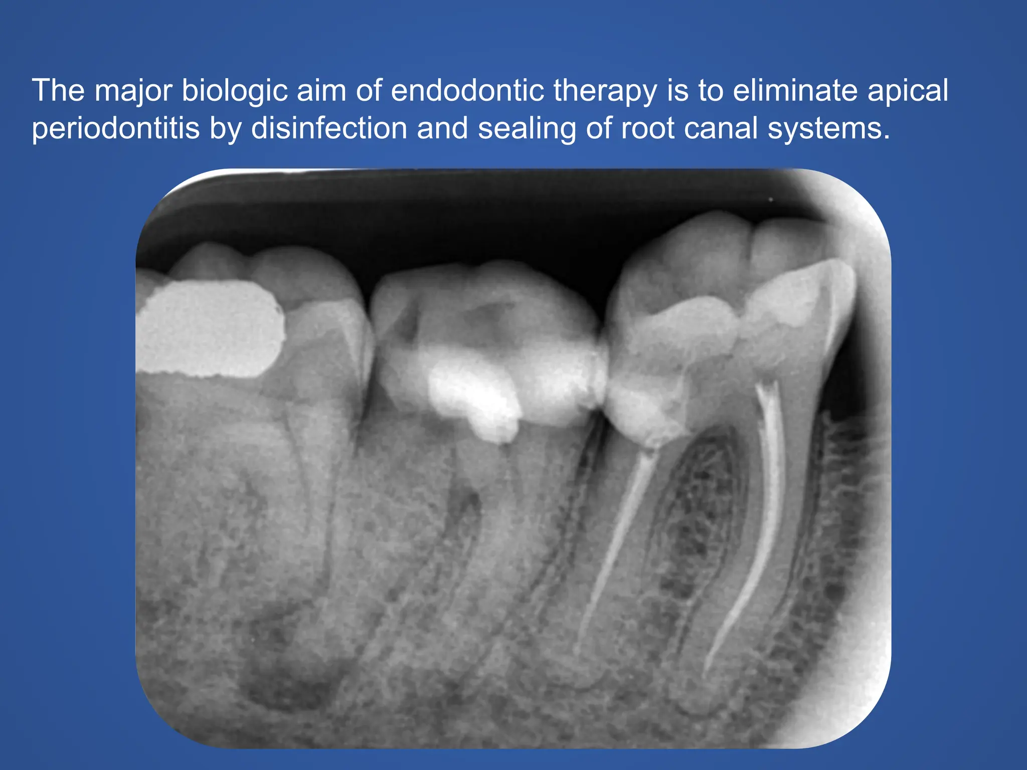
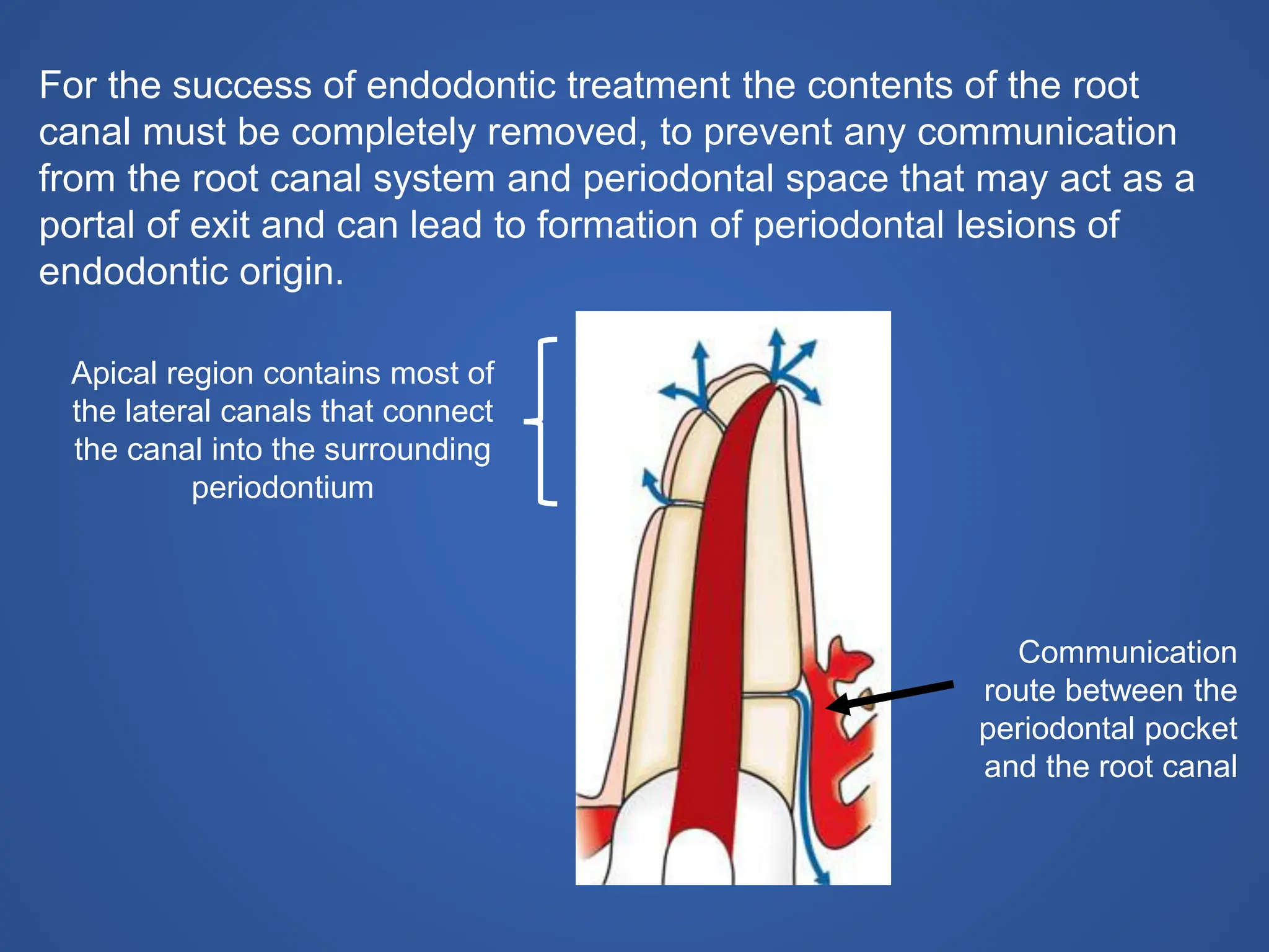
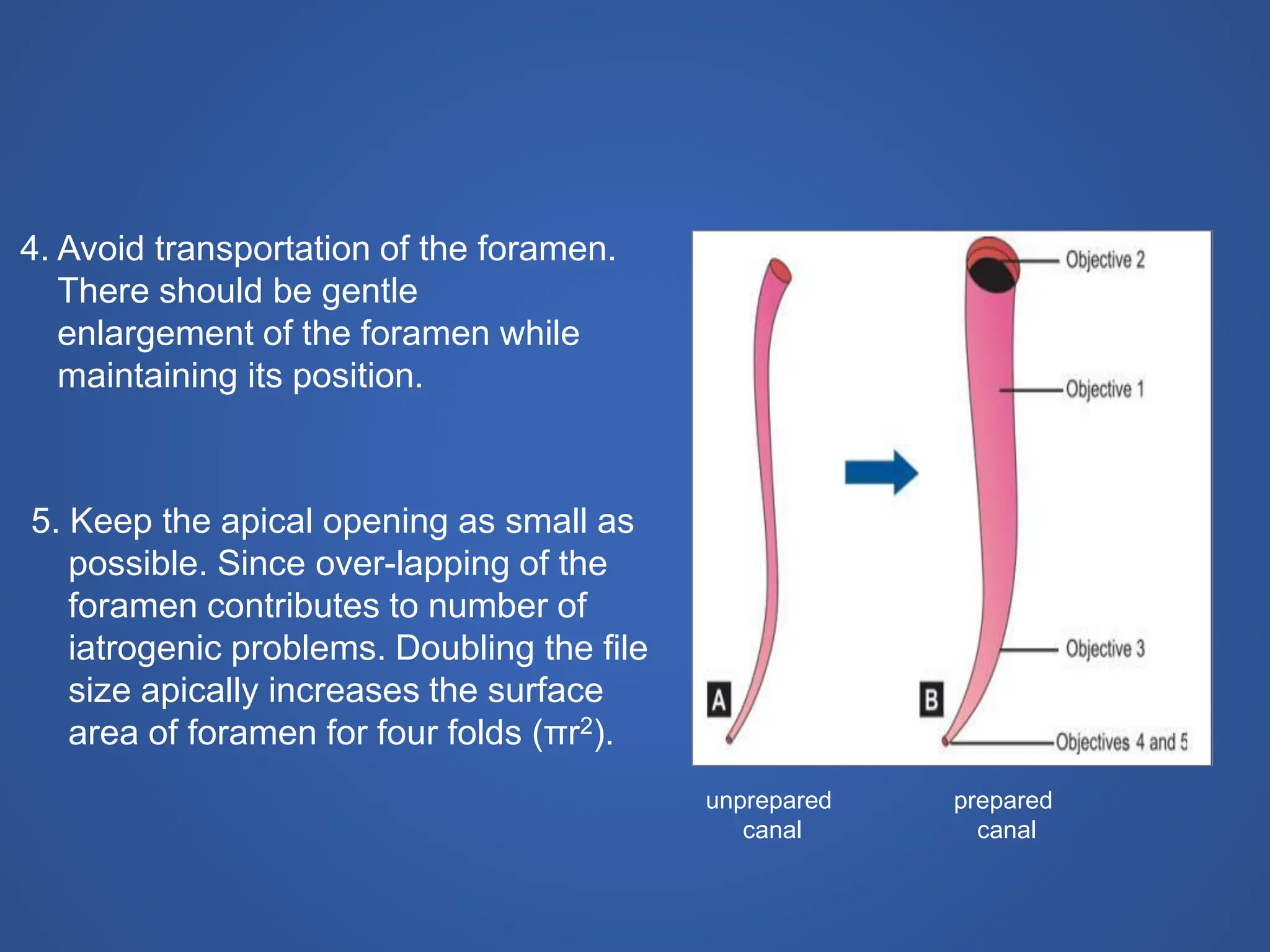
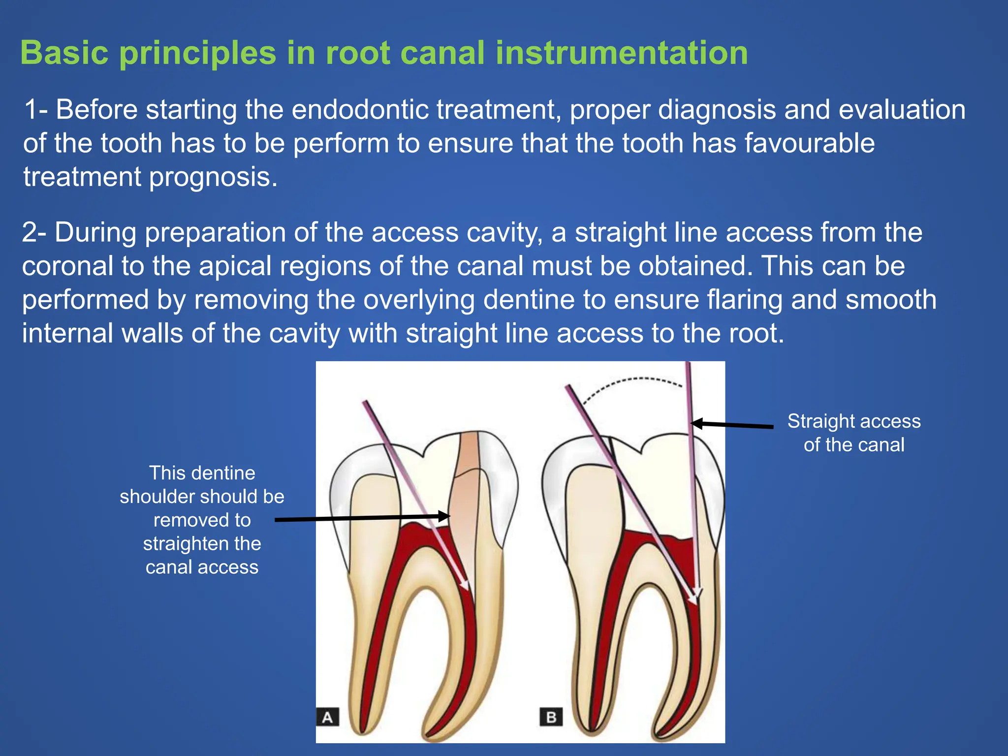
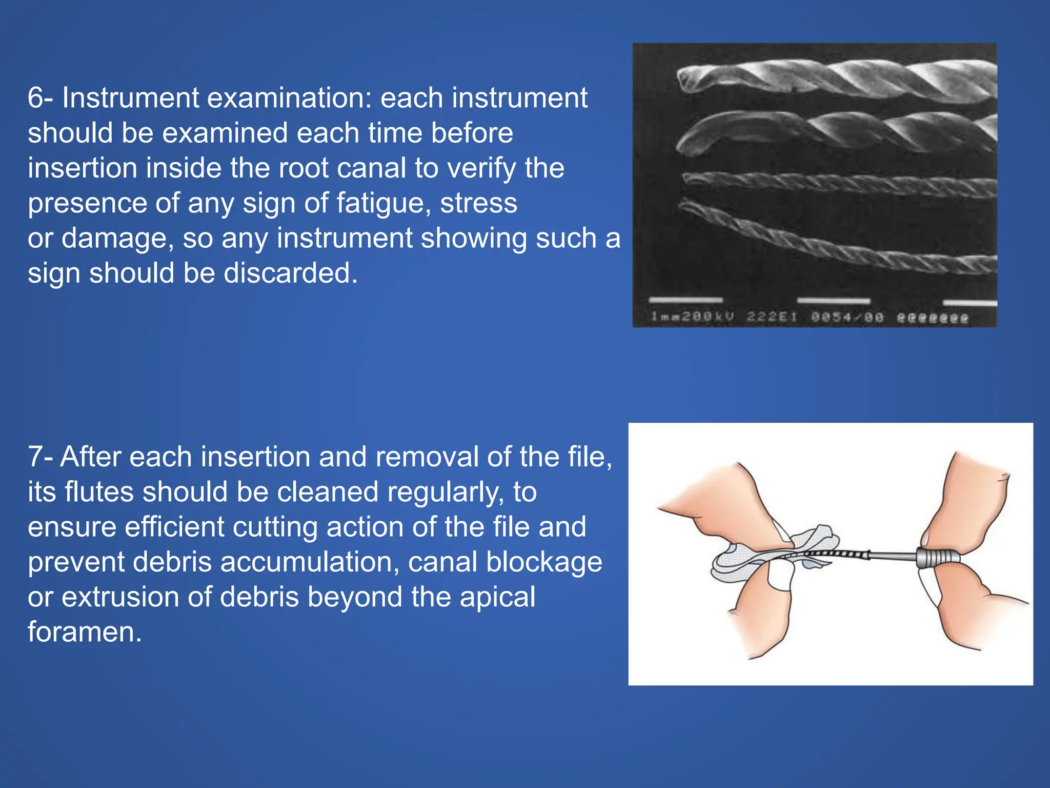
History Class XII Ch. 3 Kinship, Caste and Class (1).pptx
Lec-6-2021.pdf. ,,,,,dentistry endodontic.
1. Cleaning and Shaping of Root
Canal
Ass. Prof. Dr. ANAS F MAHDEE
BDS. MSc. Iraq
PhD. UK
2. The outcomes of this lecture
• Understand the objectives of endodontic treatment.
• Recognise the difference between cleaning and shaping of root
canal.
• Acknowledge the mechanical and biological objectives of
successful root canal therapy
• Know the basic principles of mechanical root canal
instrumentation
3. The major biologic aim of endodontic therapy is to eliminate apical
periodontitis by disinfection and sealing of root canal systems.
4. Endodontic treatment mainly consists of three steps:
1. Cleaning and shaping of the root canal system.
2. Disinfection of the canal system.
3. Obturation and 3D closure and seal of the complete canal
space.
Cleaning and shaping is one of the most important step in the
root canal therapy for obtaining success in the root canal
treatment.
5. Cleaning
It comprises the removal of all potentially pathogenic contents from
the root canal system including the necrotic pulp tissue, dentine
debris and microbes.
Shaping
The establishment of a specifically shaped cavity which maintain the
original shape and taper of the canal and allow three-dimensional
progressive access into the apical part to permit the final obturation
instruments and materials to fit easily.
6. For the success of endodontic treatment the contents of the root
canal must be completely removed, to prevent any communication
from the root canal system and periodontal space that may act as a
portal of exit and can lead to formation of periodontal lesions of
endodontic origin.
Apical region contains most of
the lateral canals that connect
the canal into the surrounding
periodontium
Communication
route between the
periodontal pocket
and the root canal
7. Schilder in early 1960s described 5 mechanical and 4 biological objectives
for successful root canal therapy.
Mechanical Objectives
1. The root canal preparation should
develop a continuously tapering cone.
This shape mimics the natural canal
shape.
2. Making the preparation in multiple
planes which introduces the concept
of “flow”. This objective preserves the
natural curve of the canal.
3. Making the canal narrower apically
and widest coronally. To create a
continuous tapers up to apical third
which creates the resistance form to
hold gutta-percha in the canal.
unprepared
canal
prepared
canal
8. 5. Keep the apical opening as small as
possible. Since over-lapping of the
foramen contributes to number of
iatrogenic problems. Doubling the file
size apically increases the surface
area of foramen for four folds (πr2).
4. Avoid transportation of the foramen.
There should be gentle
enlargement of the foramen while
maintaining its position.
unprepared
canal
prepared
canal
9. The Biological Objectives are:
1- Confinement of instrumentation within the root canals only.
2- Ensure not to force necrotic or instrumentation debris
beyond the apical foramen.
3- Optimum debridement of the root canal space.
4- Creation of sufficient space for intra-canal medicaments.
10. 1- Before starting the endodontic treatment, proper diagnosis and evaluation
of the tooth has to be perform to ensure that the tooth has favourable
treatment prognosis.
Basic principles in root canal instrumentation
2- During preparation of the access cavity, a straight line access from the
coronal to the apical regions of the canal must be obtained. This can be
performed by removing the overlying dentine to ensure flaring and smooth
internal walls of the cavity with straight line access to the root.
This dentine
shoulder should be
removed to
straighten the
canal access
Straight access
of the canal
11. 3- Ensure glide path of the canal and
apical patency before starting canal
preparation. This can be performed by
passing a small size K-file (usually a
size #8 or #10) beyond the apex. The
glide path file can help to ensure
complete opening of the canal and
facilitate working length estimation
4- Precurved instrument: In case of a
curved canal, the instrument should
be precurved to estimate the
curvature of the canal. This is true
only in case of stainless steel
instrument, but nickel titanium
instrument is flexible and cannot be
curved.
Various precurved, stainless steel glide-path file.
Compare the curves in the instruments to the
ones in a plastic training block
12. 5- The use of intracanal irrigation solutions
that serve many advantages:
• Dissolving and flushing out of the debris
from the root canal,
• Lubrication for the cutting motion of the
files within the canal,
• Antimicrobial activities.
The most popular intra-canal irrigation
solution is Sodium hypochlorite (NaOCl)
2.5-5.25%.
The irrigation solution can be delivered
inside the canal by using hypodermic
syringe.
13. 6- Instrument examination: each instrument
should be examined each time before
insertion inside the root canal to verify the
presence of any sign of fatigue, stress
or damage, so any instrument showing such a
sign should be discarded.
7- After each insertion and removal of the file,
its flutes should be cleaned regularly, to
ensure efficient cutting action of the file and
prevent debris accumulation, canal blockage
or extrusion of debris beyond the apical
foramen.
14. 9- Use of instruments in sequential order. Root
canal preparation is done gradually by using
successively larger files (never skip any size of
instrument) e.g. size 20 followed by size 25
then 30 and so on, but not size 20 then size 30.
8- Never force the instrument in the canal. Forcing or continuing to rotate
an instrument while its bind to the canal wall may break the instrument.
10- Over preparation and too aggressive over enlargement of the curved
canals should be avoided.
11- Creation of an apical stop should be maintained during instrumentation
procedures by maintaining the apical size as clean and small as possible.
Over enlarging of the apical foramen should be avoided because it destroy
the apical stop and cause apical extrusion of the irrigation and obturation
material and cause failure of endodontic treatment.
15. Manual or Hand instrumentation techniques
Standardized Technique
Step-Back Technique
Step-down Technique
Balanced Force Technique
Crown Down Technique